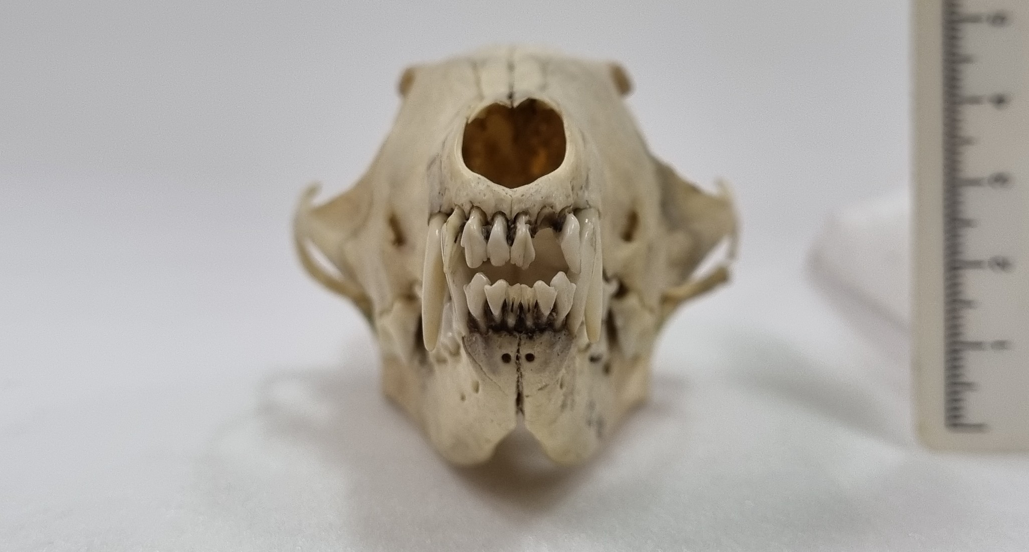
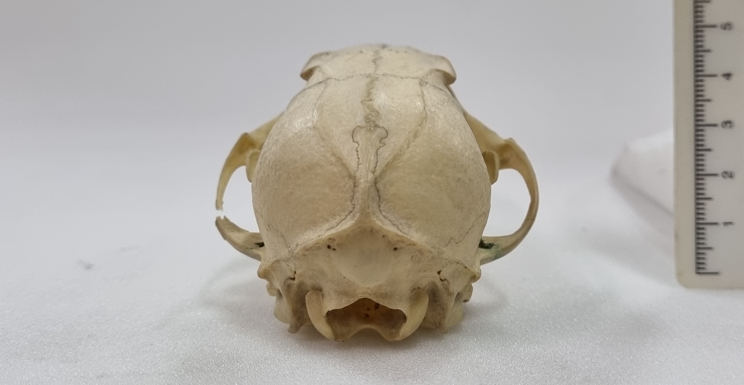
Last week I gave you this unidentified skull from the collections of the Dead Zoo:
I say it was unidentified, but in a strange quirk of coincidence I actually did identify this skull three years ago, just a month before the start of the Covid Pandemic (which might explain why I never had a chance to update the record).
I think the specimen is most likely to be the skull of a White-nosed Coati Nasua narica (Linnaeus, 1766), for the reasons I outlined back in 2020. So well done to Leon and Chris for recognising this cousin of the Raccoon from South and Central America and the southern parts of some North American states .
Coatis have quite distinctive upper canine teeth, that look almost like short tusks. These are useful for defence from other Coatis, but they are not very well adapted for subduing larger prey. This isn’t really a problem for Coatis, since they mainly feed on invertebrates, fruit and small vertebrates that they undcover during their energetic foraging.
So I apologise to everyone for repeating a specimen – this is the first time this has happened (and hopefully the last)!
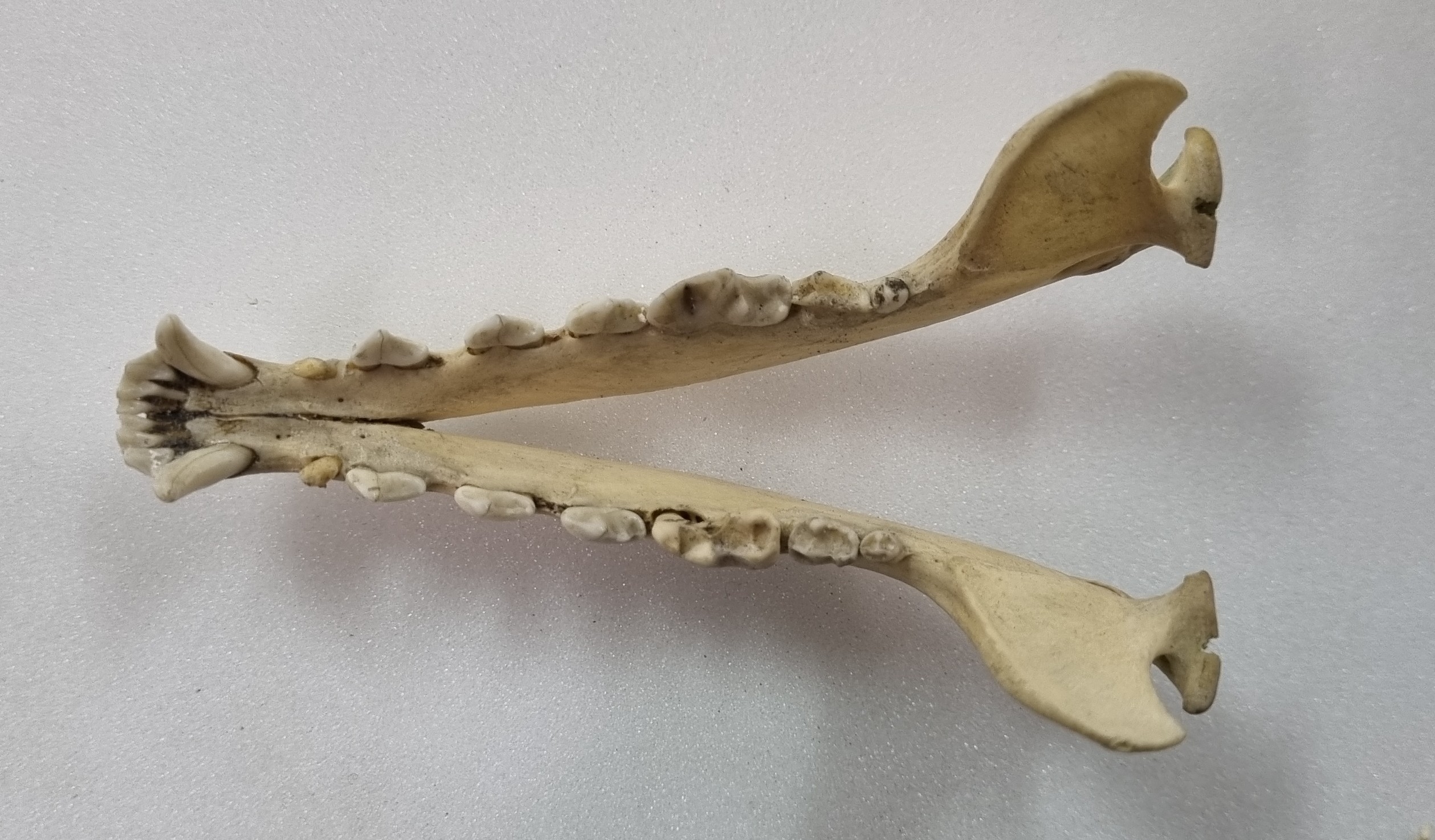
This week I’m continuing with skulls from the collections of the Dead Zoo, and this one was sitting in the “Unidentified” drawer:
Do you have any ideas what this might be? I suspect that some of you will be familiar with this, so perhaps it’s time for some cryptic suggestions in the comments. Have fun!
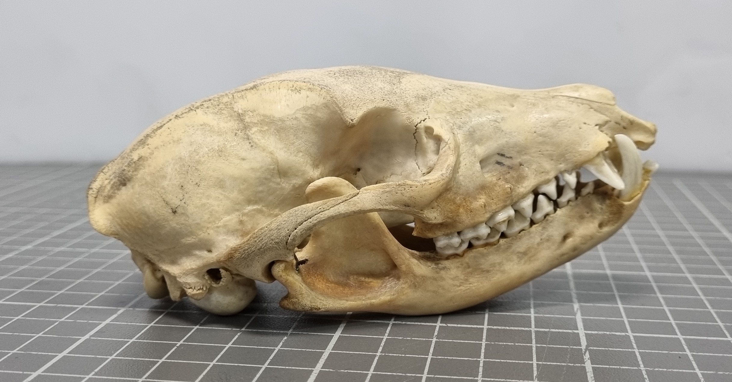
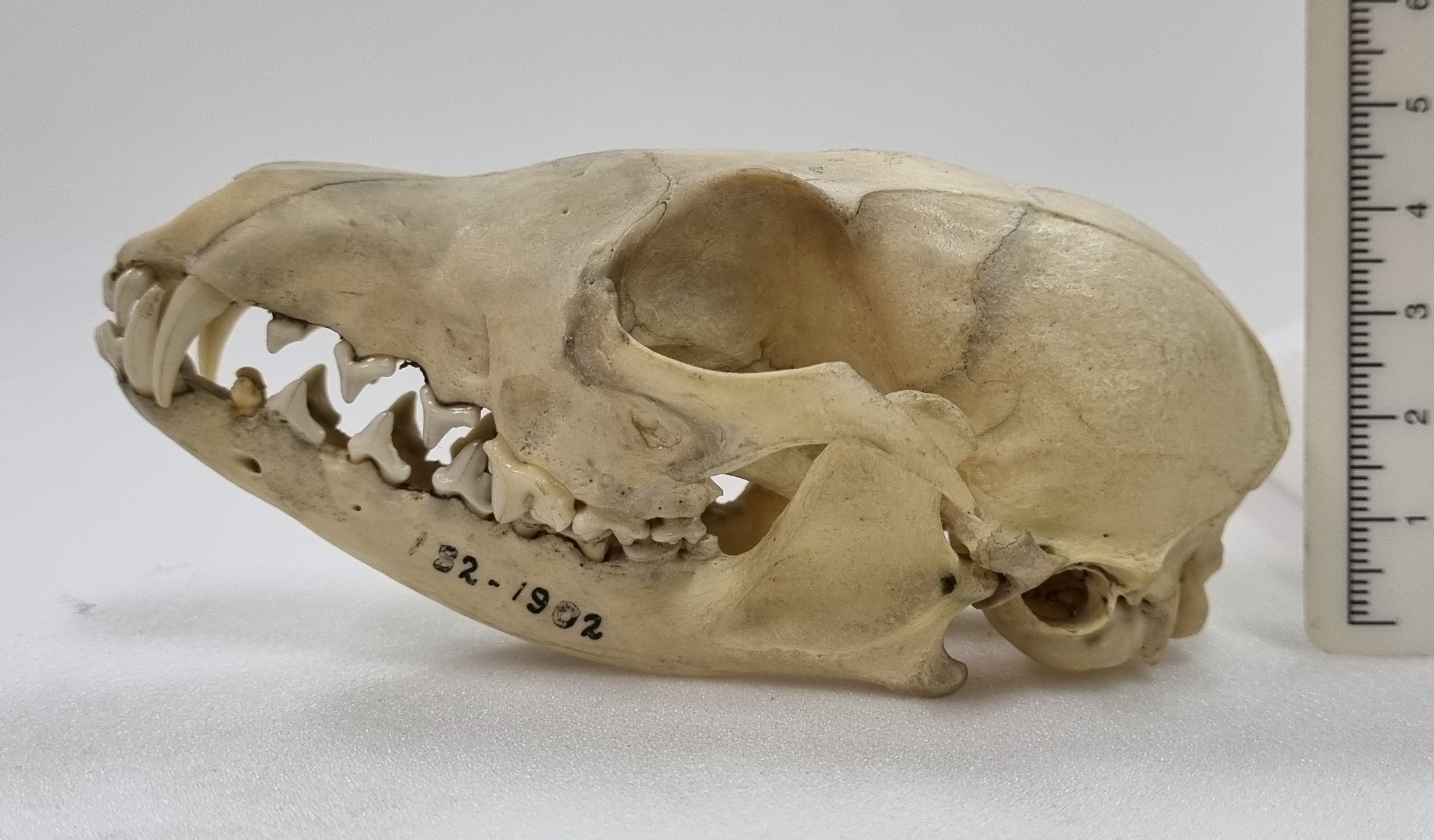
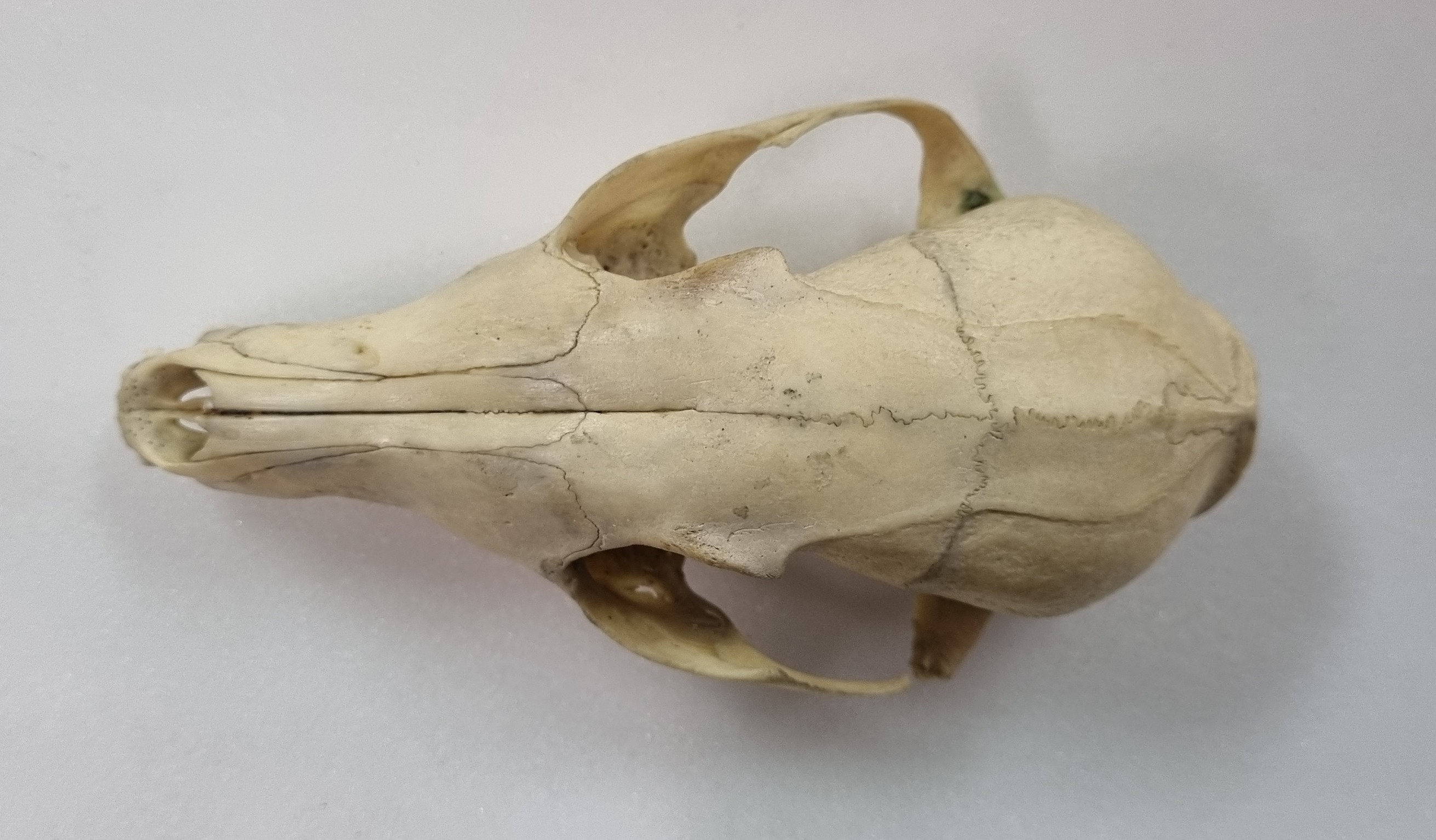
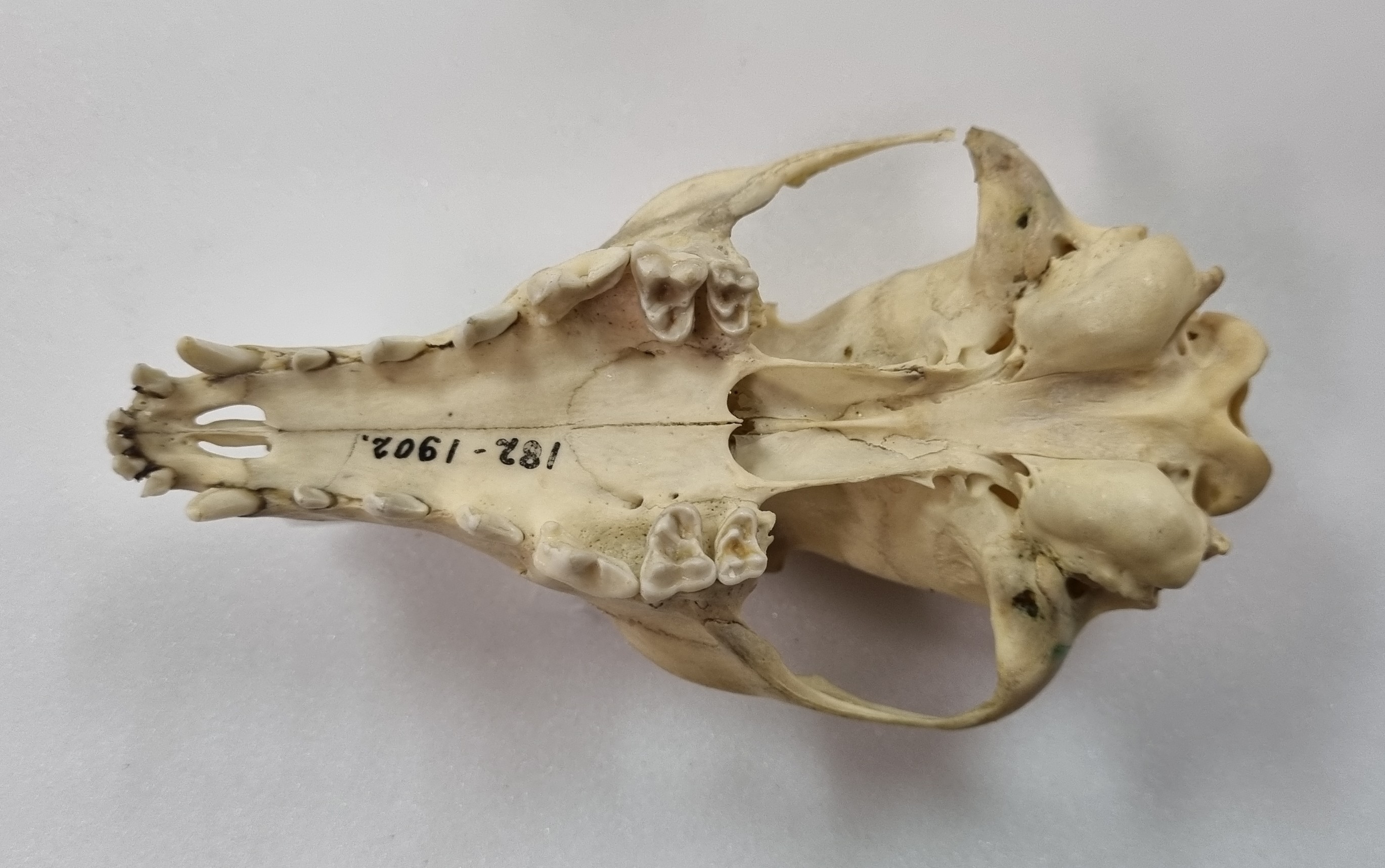
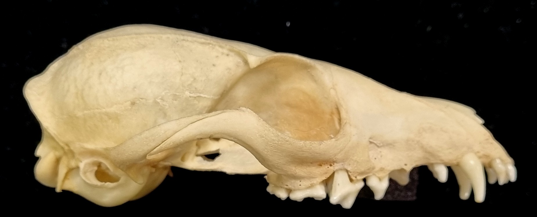
Last week I gave you this neat little skull to have a go at identifying, from research collections of the Dead Zoo in Dublin:
I wasn’t surprised that everyone in the comments worked out that this is the skull of some sort of fox, but I was equally unsurprised that nobody worked out the species. Generally speaking, most people immediately think of the near ubiquitous Red Fox or perhaps Grey Fox (or Gray Fox to our American friends), but there are plenty of others – 24 species commonly referred to as “fox” and 12 species of “true fox” in the genus Vulpes.
This particular specimen has a sagittal crest that forms a lyre-shape – normally something associated with the Grey Fox:
However, this feature can occur in other species, often in females or subadults, where the surface of the bone has not finished remodelling at the margin of the attachment of the temporalis muscles (those are the ones that connect to the lower jaw from the sides of the cranium and are responsible for the operation of the lower jaw during powerful biting).
However, in this specimen the muzzle is more tapered and the postorbital constriction is relatively broad. All of these point away from the Grey Fox.
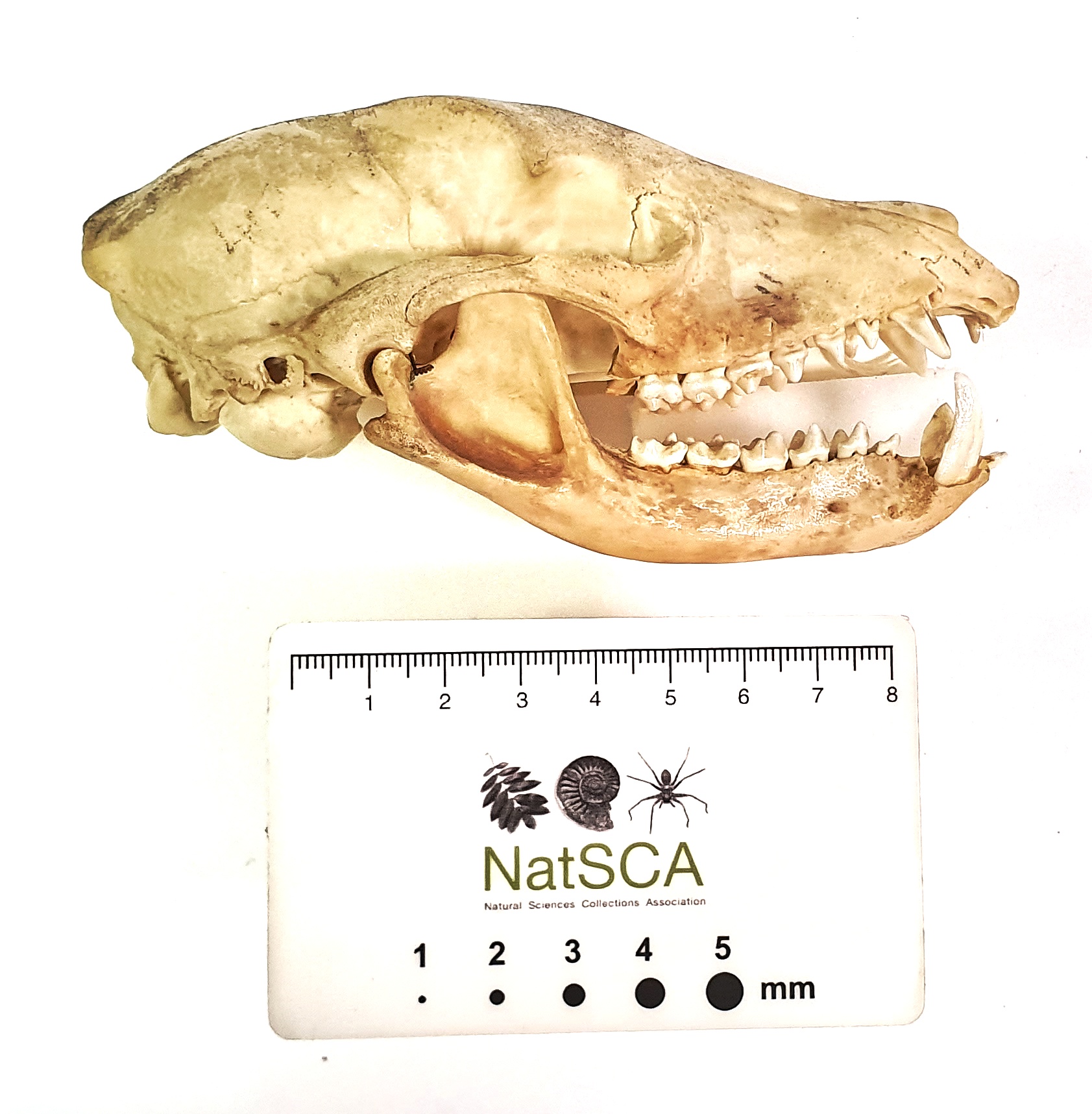
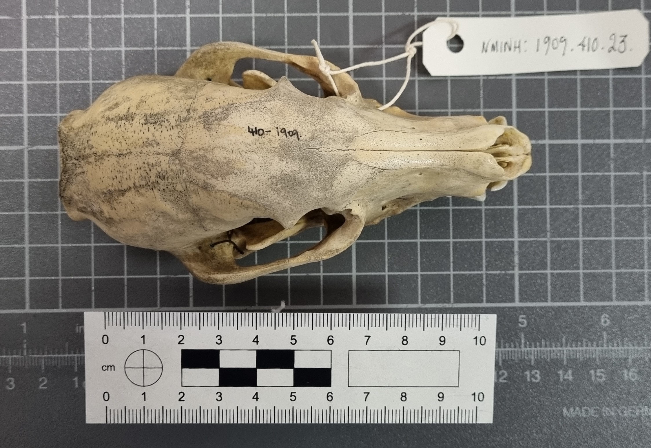
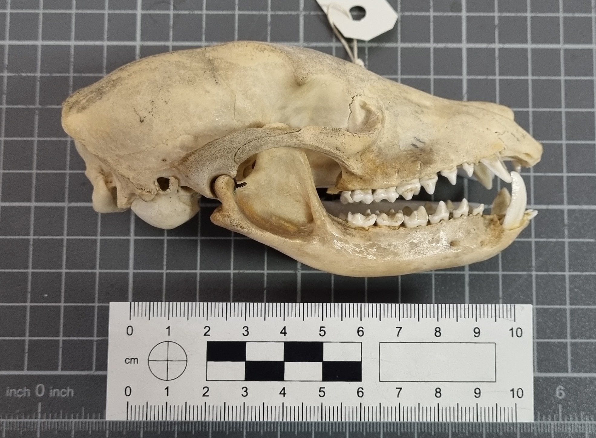
With foxes there can be a lot of similarities between the skulls of species, with all the usual compounding issues of sexual dimorphism, age and regional variation. However, size can give some clues, and things like the relative size of the external auditory meatus (also known as the ear-hole), and the shape of the auditory bulla, are useful for differentiating between species.
With a bit of patience, a bit of pattern recognition, and a resource with good images of specimens, like the Animal Diversity Web, it is possible to work out what you’re looking at.
In this case, the mystery object is a Swift Fox Vulpes velox (Say, 1823).
These North American foxes are smaller than the Red or Grey Fox, but a bit bigger than their close cousin, the Kit Fox. They live in grasslands and praries, where they prey on rodents, birds, reptiles and pretty much anything they can find – including insects, fruit and grasses.
As with many species, the Swift Fox has declined due to changing land use and the systematic persecution of predators in the first half of the 20thC. In fact, it was wiped out in Canada around this time, although it was subsequently successfully reintroduced and numbers have increased.
So watch out for those foxy skulls – there are more species to consider than you might think and they can be tricky to identify without reference resources. I hope you enjoyed this little detour down the fox hole!
For the first time in quite a while, I managed to escape from my desk and spend a little time in the collections of the Dead Zoo. The main reason was to facilitate access for researchers doing some really cool projects, but it also gave me a chance to spend a little time exploring the collections I’m responsible for.
In one of the cabinets I spotted this skull, and I thought it might make a good mystery object:
So, do you have any thoughts on what this might be? As ever, you can leave your questions, observations and suggestions in the comments section below. I hope you enjoy this specimen as much as I did!
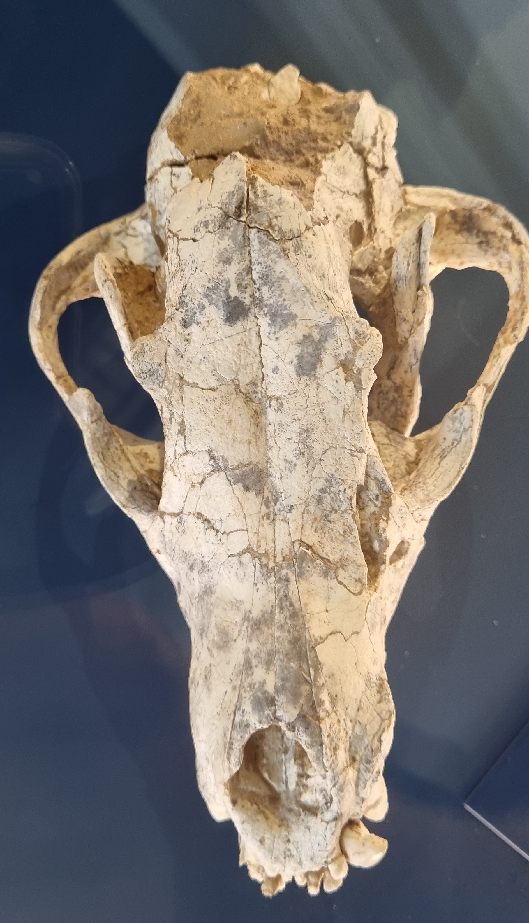
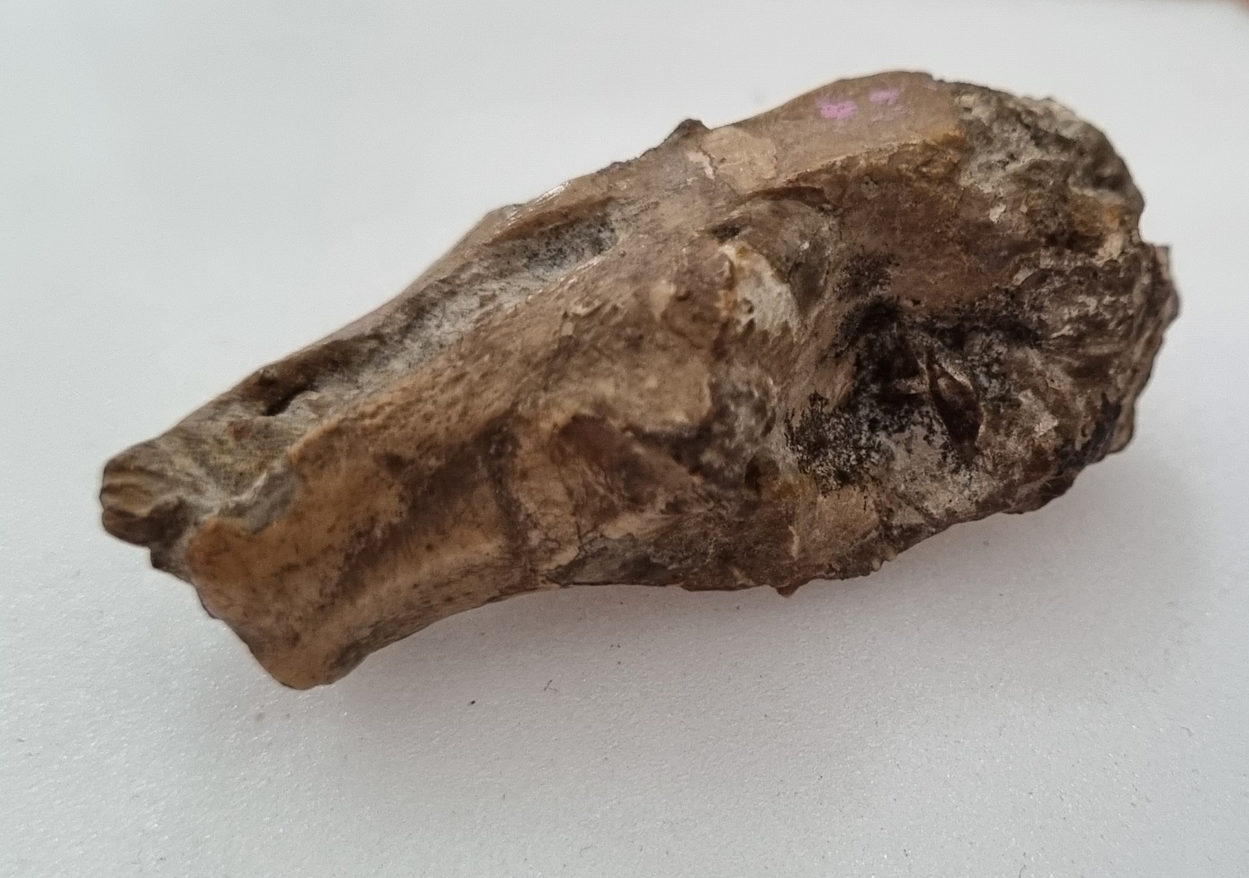
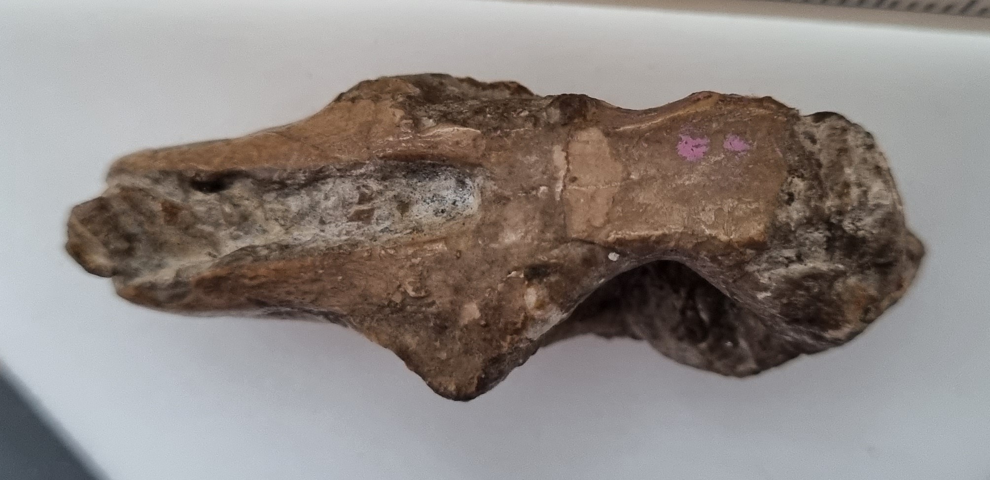
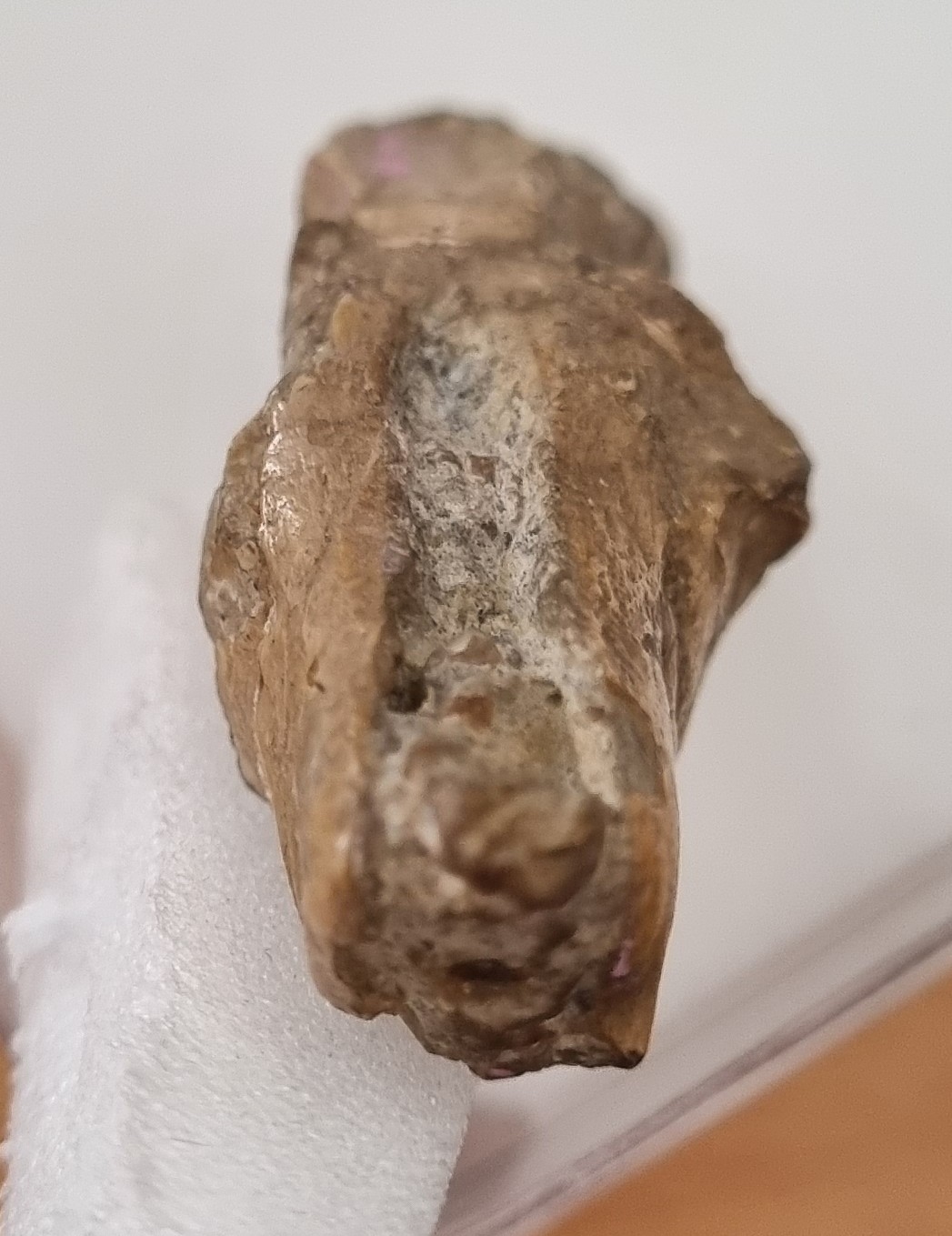
Last week I gave you this 6 million year old fossil skull to have a go at identifying:
The specimen is on display in the geology galleries of the Natural History Museum at the University of Oslo (which is well worth a vist). However, this did mean the photos provided weren’t quite as good as I’d like, particularly notable being the lack of a scale bar (sorry!)
Even without a scale, consensus shifted towards this being some kind of hyena, thanks to the curved mandible and (hint of) robust molars and shorter toothrow than you might expect to see in a canid. The broad and flat profile of the frontals between the eye sockets probably helped too:
Hyenas have an interesting evoloutionary history, branching off from the basal feliforms around 22 million years ago and adapting to fill a terrestrial carviore niche in Eurasia and becoming quite diverse. In America the canids were filling that same niche, which led to some competition when the canids made it to Eurasia (spoiler alert – the hyenas lost that competition, leaving us with just three highly specialised bone crushers and the decidedly weird Aardwolf living today).
Four live specimens of hyenas (clockwise from top left): spotted hyena (Crocuta crocuta), brown hyena (Parahyaena brunnea), aardwolf (Proteles cristata), striped hyena (Hyaena hyaena). Image by Termininja, 2020CC BY-SA 4.0
The mystery specimen was labelled as Thalassictis wongii (Zdansky, 1924), a species described from China and originally placed in the genus Icititherium, but reassessed by Kurtén (1985). A cladistic treatment of the Hyaenidae by Werdelin & Solunias (1991) later placed it in the genus Hyaenotherium, but that may not have been accepted by the curatorial team in Oslo without an accompanying formal taxonomic treatment.
These are the sorts of decisions that need to be made when considering something as simple as a label stating a species name, so you can imagine my sense of trepidation as we are about to embark on a major project at the Dead Zoo, which will allow us to reassess the information with our 10,000 or so display specimens. Fun times ahead!
This week I have a mystery object for you from a recent visit to Oslo:
I apologise for the poor image quality – I was using my phone and this specimen was behind glass, so it was tricky getting a decent photo without a lot of reflections. Given the poor images I’ll drop in a clue – this is a fossil specimen from around 6 million years ago.
Any thoughts on what it could be? As ever, you can leave a comment down below. Have fun working this one out!
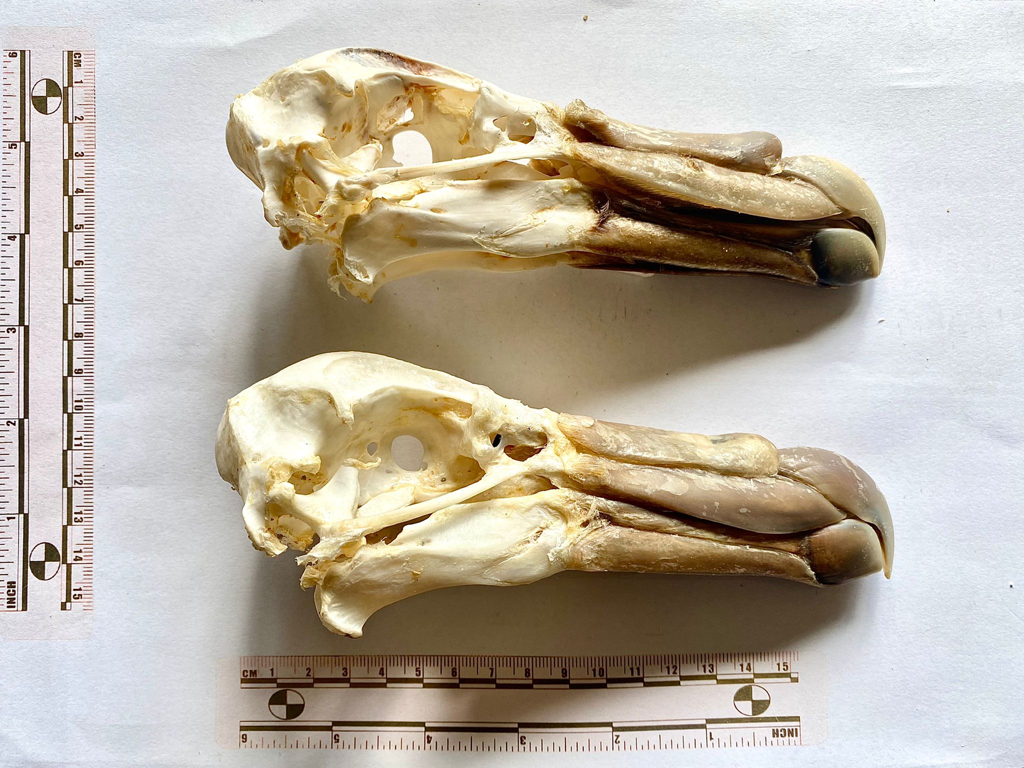
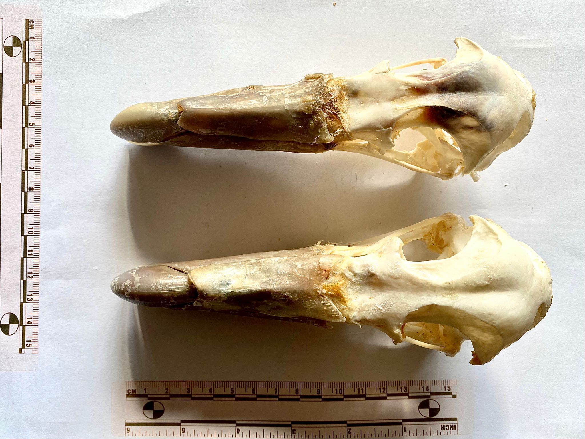
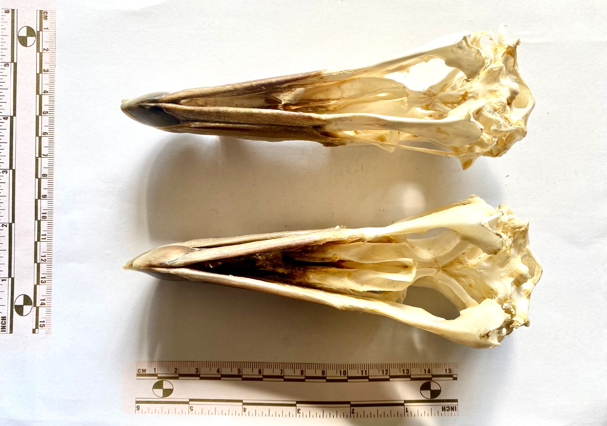
Last week we had these two skulls from Andy Taylor, FLS to have a go at identifying:
Everyone recognised that these are the skulls of Tube-nosed birds in the Order Procellariiformes – very large Tube-nosed birds.
Usually, you’d think of albatrosses when considering large Procellariiformes, but they have proportionally longer bills than this and while they have the nose-tube characteristic of the group, the tube is quite small and to the sides and rear half of the bill. In the mystery specimens, the tube is large and located on the top, and in the front half of the bill.
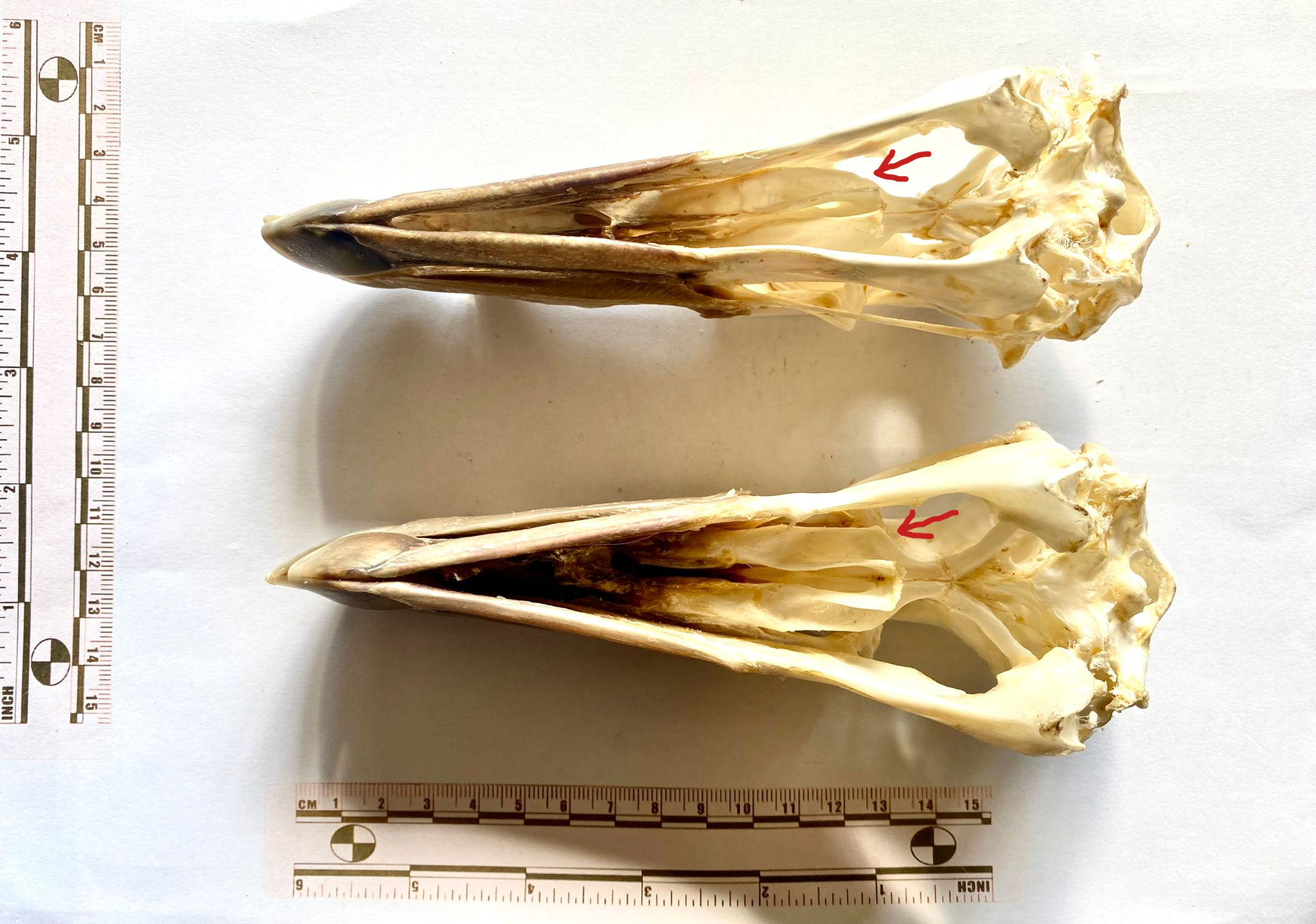
As Wouter van Gestel recognised, these skulls are from Giant Petrels in the Genus Macronectes. They actually represent both species in the Genus – the top one is the Southern Giant Petrel Macronectes giganteus (Gmelin, 1789) and the lower one is the Northern Giant Petrel Macronectes halli Mathews, 1912.
They’re quite hard to tell apart, and the best feature I noticed for distinguishing them is the shape of the palatine, with the Southern having a very gentle curve to the rear section – as indicated below (in a very rudimentary way):
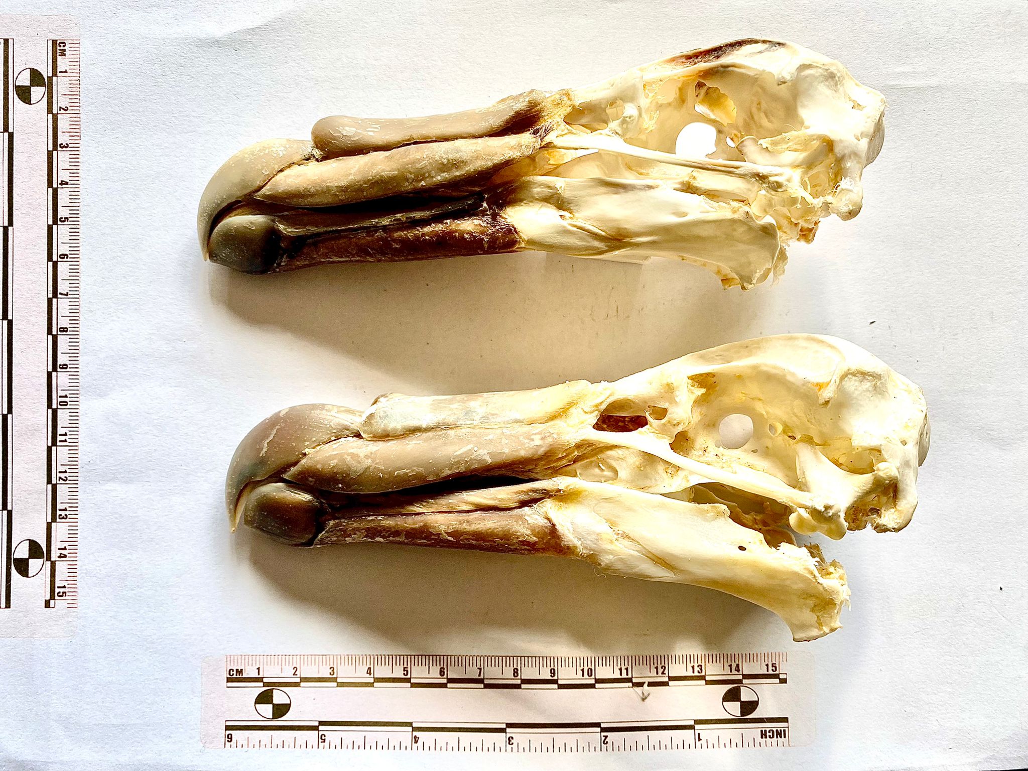
This week I have a guest mystery object – or two – for you to test your skills on:
I’d love to hear your thoughts on the identification of the skulls here – keeping in mind that any differences could be due to individual variation, sexual dimorphism or they may even be different species. Looking forward to hearing your thoughts!
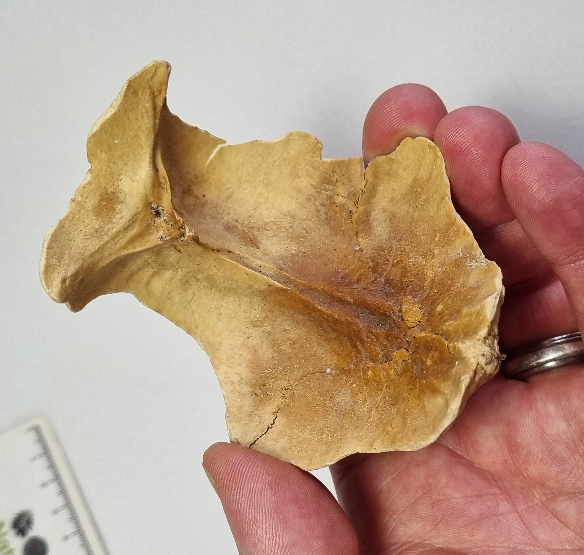
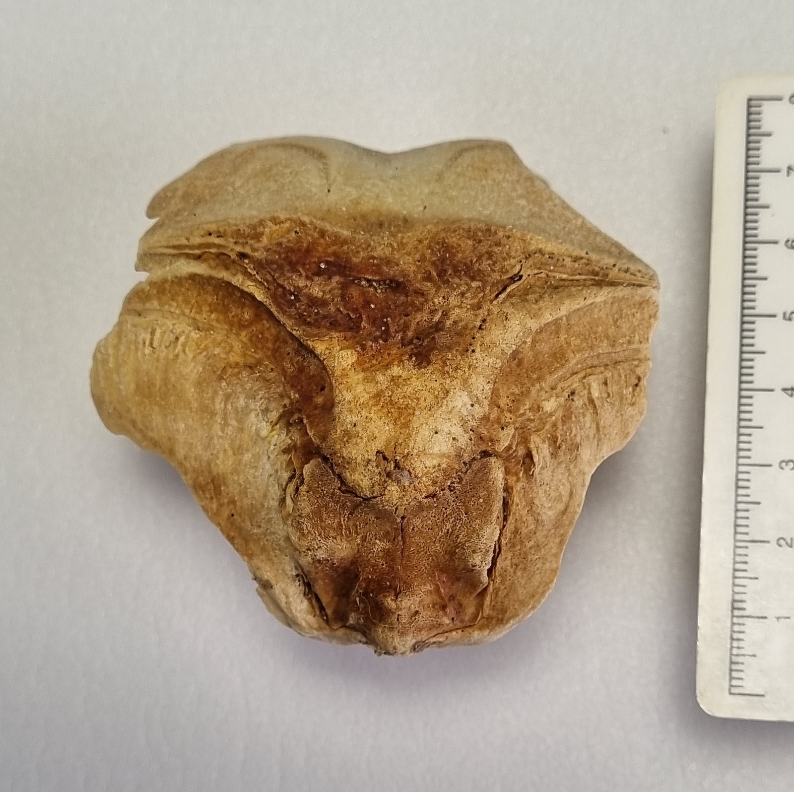
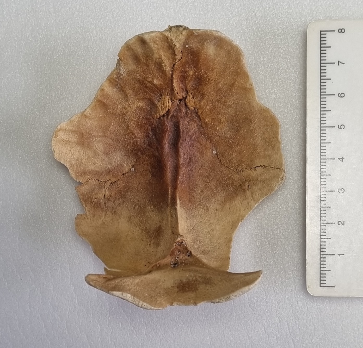
Last week, I gave you this devilishly difficult genuine mystery object to have a go at identifying:
At first glance, it looks like it should be the occipital (the bone at the very back of the skull) of an Ostrich, or other very large bird. The bone is thin and dense (typical for a bird) and the overall shape and size looks like it might fit. However, none of the details of the bony sutures fit that possibility, for any large bird. Also, this came in as an enquiry, and was almost certaily found in Ireland, making a big bird even less likely,
With birds ruled out, I looked into the mammals. Generally it’s helpful to start with common species, to start ruling out the more frequently encountered species. There are some unfused sutures, so I began with looking at some common large mammals, keeping in mind the developmental differences that occur, making the skulls of juveniles appear quite different to adults of the same species. This is especially the case in relation to skull shape and presence of unfused sutures that can vanish in adults.
Sticking with the occipital, since the shape looks right and several people converged on the same idea (although the species suggested varied quite considerably), for me, the nuchal crest (the area of bone where the ligaments for the neck muscles attach to the back of the head) is very similar in shape to that of a sheep:
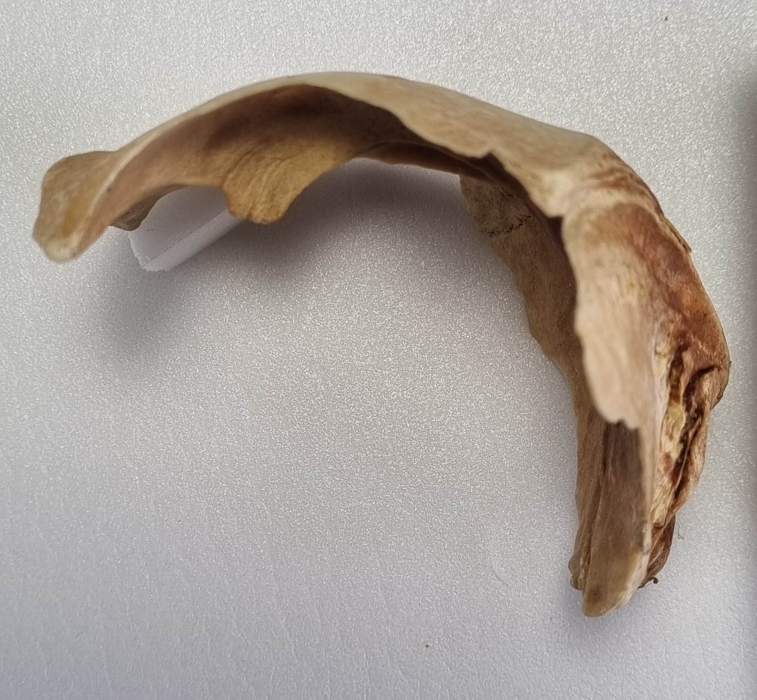
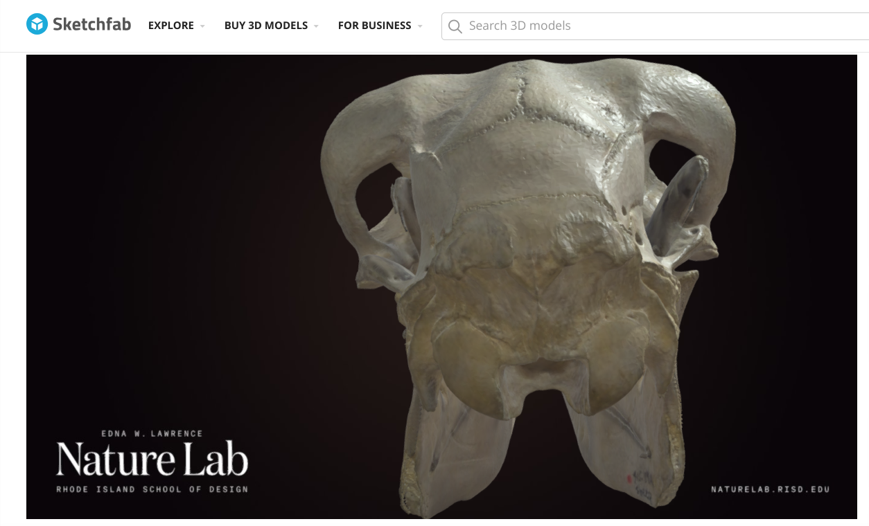
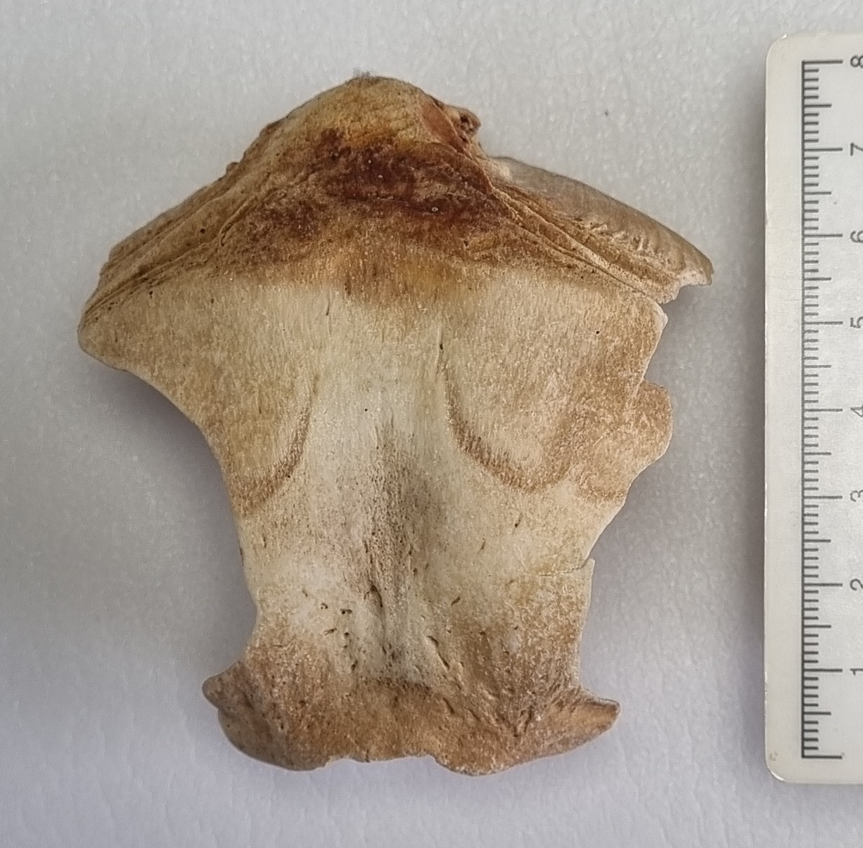
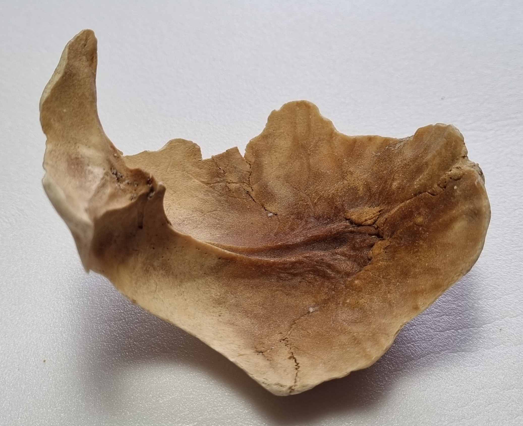
This would have been a nice and simple way to wrap things up, but unfortunately I’m still unsure. Mainly this is because the shape doesn’t match so well from other angles:
Of course, this may simply be an artefact of comparing a juvenile animal skull to an adult – so I’ll need to check with a range of specimens of different ages to be more certain.
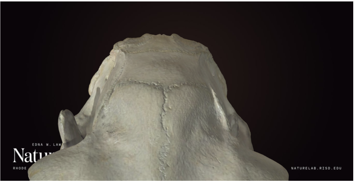
However, there was also a suggestion of Porpoise (or other cetacean) by Adam Yates and Kat Edmonson came up with an intriguing suggestion that I am quite taken by. It is possible that the raised region is not the nuchal region at all (in Porpoises and many other cetaceans there’s actually a depression rather than a raised ridge in that area of the back of the skull), it may actually mark the junction between two very short nasal bones, a very compressed frontal region and the occipital at the back of a cetacean skull:
So it may be that I was looking at the bone upside down the whole time. I’ll need to do some more comparisons to narrow down species if that is what it is, but huge thanks to Kat for getting me to see this object from a new perspective!
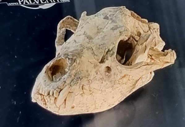
Last week I gave you this really difficult, but incredibly cool mystery object to identify:
Definitely not a simple one for the uninitiated, but most of you got impressively close.
It looks a bit like ancient chewed gum at first glance (hey, it’s a thing!):
A nearly 6,000 year old piece of chewing gum, found in Scandinavia, perfectly preserved the DNA of its chewer. She was a young, hunter-gatherer girl, with dark skin, brown hair & blue eyes, who had recently eaten a meal of hazelnuts & duck. Archaeology is amazing. pic.twitter.com/kfav9PkonE
However, on closer inspection, some of the features start to emerge – including teeth:
Obviously, this is the fossilised skull or some critter, but what kind of critter is harder to determine.
The length suggests it’s something about the size of a rabbit:
And if you’re looking for a good fossil rabbit, you can’t beat Palaeolagus:
Palaeolagus skull. Image by Smithsonian Institution, 2019
NotPalaeolagus skull.
As you can see, the mystery object has a few differences, but due to the various missing parts, it’s a little hard to be confident exactly how different they are – although the shape of the orbital margin (the front of the eyesocket) gives a bit of a hint.
But, even more useful, is the curve in the maxilla (the upper jaw bone) that traces the root of the first incisor. In lagomorphs (rabbits, hares and even pikas), the incisor roots terminate with quite a big gap before the orbital margin, often with a triangular fenestrated region of cancellous bone (a sort of window of bony struts) in between.
The mystery specimen doesn’t have that – in fact the end of the incisor root is very close to the orbital margin. This is something you see in rodents.
I would have been impressed if you got that far, since the overall shape and size of this specimen definitely gives off a rabbity vibe, but believe it or not, this a dormouse. More specifically, it’s the Gigantic Dormouse Leithia miletensis (Adams, 1863) or if you want to go with the commonly used and more technically accurate, but nomenculatorily incorrect, L. melitensis, since Adams made a spelling error in his original description.
In fact, this is one of the specimens collected and figured by Adams in that original work describing the species, making this part of the type series for the species (although the holotype is more likely to be a very well preserved half mandible from the same site).
The fact that this is a fairly large and intact part of the type series means that it is of great interest to researchers. The reason I had this specimen to hand for the mystery object, is because I was preparing it for a research loan to some of my old colleagues in UCL, where it’s being MicroCT scanned.
This research will help refine an understanding of the morphology of the Gigantic Dormouse and offer some clues to what happens on islands that leads to the development of giants, building on work that they’ve been doing on this fascinating species, which is an interesting read that you can find here (you may even recognise Fig. 1B).
Virtual Cranial Reconstruction of the Endemic Gigantic Dormouse Leithia melitensis (Rodentia, Gliridae) from Poggio Schinaldo, Sicily, By Jesse J. Hennekam , Victoria L. Herridge, Loïc Costeur, Carolina Di Patti, Philip G. Cox – CC BY 4.0, https://commons.wikimedia.org/w/index.php?curid=92037373
This week I am have a great guest mystery object from Andy Taylor for you to have a go at identifying:
Image by Andy Taylor, 2023
Image by Andy Taylor, 2023
Image by Andy Taylor, 2023
Image by Andy Taylor, 2023
Here’s what Andy says about the specimen:
On Sunday, myself and Sophie Bagshaw were working through specimens that were donated to me from a person who had been given them by a zoological park. The specimen in question was part of a huge shipment of almost 140 frozen specimens that were in various states of preparation and were mostly head specimens. … I have a large rodent skull that I’m struggling to ID
Andy and Sophie have been doing great stuff with osteology for educational purposes for a while now, so it was a real pleasure to get a question like this, and it seems like a perfect opportunity for the community here to add their thoughts.
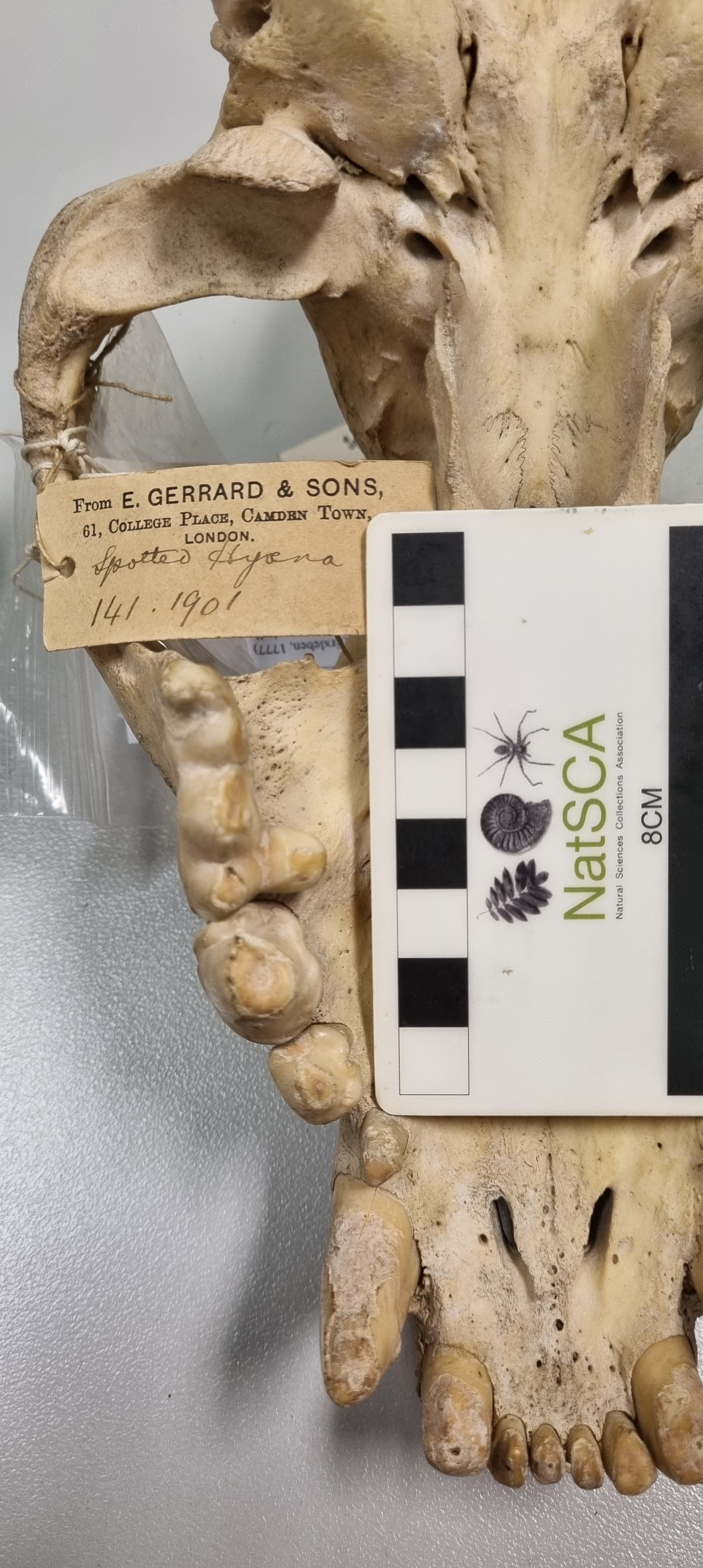
Last week I gave you this skull from the collections of the Dead Zoo, which had been misidentified and that came to light when Dr George Argyros was doing some research on the carnivore skeletons:
The label attached to this specimen indicated that this is the skull of a Leopard, which is clearly wrong. The label also identified the specimen as having been collected in East Africa by Major A.W.V. Plunkett.
Labels like this worry me. Not because they contain a misidentification, but because they may indicate that someone in the past has mixed up the specimen labels. This is a much bigger problem than a simple misidentification, as it can mean the real specimen has become dissociated from its information.
The huge, robust teeth of this specimen should make it fairly clear that it belongs to one of those specialist bone-crushers – the hyenas:
However, there are three species of hyena to choose from (I’m leaving the Aardwolf of this, since they don’t match this dental morphology even remotely).
My first thought was that this specimen is on the small side for a Spotted Hyena:
Striped Hyena on left, Spotted Hyena on right
Size is seldom a definitive feature, especially in species that display sexual dimorphism, but what is more useful is the detail of the tiny molar at the back of the maxillary toothrow. This is absent in Spotted Hyenas, but it occurs in both Striped and Brown Hyenas.
So you might ask, how do we distinguish between Striped and Brown Hyenas? This is a good question. For starters, it’s hard to find enough reliable good images of the Brown Hyena’s skull online that show the details needed to distinguish between the species.
However, a bit of searching highlighted that the Brown Hyena has a shorter and more robust angular process of the mandible than the Striped – and the mystery object.
Image of Brown Hyena skull by David J. Stang, 2005.
This long angular process was spotted by katedmonson, but Adam Yates was the first with the identification of Striped Hyena Hyaena hyaena (Linnaeus, 1758).
This one proved a little trickier than I thought at first, due to the similarities between the Brown and the Striped species. But I’m a little relieved that the consensus fell on Striped, both here on the blog, and between myself and George, since the Striped Hyena is found in East Africa, whereas the Brown is limited to South Africa.
This at least agrees with the locality on the label, so it may well have simply been misidentified when the specimen was acquired – especially since it looks like it was skeletonised naturally, so it may have been found dead and already defleshed, making it harder to identify.
Since everyone seemed to have fun with last week’s mystery skull, I have another that was misidentified in the Dead Zoo’s collections and which came to light during Dr George Argyros’ recent research visit:
Do you recognise this species from its skull?
As usual, you can ask questions or leave suggestion in the comments box below. If you do know what it is, then please try to keep your answers cryptic, so everyone can have a go at working it out. Have fun!
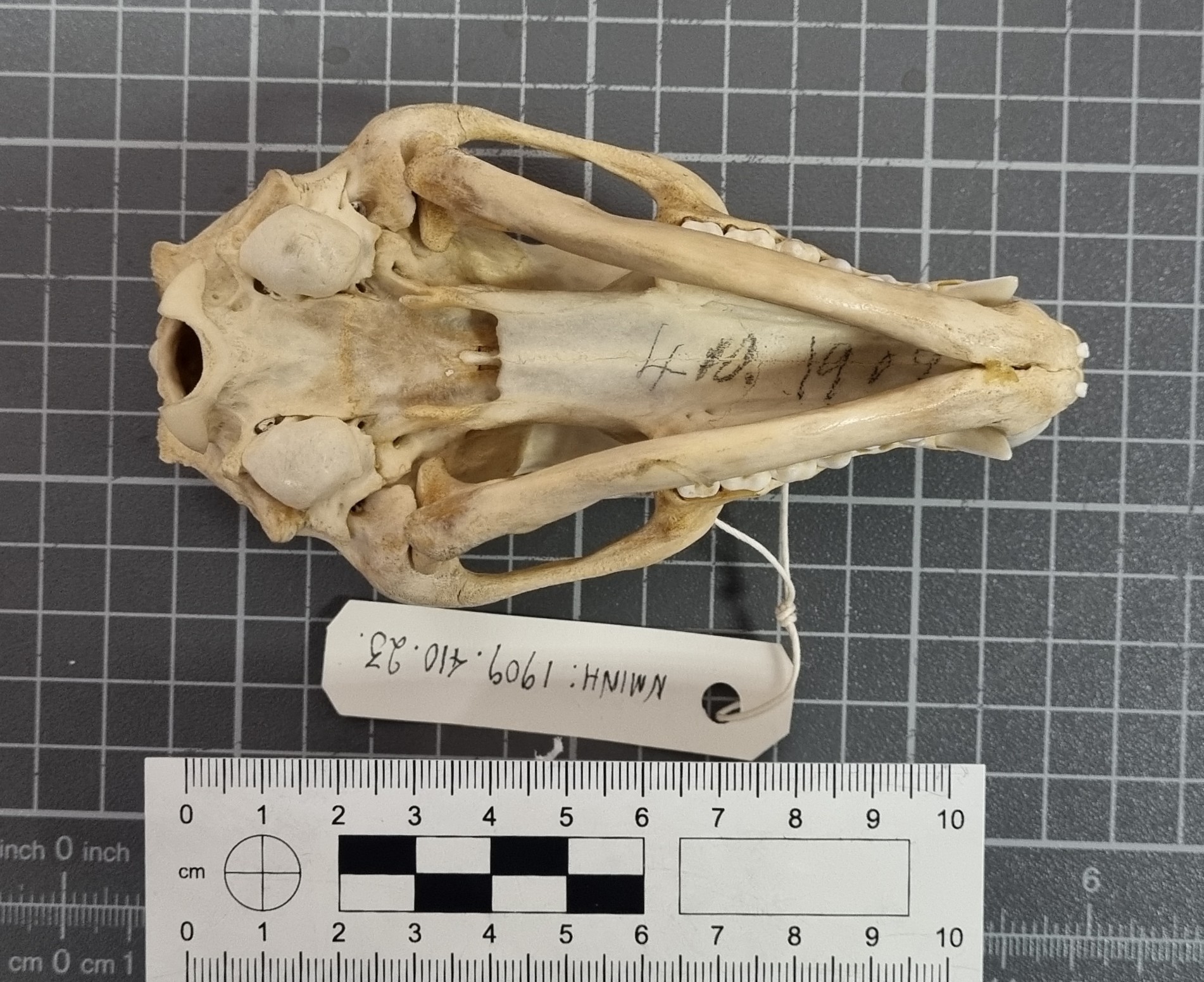
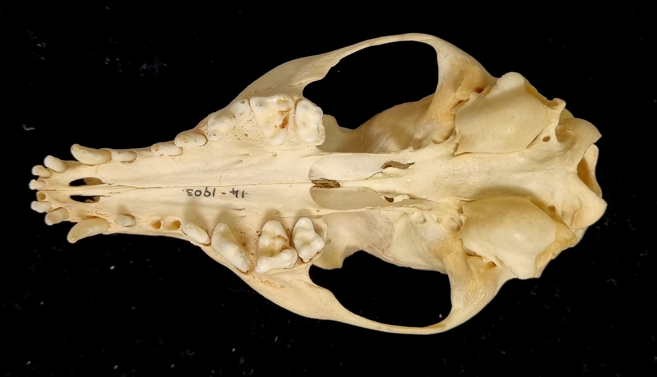
Last week I gave you this skull from the collections of the Dead Zoo to have a go at identifying:
This specimen came to light during some research being carried out on carnivore bones by Dr George Argyros, a Professor visiting us from Emory & Henry College, Virginia. It was identified as Vulpes on the label, but both George and myself were doubtful.
The specimen’s spurious identification can be tracked back to when it was named in the Museum’s register as Vulpes fulva argentata or Silver Fox. This identification was assigned to the specimen when it was given to the Royal Zoological Society by N.H.P. Vickers in March 1900 (see page 127 of the monthly Irish Naturalist covering March 1900):
The Museum bought the specimen in skeletal form from the Royal Zoological Society of Ireland in 1903 and the name Silver Fox was kept until a later review of the taxonomic hierarchy in our database, which ‘corrected’ the name to Red Fox Vulpes vulpes.
However, this name change was not based on the morphology of the specimen. The characteristic lyre-shaped sagittal crest1 immediately made both myself and George think Urocyon and the small size of the specimen made both of us converge on an identification of Island Fox Urocyon littoralis (Baird, 1857) after independent bouts of measuring.
So I offer a hearty congratulations to everyone who spotted that this skull is from the genus Urocyon, although I think most people were thinking of the Grey Fox, Urocyon cinereoargenteus.
1It probably shouldn’t really be referred to as that, since it isn’t actually sagittal, except perhaps where the two ridges meet at the very back of the skull – but you know what I mean.
This week I have a nice skull from the Dead Zoo for you to have a go at identifying:
This specimen came to light as being misidentified when a visiting researcher was taking a look through the collection. We both agreed on what we thought it was, but I’d love to hear your thoughts.
I suspect that this may be easy for some of you, so as ever, please try to keep your answers cryptic, to give everyone a chance to work out what it is. Have fun!
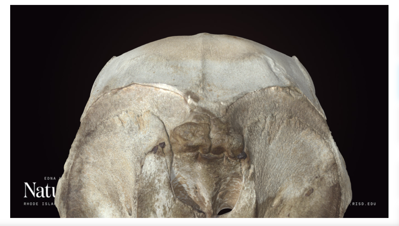
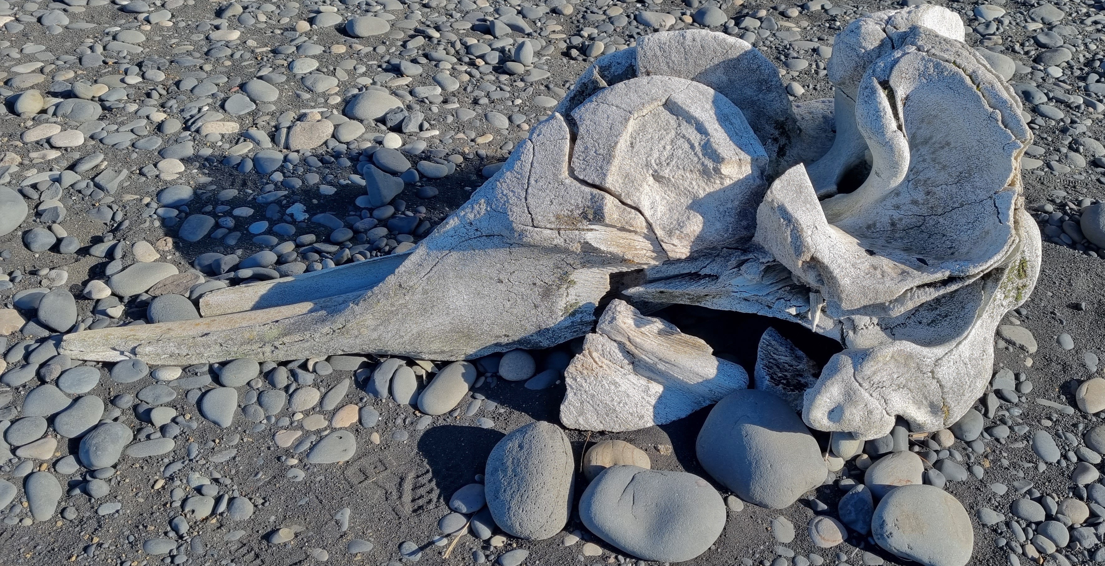
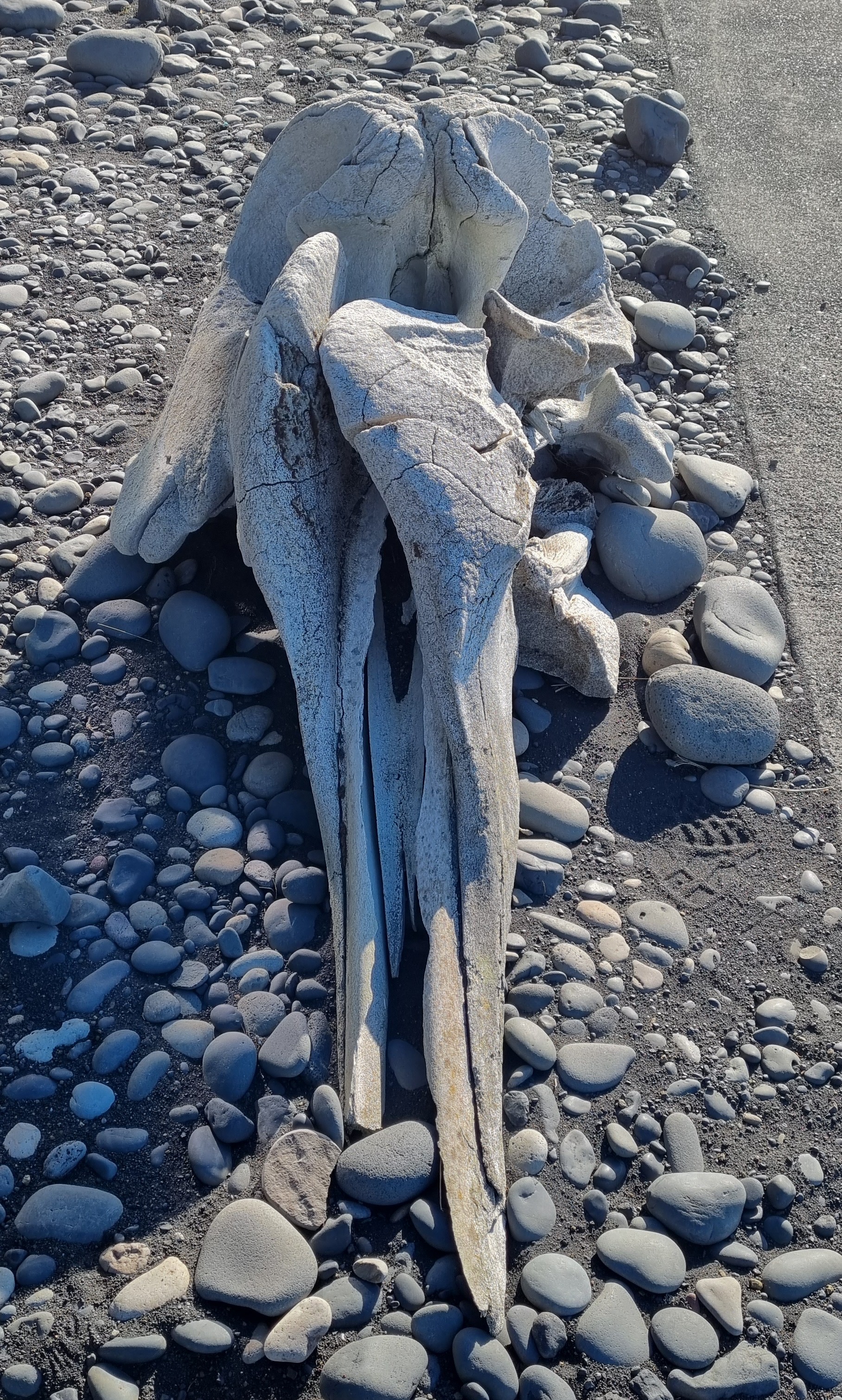
Last week I gave you this rugged skull, from a rugged place, to have a go at identifying:
As everyone spotted, this is a whale of some sort (what else has a skull that weird-looking?), but the question is, which species?
The location led to a few suggestions of Arctic / sub-Arctic species like Narwhal or Beluga, but they have a much flatter top section of the skull. In fact, those huge vertical lobes of the maxillae seen here is pretty unusual and quite distinctive (even if it is a ittle weathered and broken):
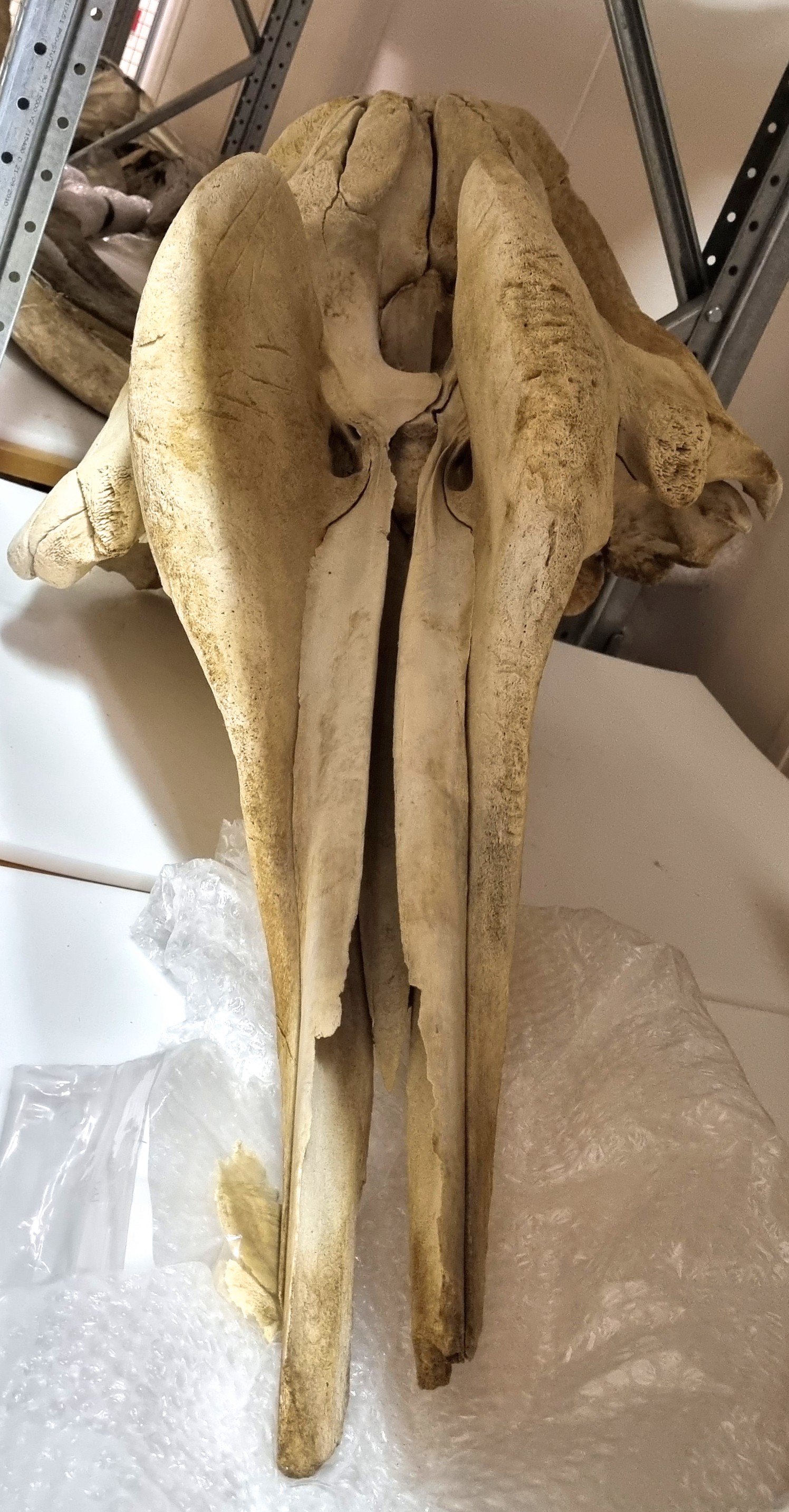
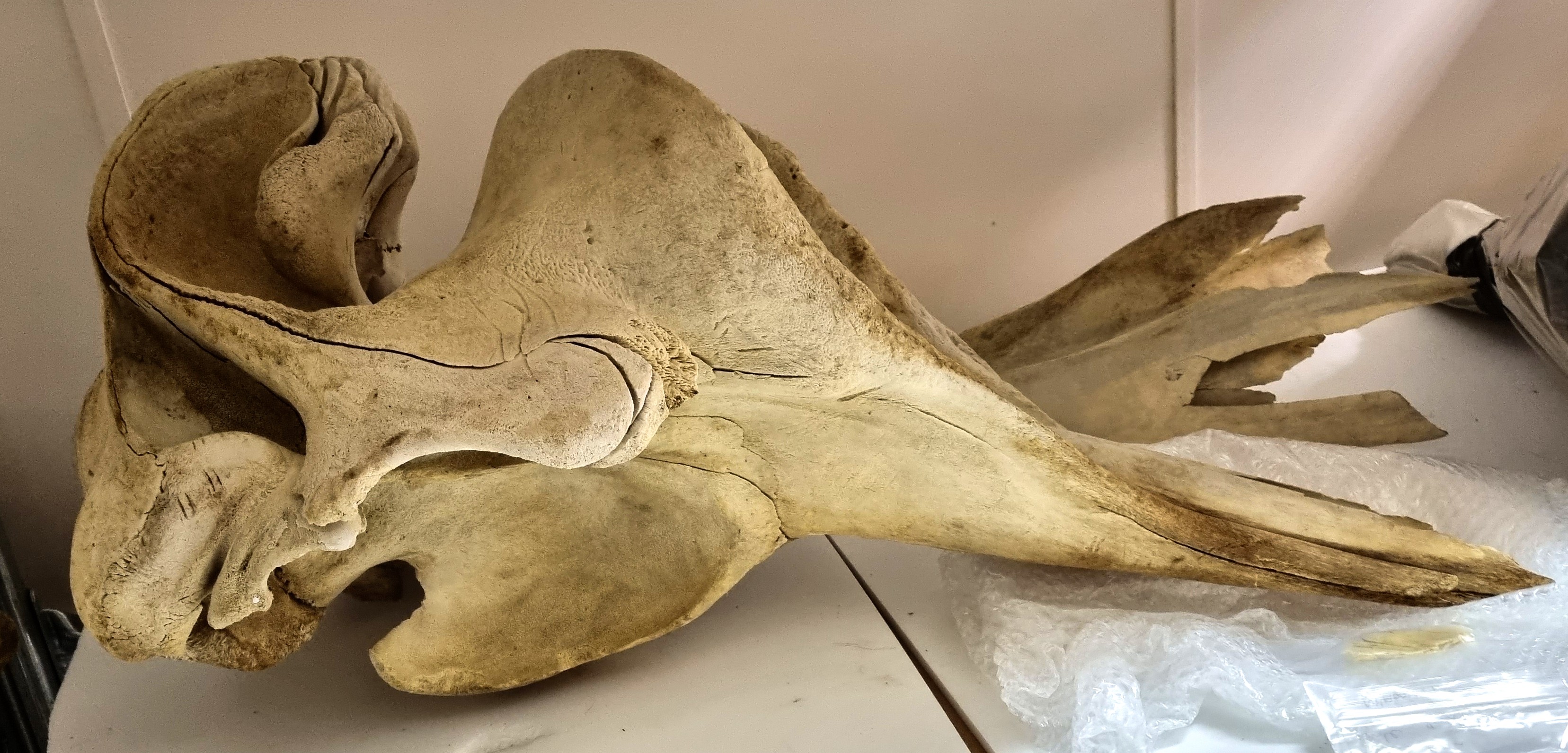
This reminded me of a specimen in the collections of the Dead Zoo and which I had to check, just to be sure of my identification:
As spotted immediately by Chris and not too long afterwards by Adam Yates and Wouter van Gestel, this is the skull of a Northern Bottlenose Whale Hyperoodon ampullatus (Forster, 1770).
This sub-Arctic species has a distribution across much of the North Atlantic. They tend to stick to quite deep water, which makes sense in the case of the specimen I shared from Iceland, since the Reynisfjara beach is infamously dangerous because it shelves off very steeply into very deep water, making the waves that break along the beach behave in an unusual (and frankly terrifying) way.
Occasionally this species will come into shallower waters, in one (somewhat tragic) case a female Bottlenose Whale swam up the Thames (and is now in NHM, London). Our specimen came from an animal stranded on the Irish coast and there are theories that maritime sound pollution is connected to them being driven into shallower waters.
Well done to everyone who worked out which species this skull is from – hope you’re ready for another mystery next week!
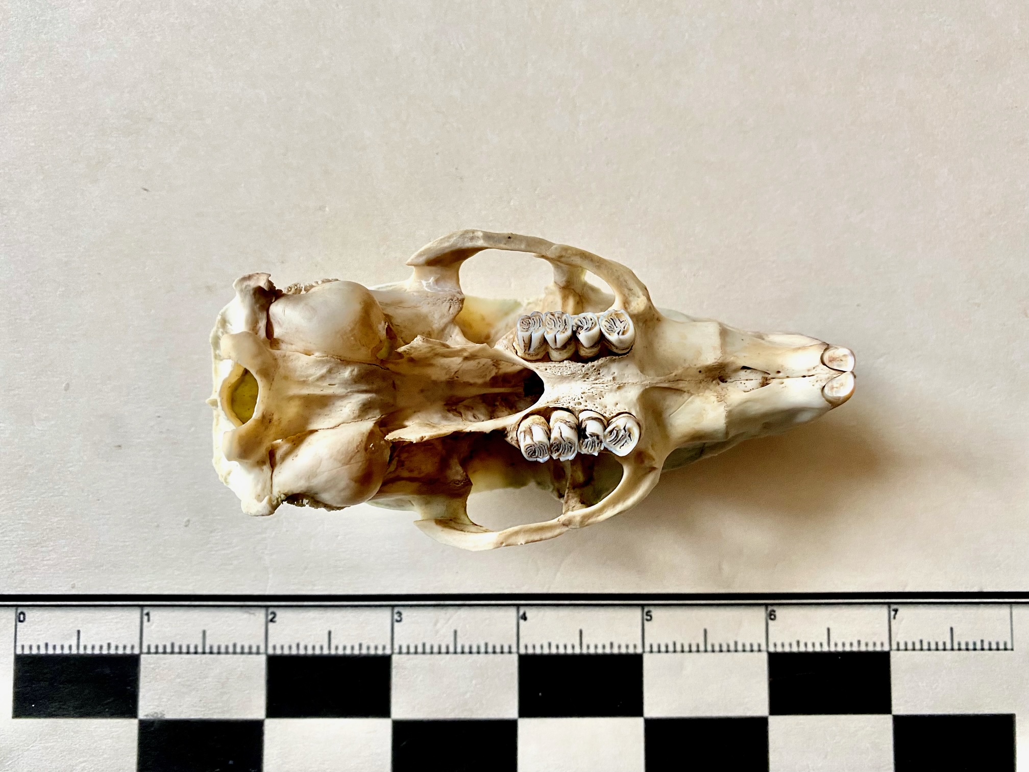
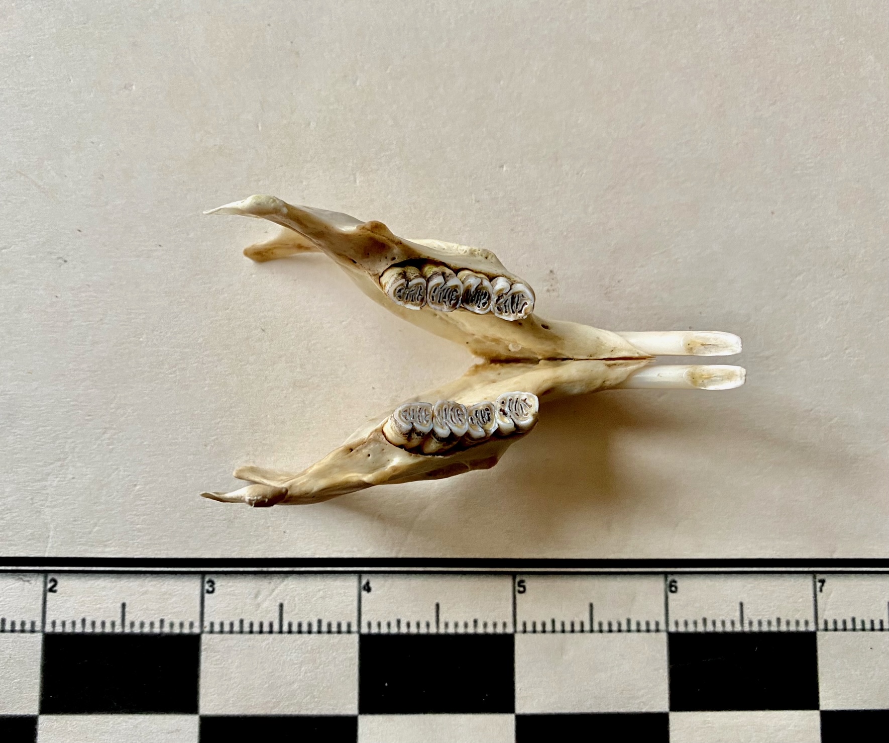
Last week I gave you a challenge to get your teeth into:
As I suspected, everyone managed to figure out what type of animal this is, since these teeth are quite distinctive (as mammal teeth often are).
To start with, there are canines and incisors in the premaxilla (the top jaw). These are missing from things like cows, sheep and deer. So it’s not one of them. The premolars are adapted to grinding rather than cutting, so it’s not some kind of pig or carnivore.
The molar teeth are low-crowned, unlike the teeth of grazers like horses which are high crowned, to cope with the wear and tear of silica-toughened grasses. This suggests an animal that browses on softer vegetation. Also, the lophs (those ridges of enamel that join the tooth cusps) are well defined and quite distinctive in their shape. That rules out most other herbivores, including the camels and their relatives.
I think it’s understandable that nobody got the correct species, since the specimen is a subadult (check out the molar in the jaw that’s still developing) which will somewhat alter the proportions compared to an an adult – especially considering the photos I gave you were restricted to the teeth and missed all the useful features of the rest of the skull.
So well done to everyone who worked out that the teeth belonged to a tapir!