This week I have what I think is a nice and simple mystery skull for you:
Any suggestions about which species this comes from? Cryptic clues are welcome, just to let people who aren’t sure have a chance to work it out. Have fun!

This week I have what I think is a nice and simple mystery skull for you:
Any suggestions about which species this comes from? Cryptic clues are welcome, just to let people who aren’t sure have a chance to work it out. Have fun!
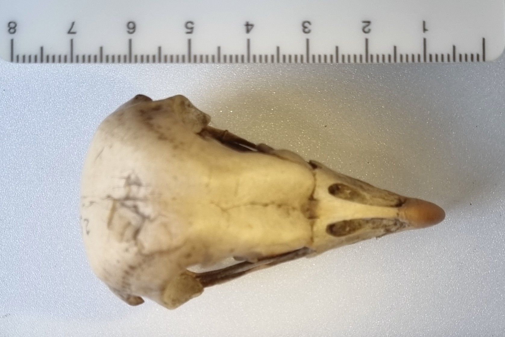
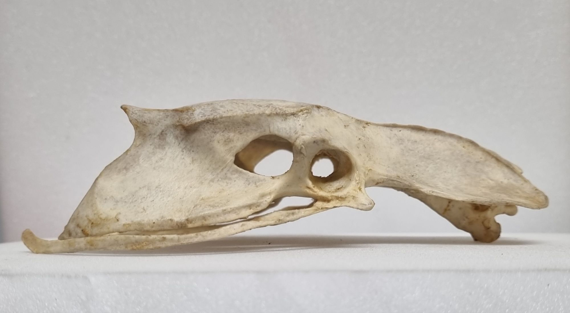
Last week I gave you this skull to try your hand at identifying:
It’s not the easiest of tasks, as there are quite a lot of birds in this general size range and with this general shape of skull, so the overall outline from the top only helps narrow things down a little. It is worth noting that general proportions all suggest some kind of Passeriform.
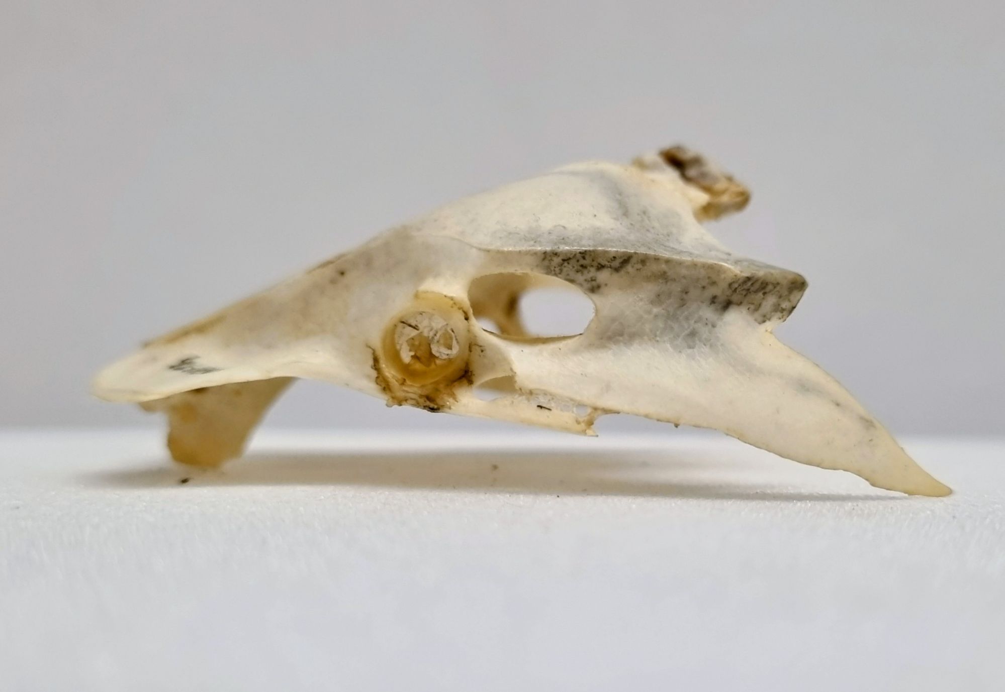
The exact length of this specimen is 49mm, but as with all species, there is a bit of variation depending on subspecies, age, sex, quality of diet and other environmental factors. I suspect this one is a little on the small side for the species.
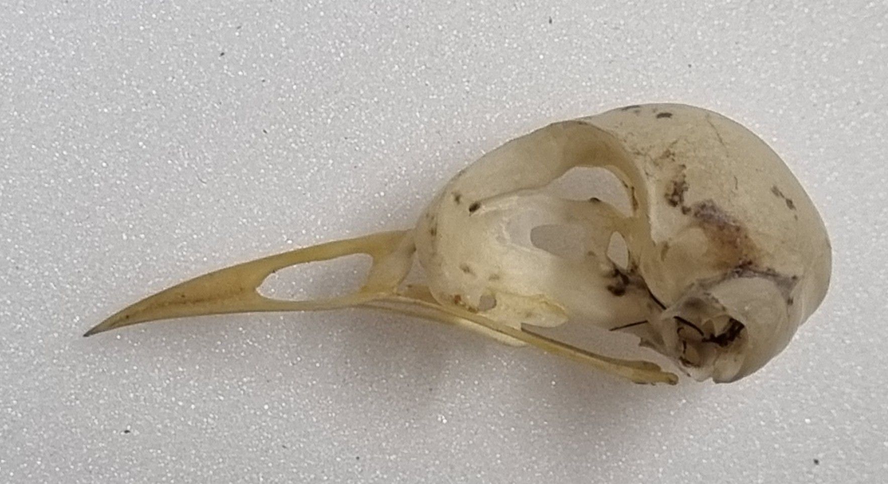
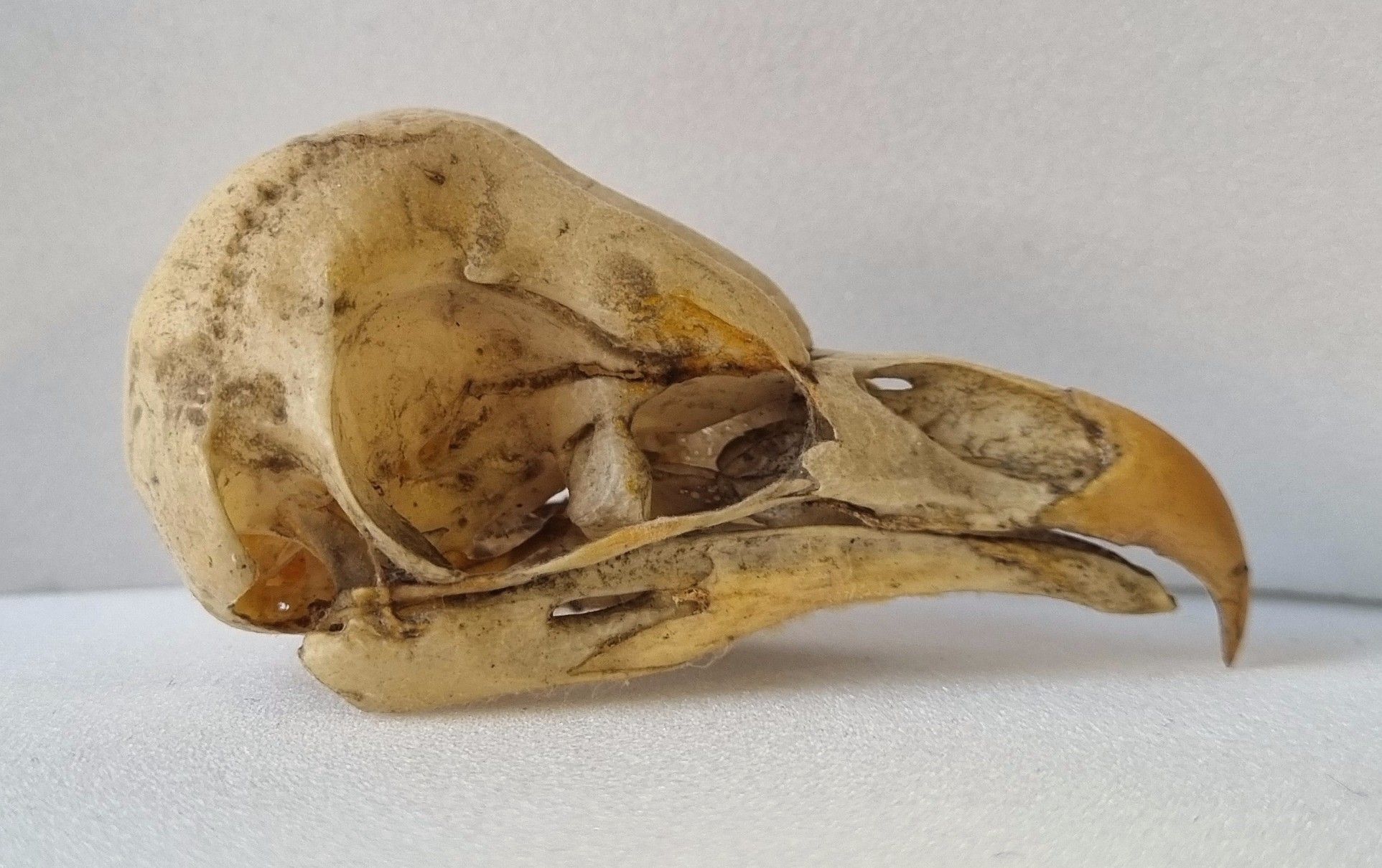
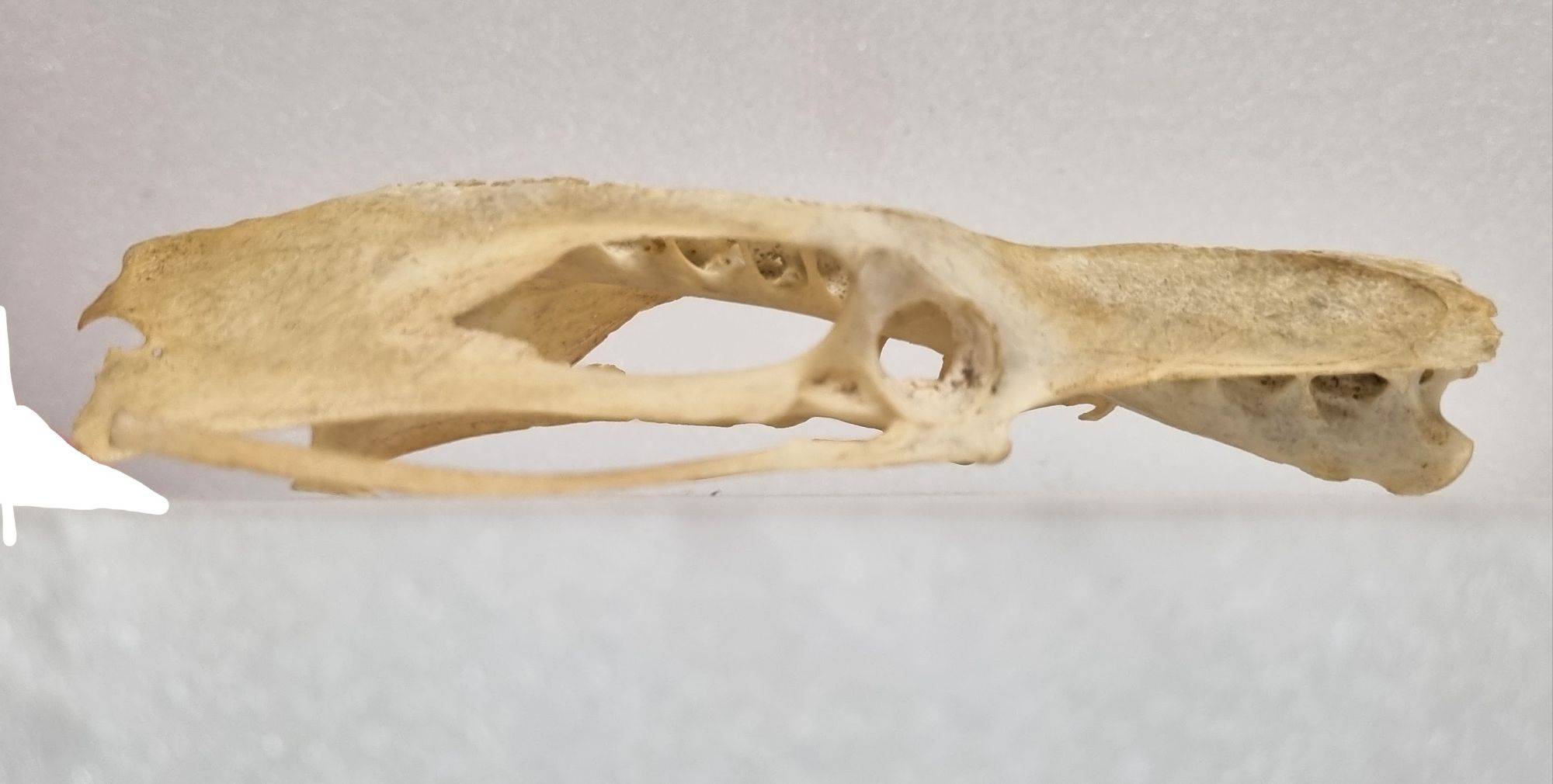
The side view offers another perspective on the bill morphology, which is looks quite generalist, perhaps a little on the gracile side, but not overly skinny. Given the size that puts me (and many who left a comment) in mind of something in the Thrush family.
It’s also worth noting that the bill sheath, while not very apparent, is still present (if it wasn’t you’d see small holes along the side of the bill, which are the blood vessels that supply the tissues that secrete the keratin of the bill sheath). This is a big clue, as the bill sheath is yellow.
For me that immediately put in my mind a very specific type of Thrush – the male Common Blackbird Turdus merula Linnaeus, 1758.
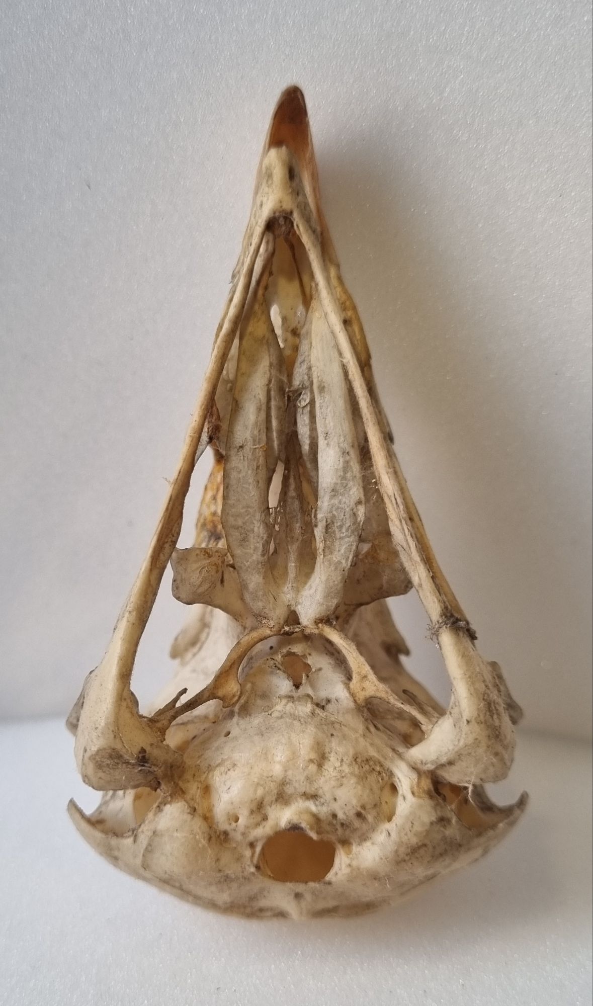
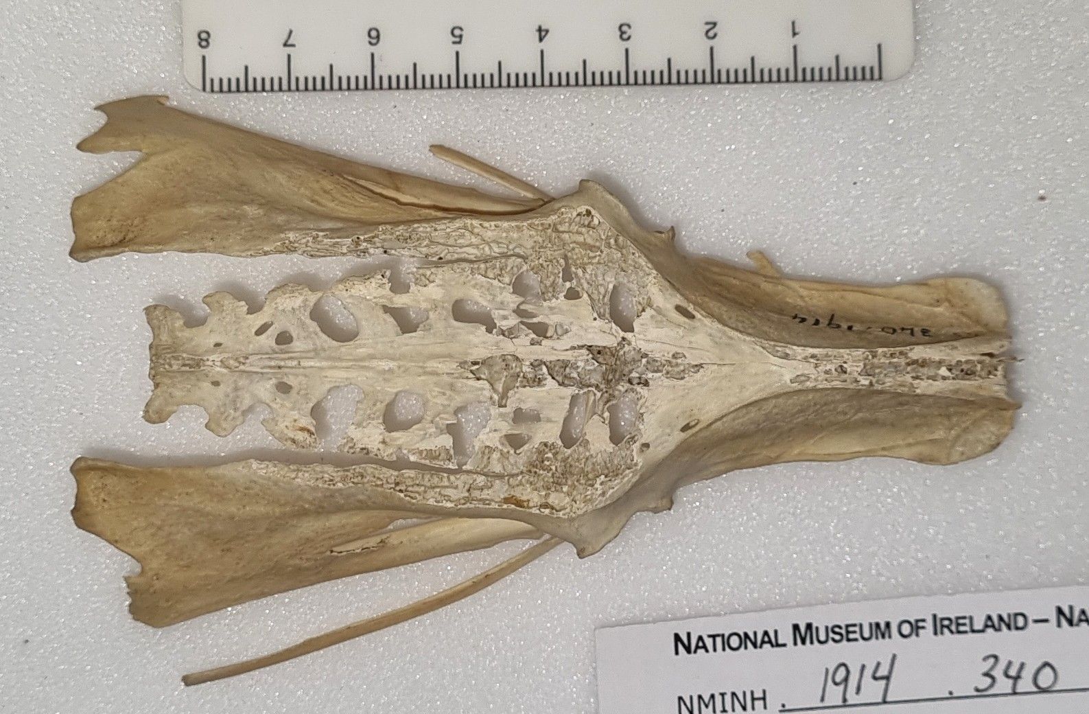
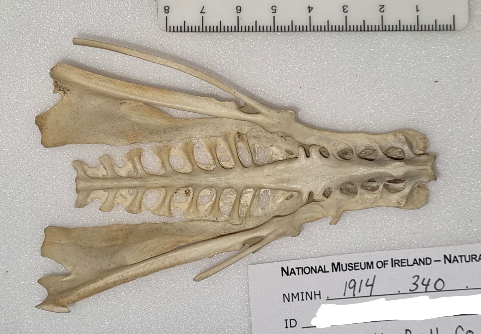
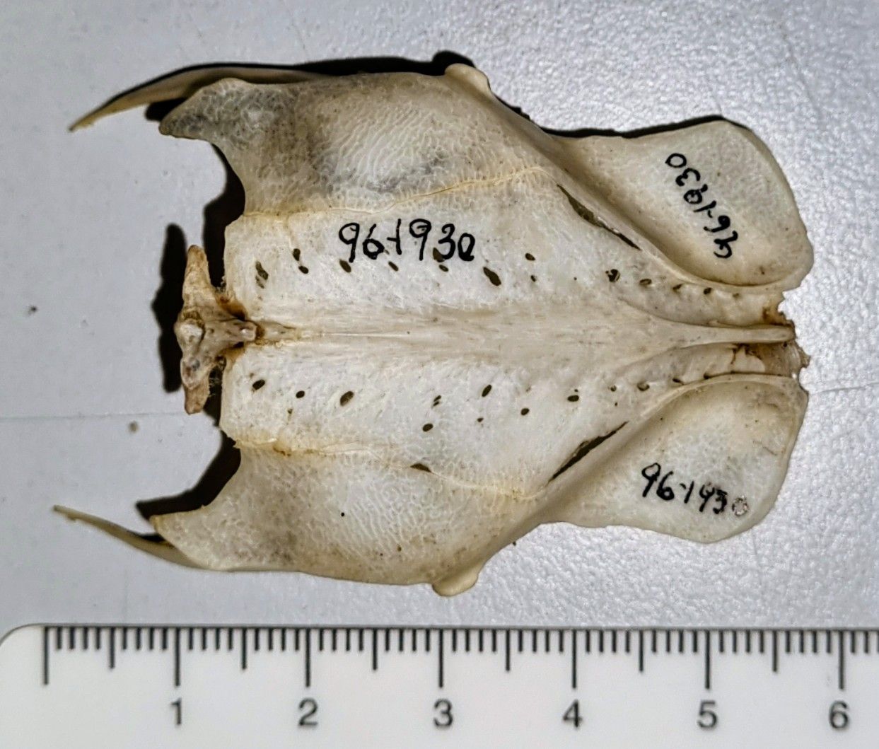
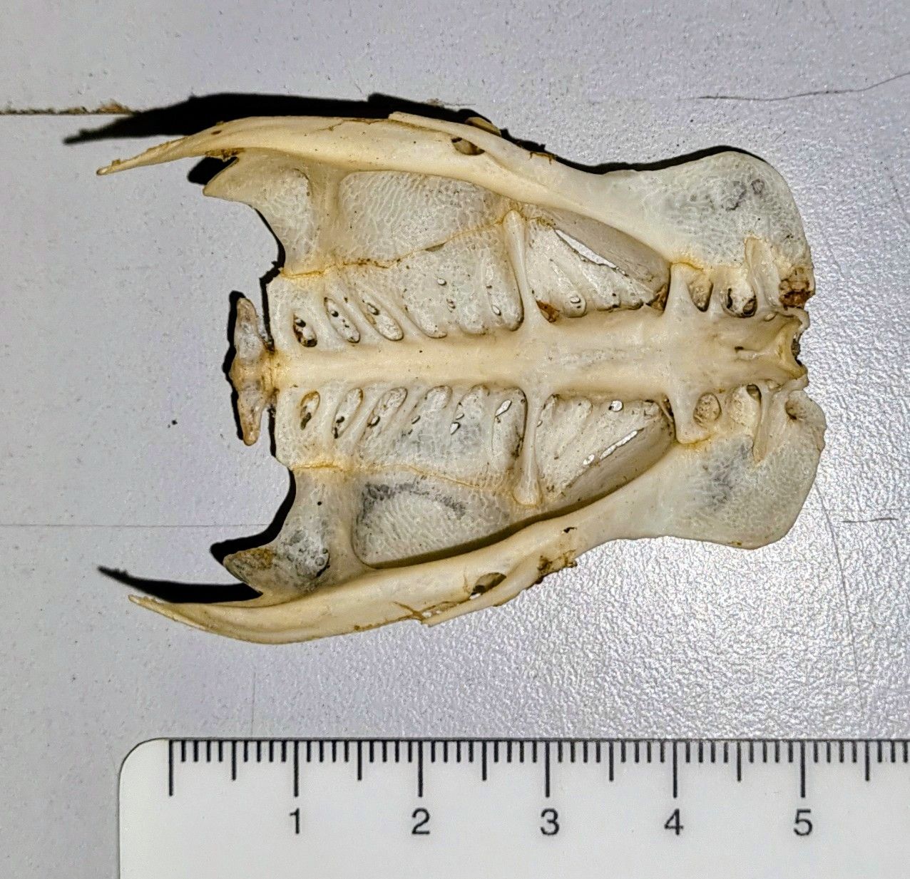
This can be checked with reference to the excellent Skullsite resource, which provides a fantastic range of bird skulls with images from several different angles. Take a look at the underside of this specimen:
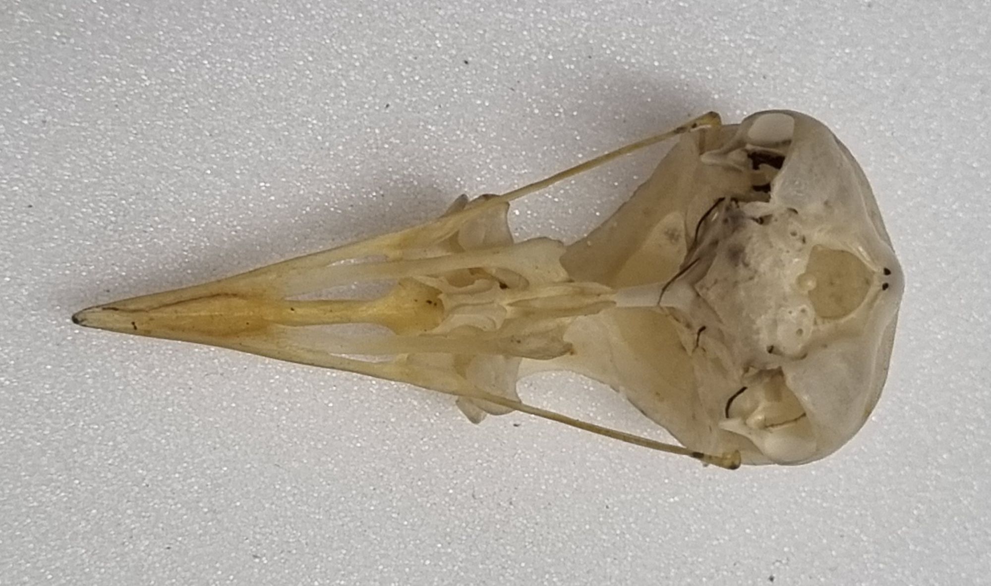
The posterior region of the palatine bones have quite a distinctive shape – flared and with a broad shallow notch at the posteriormost margin. If you compare this to other skulls in the genus Turdus, you’ll notice that the notch is almost absent in the Redwing, it has a much sharper outer angle in the Songthrush, it’s a lot deeper in the American Robin, it’s deeper and sharper in the Fieldfare, and so on. The important thing is that the palatine of the Blackbird on the Skullsite is still consistent with that of the mystery object.
While this feature takes a while to get familiar with, it can be quite helpful when narrowing down similar skulls like these. I hope that pointer proves useful!
Finally, I offer up congratulations to Adam Yates who not only got the correct identification, he also left a great cryptic clue to share that information. Bravo!
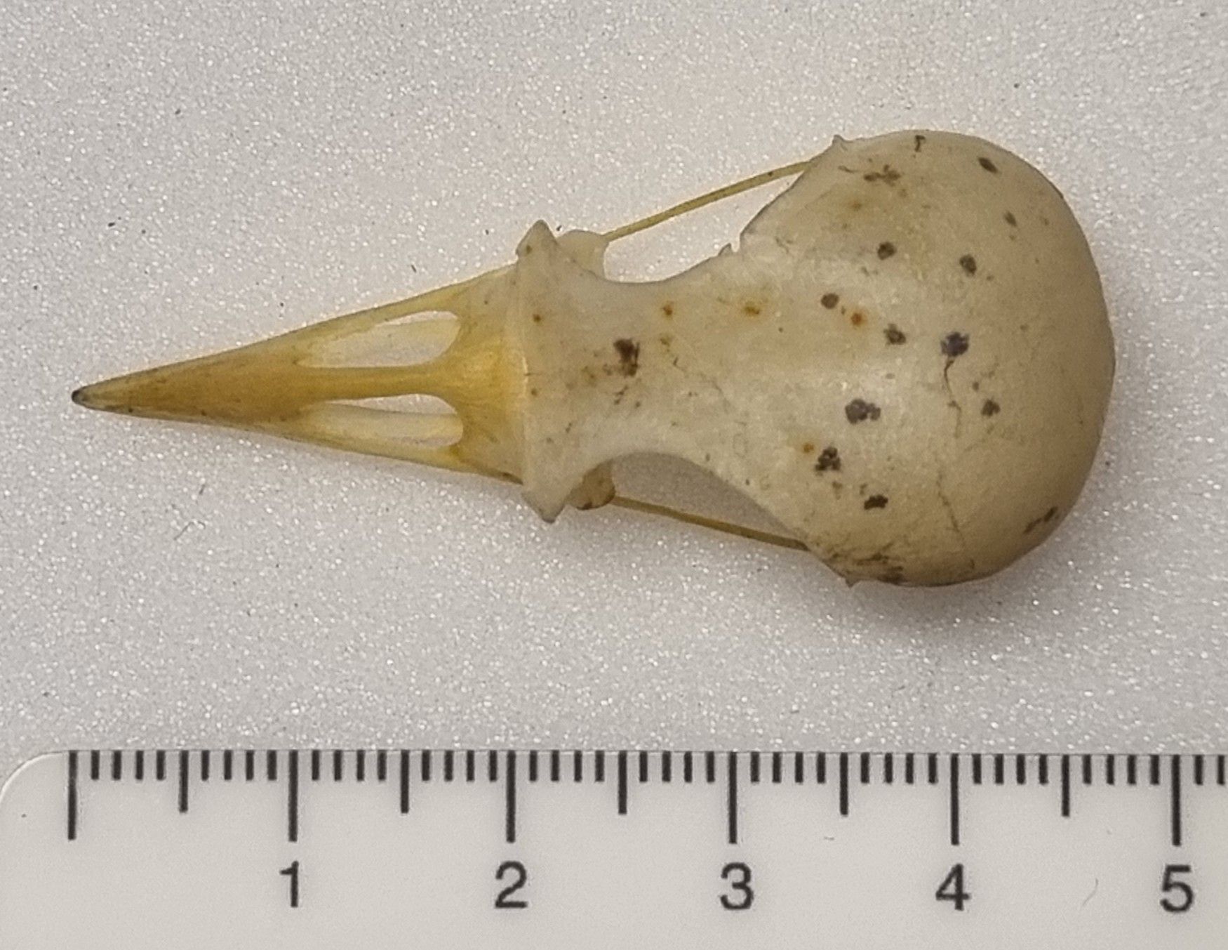
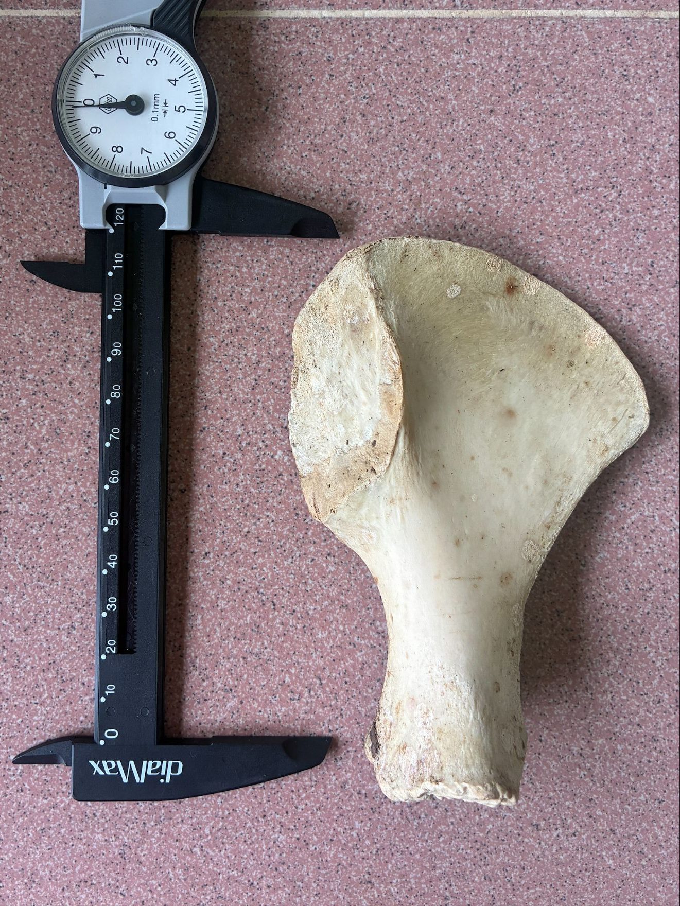
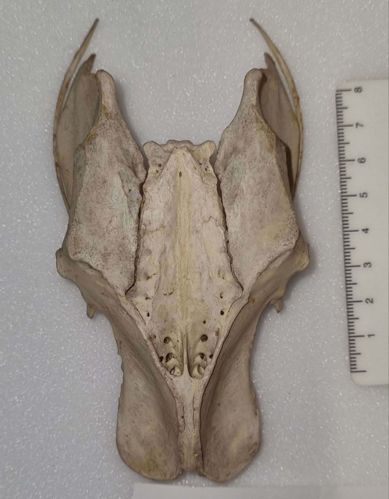
This week I have a bony mystery object for you, measuring 49mm long:
If you have any idea what this might be, you can leave your suggestions in the comments below. Have fun with it!
This week I have a mystery object for you that I saw when visiting Conservation Letterfrack to talk about some work being done on historic display cases for the Dead Zoo:
It’s an interesting specimen and I’d love to hear your thoughts on what you think it is. Have fun!
Last week I gave you this sad scene with a mystery victim to try to identify:
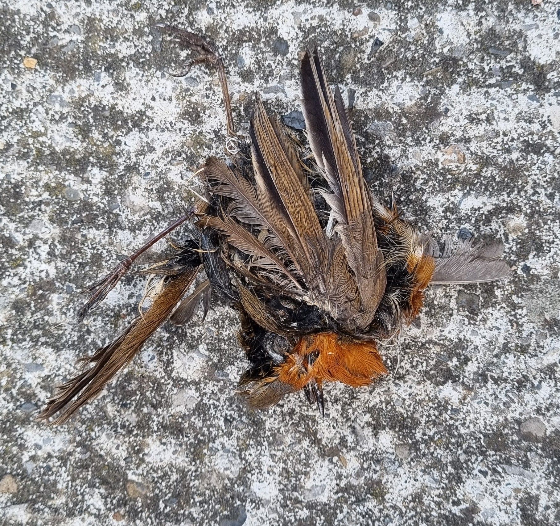
I thought it might be an interesting one, because when I first saw it my mind took me around the houses thinking about what it was.
My first thought was actually that it had some features I associated with a European Stonechat:
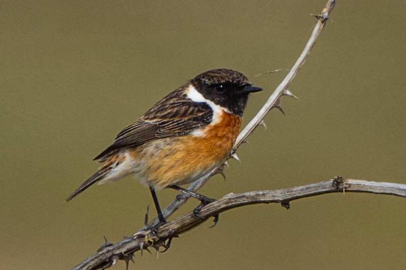
But on closer inspection I realised that the bill was surrounded by red feathers rather than black, and the “black” feathers were probably not originally black, and were as a result of staining from the products of decomposition.
A similar sized chat that’s more commonly encountered in the area fits the “bill” more closely – or more to the point the feathers around the bill fit its plumage more closely. The European Robin Erithacus rubecula (Linnaeus, 1758).
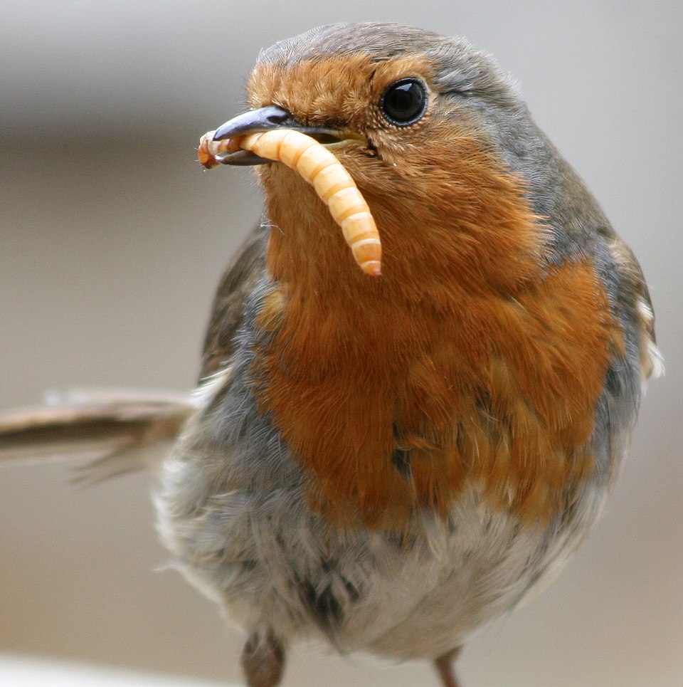
So well done to Hilary, Chris and Joe who all saw through the feather discolouration and recognised the bold and beautiful bird that this sorry specimen started out as. Now all remains is to do the CSI work to figure out if this gruesome death was the result of a cat, a car or a window – three of the most common terminators of urban birds…
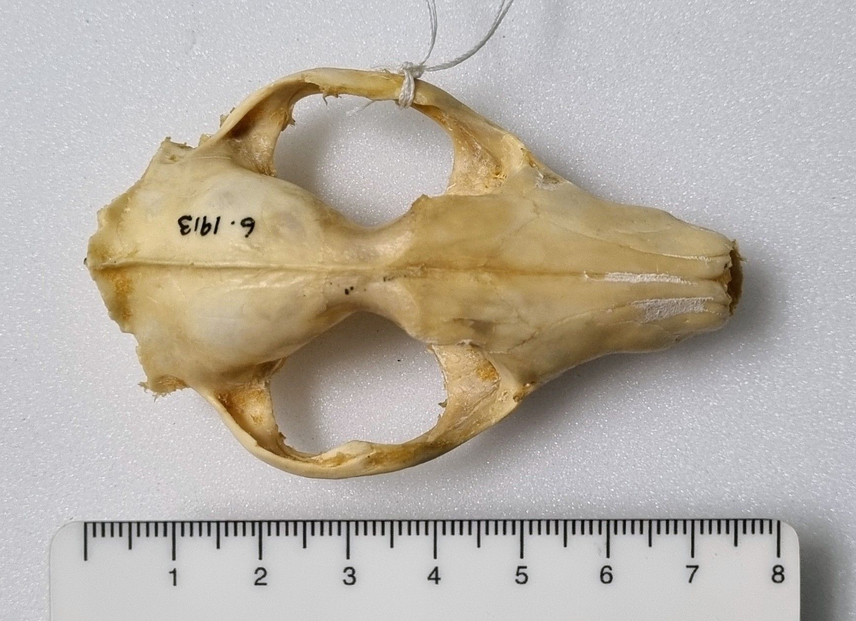
Last week I gave you this mystery skull to try your hand at identifying:
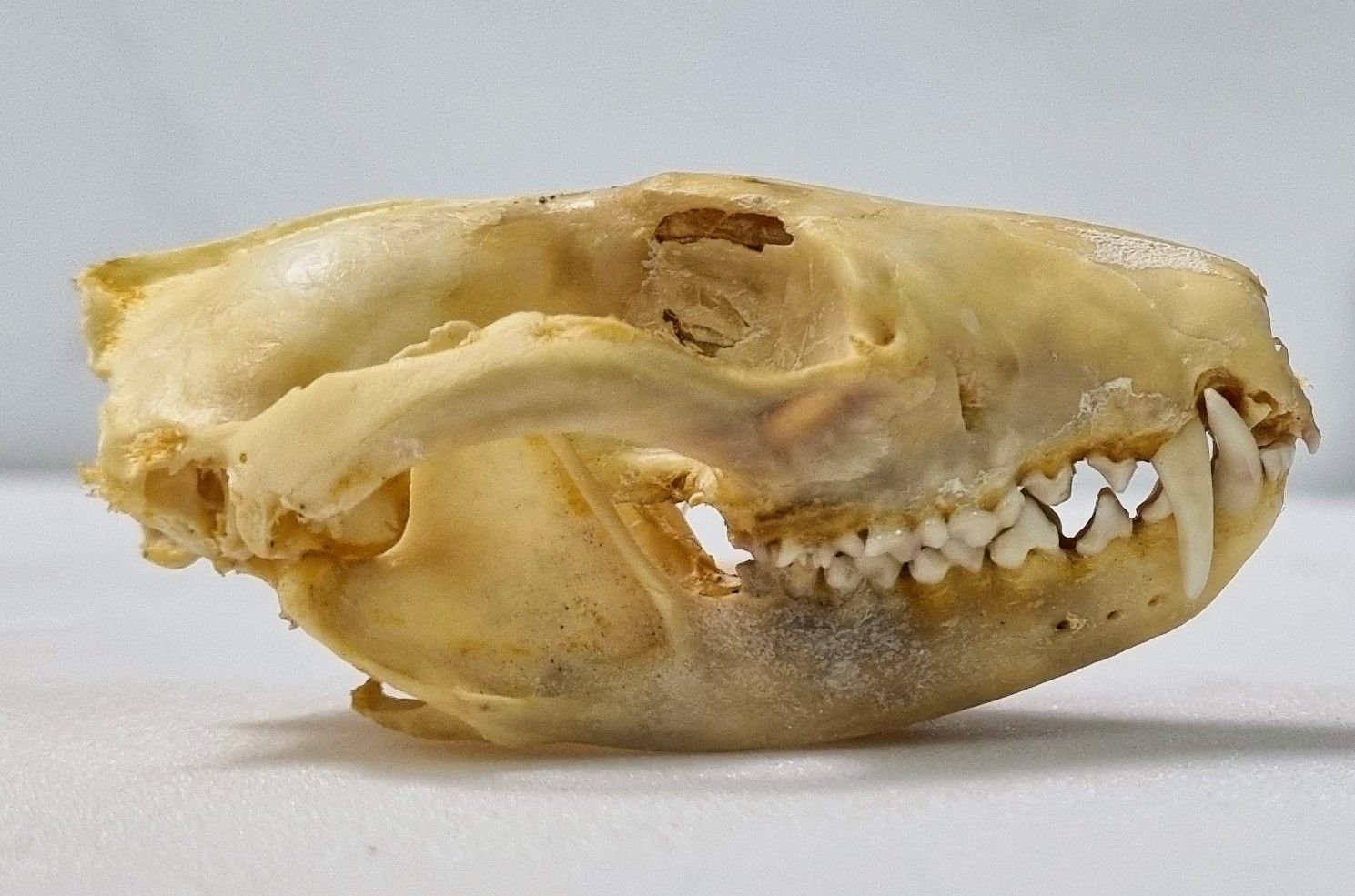
It wasn’t quite as easy as it might at first seem, and it led to a great discussion in the comments section.
Several people were initially taken in by the shape, perhaps without considering the scale, as this looks very similar to our old friend the Tasmanian Devil:

However, the mystery specimen is almost half the size of Tasmanian Devil. The similarities do offer a good clue of where to look for alternative identifications though. The Dasyuridae is a family of Marsupial carnivores that includes the Tasmanian Devil, a variety of small mouse-or-shrew-like forms (which a smaller with more pointy snouts that this), plus the members of the Genus Dasyurus – the Quolls.
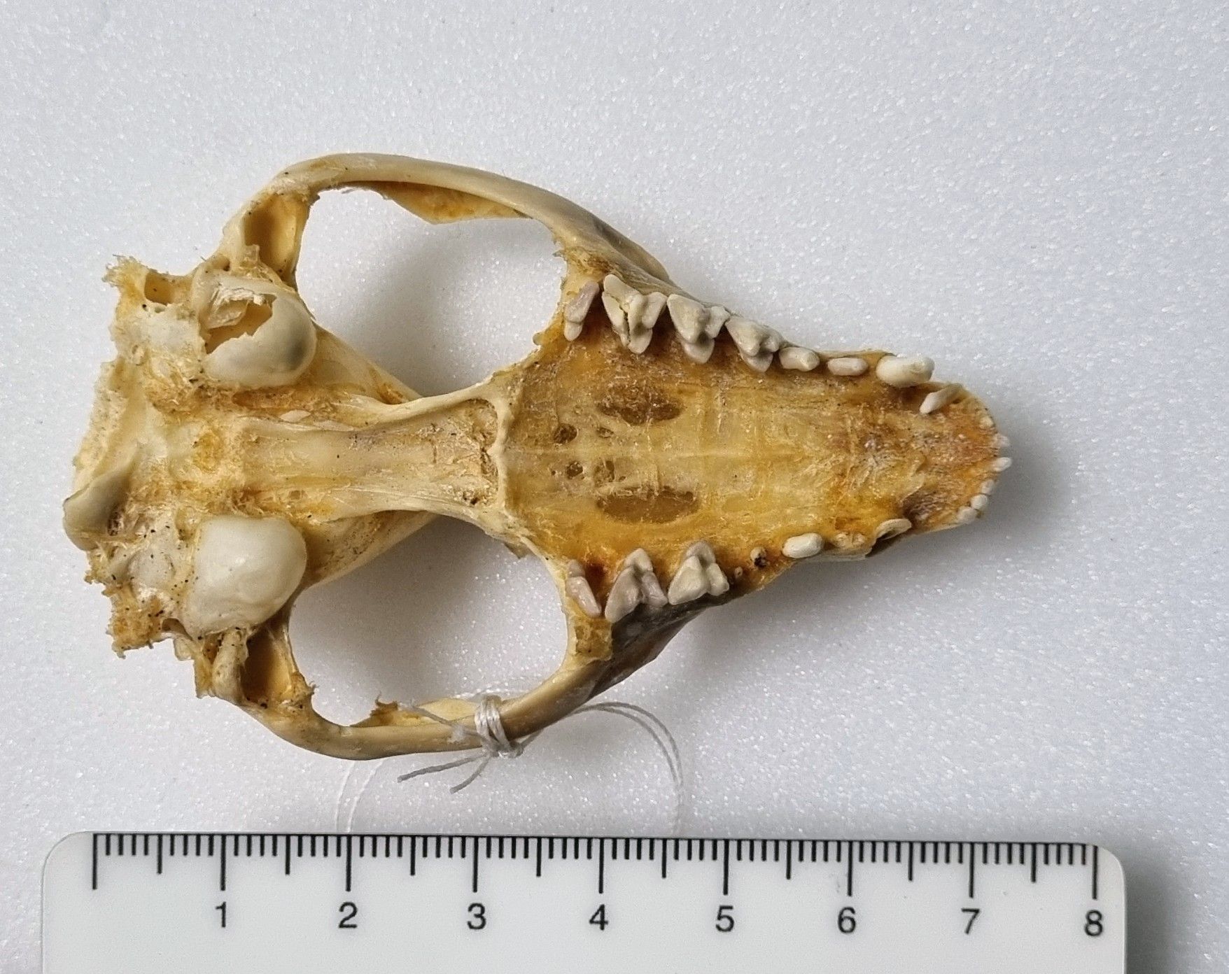
The comments moved on to the Quolls quite quickly, with some discussion around whether this was the Spotted-tailed (or Tiger) Quoll or if it could be the Western Quoll. In the end, Kat Edmonson noticed a feature of the nasals to help distinguish which it was, and Adam Yates noticed a useful feature of the angular process of the lower jaw:
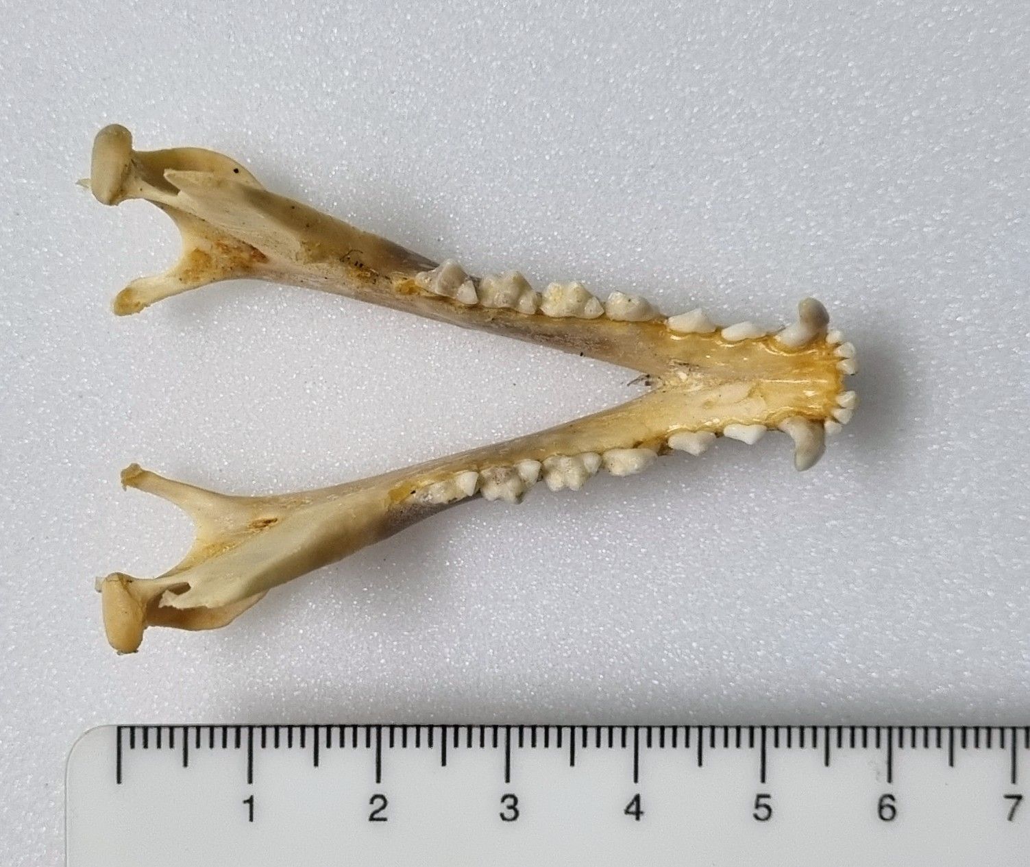
In the mystery specimen those processes that project inward on the mandible are “pronglike” but the Spotted-tailed Quoll they are more triangular.
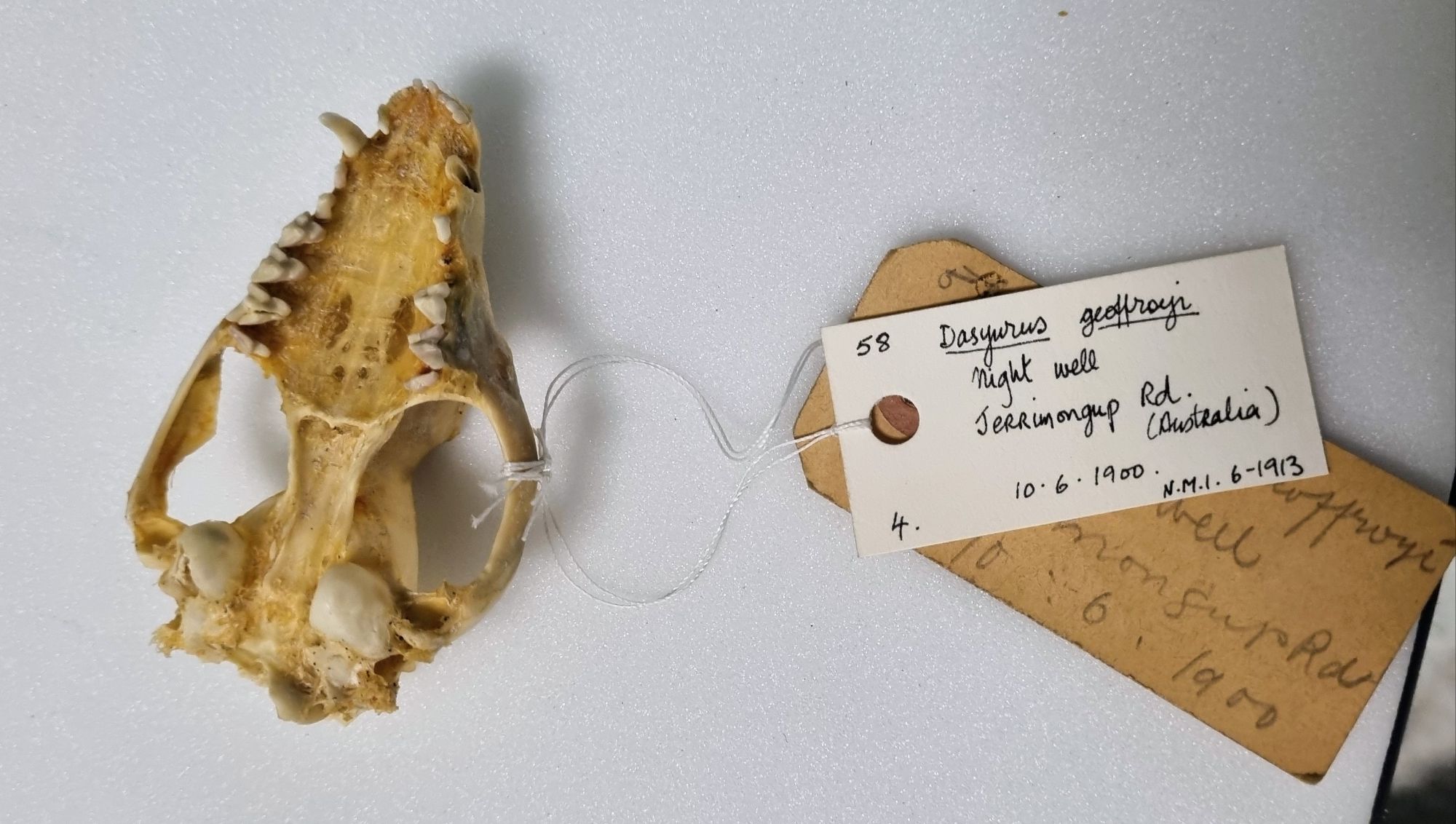
So well done to everyone who worked out that this is the skull of a Western Quoll, Dasyurus geoffroii Gould, 1841. I hope you enjoyed the challenge!
Just to note, I’m currently at the Ulster Museum for the NatSCA conference – my annual opportunity to connect with other natural science collections and one of the highlights of my year! While I’m here I may see if there are any interesting obects I can find to use for future mystery objects – let’s see what I can find!
Before I share this week’s Friday mystery object, I’d like to just take a moment to mark the 100th birthday of Sir David Attenborough.
I think it’s fair to say that he is a national hero in the UK and the source of inspiration for generations of naturalists, thanks to his documentaries. I’ve been fortunate enough to meet the legend himself a couple of times, and he was even generous enough to lend his Elephant Bird egg to an exhibition I was involved in at the Horniman Museum over 10 years ago.
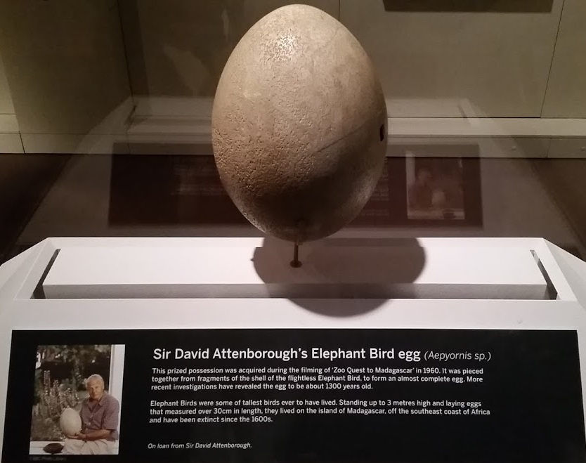
So here’s wishing a very happy century celebration to Sir David!
With that said, here is a nice skull for you to have a go at identifying – I’d love to hear your thoughts on what it might be:
Let me know what you think in the comments below, and I hope you have fun working it out!
Last week I gave you this rather beautiful mystery object to have a go at identifying:
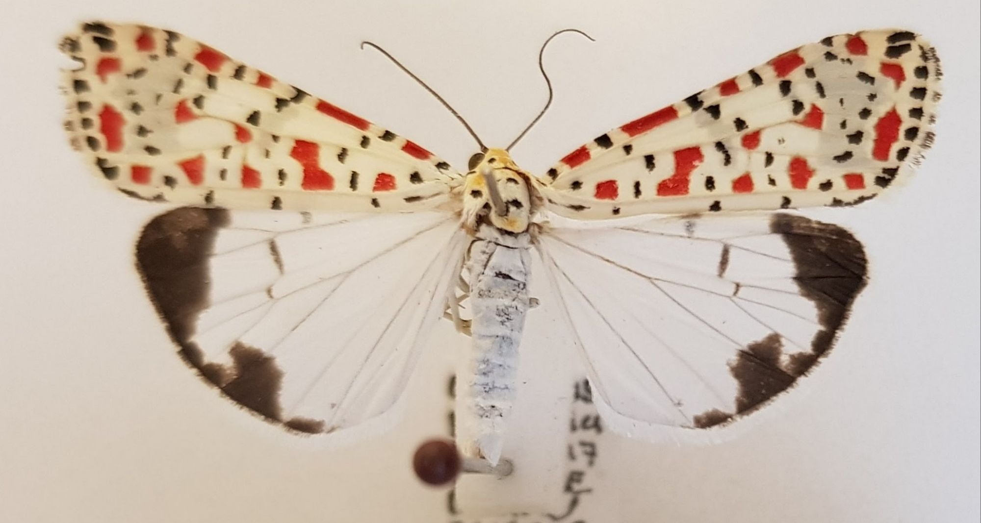
This is a pretty distinctive species, but not without some very similar relatives who can complicate things.
The speckled crimson pattern on the forewings of this species provide all the clue you really need for the identification – it’s a Speckled-crimson Moth (or Crimson-speckled Flunkey, or Crimson-speckled Footman) Utetheisa pulchella (Linnaeus, 1758). I even dropped a hint when I described it as a “rather beautiful beastie” as the Latin species name pulchella translates to “beautiful”.
This species occurs across Africa, Asia, and Europe, with occasional migratory visitors turning up in the UK and Ireland – in this case in Tramoe, Co. Waterford.
However, there is a very similar looking species in the same Genus that goes by the name Utetheisa pulchelloides – which literally means “resembling Utetheisa pulchella“. The common name for that species is the Heliotrope Moth, which mainly occurs in the Southern Hemisphere – mainly New Zealand, Australia and up into Borneo and Singapore.
Here’s a comparison between specimens of these two species from other collections:
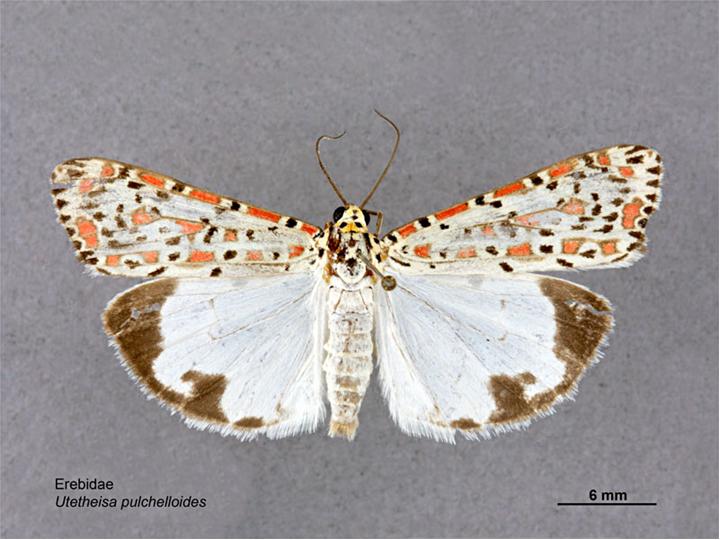

You can see how similar they are – and if you look at the underwing the U. pulchelloides specimen looks more similar to the mystery specimen – which I suspect may have led Adam Yates down the wrong path.
If you look at the forewing you might notice that U. pulchelloides has small black triangles along the margins, whereas U. pulchella (and the mystery specimen) has truncated triangles or rectangles. You may also notice that the crimson speckles of U. pulchelloides are more faded and have a dark margin.
So well done to Chris Jarvis, Katenockles, and Jonathan T who all spotted the subtle differences either in the comments or on Mastodon.
This week I have an invertebrate for you to have a go at identifying for a change:
Any idea what this rather beautiful beastie might be?
Let me know what you think it might be in the comments below. I hope you have fun working it out!
Last week I gave you this genuine mystery object from the forests of Borneo to get your thoughts on:

This was sent to me by Dave Hone (who runs Archosaur Musings), but it was found and photographed by his colleagues Lauryn and Tom of Queen Mary University of London, who are doing research in Borneo.
I had an idea of what it might be, and I’m pleased to say that everyone who responded – both here and on social media – came to the same conclusion as me.
This is the left ilium crest from the pelvis of a juvenile animal. That much can be seen from the unfused sections where this would have connected to the sacral vertebrae and the pubic bone:
An unusual thing about this ilium is the position of the point of fusion with the sacrum:
For most species the iliac crest (that’s the curved bit at the top) extends upward quite a bit before flaring out – and that flaring is often quite squared off and tends to be narrow and more blade-like. That configuration is what you would expect to see from most quadrupedal mammals – a notable exception is in the form of the human pelvis, due to our bipedalism.
However, this pelvis doesn’t quite conform to the human shape, as it’s a little bit less curved and a little more blade-like. This is consistent with one of our close cousins – there are some useful comparisons of disarticulated primate pelvises on the Bone Clones website and while this is very similar to a female Chimpanzee that’s shown, the location of Borneo suggests a more likely option – a Bornean Orangutan Pongo pygmaeus (Linnaeus, 1760).
On BlueSky, osteoarchaeologist Terry O’Connor spotted this straight away, as did Adam Yates and Chris Jarvis here in the comments. 10 year old Viren, who is a new visitor to Zygoma, was also spot-on. So well done to everyone, and thanks for your thoughts – it’s great to have my conclusion supported!
This week I have a genuine mystery for you to help solve, courtesy of Lauryn and Tom – researchers working in the forest in Borneo, and passed on by Dave Hone (an old friend and fellow blogger who runs Archosaur Musings):
I think I know what this is and what it’s from, but I’d love to hear your thoughts on this Bornean bone in the comments below!
Last week I gave you this bird pelvis to have a go at identifying, as part of a series of posts on that particular feature across birds utilising different environments:
It probably wasn’t the easiest challenge, since this pelvis is damaged. It’s missing part of its dorsal surface and with some small amount of damage to the posterior margin of the right ischium (that’s the broad bit that flares out on the bottom left of the image) and with both of the pubic bones snapped (those are the long skinny bits that stick out to either side).
Still, the overall shape still gives some useful clues. The long, narrow, triangular form offers some ideas about the distribution of mass and use of the legs, to give a sense of possible locomotion habits.
The pelvis is very flat and the points of articulation with the femur are quite far forward:
At least in comparison with something like a Chicken:
This suggests something that has a centre of mass that’s quite far forward compared the foot position. This immediately raises the question of why? The obvious answer for me is that this is a species that doesn’t brisky strut around, relying on more of a low-speed waddle to get around – or perhaps a paddle.
Waterbirds have legs located quite far back in the body to aid propulsion in the water. Perhaps a bit confusingly this requires the articulation of the femur with the pelvis to be pushed further forward due to the more horizontal orientation of the spine to allow the correct orientation of the legs to push water backwards and the body forwards – unlike terresrial species that are more interested in pushing the body upwards against gravity.
There are of course a lot of waterbirds, from the Pelecanimorphae mentioned in the comments by Adam Yates, to the Anseriformes. That’s what we’re looking at here.
There are a lot of ducks and geese, so narrowing it down isn’t easy. The size of this mystery specimen is in the right range for a large duck or small goose, but if you look at the left ischium (the undamaged one) you’ll notice a deep notch – this is something seen in geese.
You may also notice an additional extra notch – which I think offers a great clue to hint at the species. This is the pelvis of a Brent Goose Branta bernicla Linnaeus, 1758.
I’m not sure if the thoughts I’ve laid out here, and over the last couple of mystery objects are helpful for guiding future identifications of bird pelvises, but I hope they may be of use. Let me know your tips on working these things out – more ideas are always welcome!
This week I have a third mystery bird pelvis for you to try your hand at identifying:
Let me know what you think this might be from – I’d be keen to hear your reasoning!
Last week I gave you the second of a series of bird pelvises to try your hand at identifying:
This one proved a bit more difficult than the previous example I offered up, which was from a Chicken – here’s that one for comparison:
As you can see, our mystery pelvis is a good bit smaller than that of the Chicken, and in terms of the shape, while it has some similarities, but it’s more compact and almost square. I’m not 100% sure why, but that makes me think of something that has a more upright body orientation than a Chicken.
Part of the reason it’s more square is that the areas of muscle attachment for the iliotrochantericus caudalis muscles (which I talked about a couple of weeks ago) don’t extend far forward, which suggests its femur isn’t being stabilised to enable a long stride. The attachment is quite wide though, so I suspect it may be optimised to cope with large forces in a burst instead.
There are a few species that would fit the bill (if you’ll excuse the pun) including Grouse – which was suggested by Chris Jarvis and it is remarkably similar – but this is the pelvis of a Rock Dove (in this case the domestic version) Columba livia Gmelin, JF, 1789 which was correctly spotted by Adam Yates.
I thought this one might be nice to use, as it lets me share a blast from the past that looks at the explosive take-off of a Pigeon, from Ben Garrod’s TV series Secrets of Bones, which I had the privilege to be scientific advisor for back in 2014. I hope you find the clip interesting!
The last mystery object was a bit of bird bone that I personally found really interesting, and it’s inspired me to offer up the same bit of bone, but from a different bird:
Any idea of the species that this came from? I’d love to hear your thoughts in the comments box below!
Last week I gave you this bony structure to identify:
I didn’t think this one would prove too difficult, since I went with something that most people have probably encountered on their dinner table or thrown in their domestic waste at some point. I wasn’t wrong and Chris Jarvis was first to drop a hint, with reference to Elvis the Pelvis and a famous brand of fried foodstuff from Kentucky.
This is of course the pelvis of a Chicken Gallus gallus domesticus (Linnaeus, 1758).
Bird pelvises are interesting structures, that are a bit more extensive than the usual mammalian equivalent1 due to the extended and fused vertebrae around the sacrum – the sacrum being the area of fused vertebrae where the hip bones attach to the spine. This extended region of fused vertebrae along the midline of the pelvis is referred to as the synsacrum.
In birds the fusion of the pelvis can be very extensive, and provide large areas for muscle attachment. If you look at the bottom of the photo above you can see where there are two scooped-looking sections, and this is where the “oysters” would be found in a roast Chicken. Those “oysters” are more technically referred to as the iliotrochantericus caudalis muscles and they attach to the femur and help stabilise the bird while walking.
The highly sculpted form of a bird’s pelvis creates quite a distinctive locomotor unit that reflects the way in which the bird uses its legs to walk, perch, paddle, swim or whatever else it may get up to. This means that the pelvis of a bird will usually reflect function very well and it will also carry a strong taxonomic signal since birds that are closely related will often share similar locomotion habits, lay similar sized eggs (that have to pass through the pelvis) and so on.
To my mind, the synsacrum provides an evolutionary mechanism to allow effective bipedalism while maintaining a horizontal spine – as opposed to the upright stance used in primates, which seems to come with some issues if my back is anything to go by. My background is in biomechanics and anatomy, so for me this is a topic that I find very interesting. So interesting that I may see if I can find another bird pelvis from a species with different habits to test your skills next week – let me know what do you think of that idea in the comments!
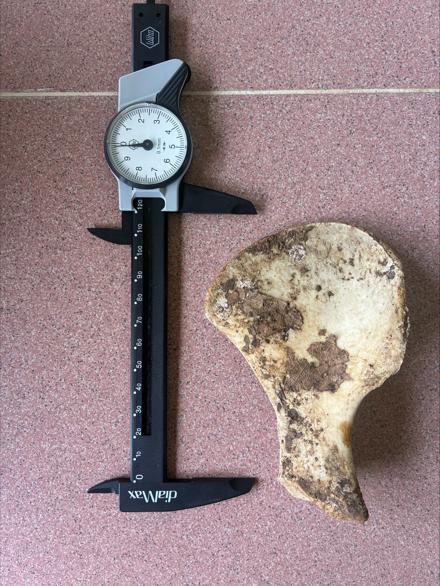
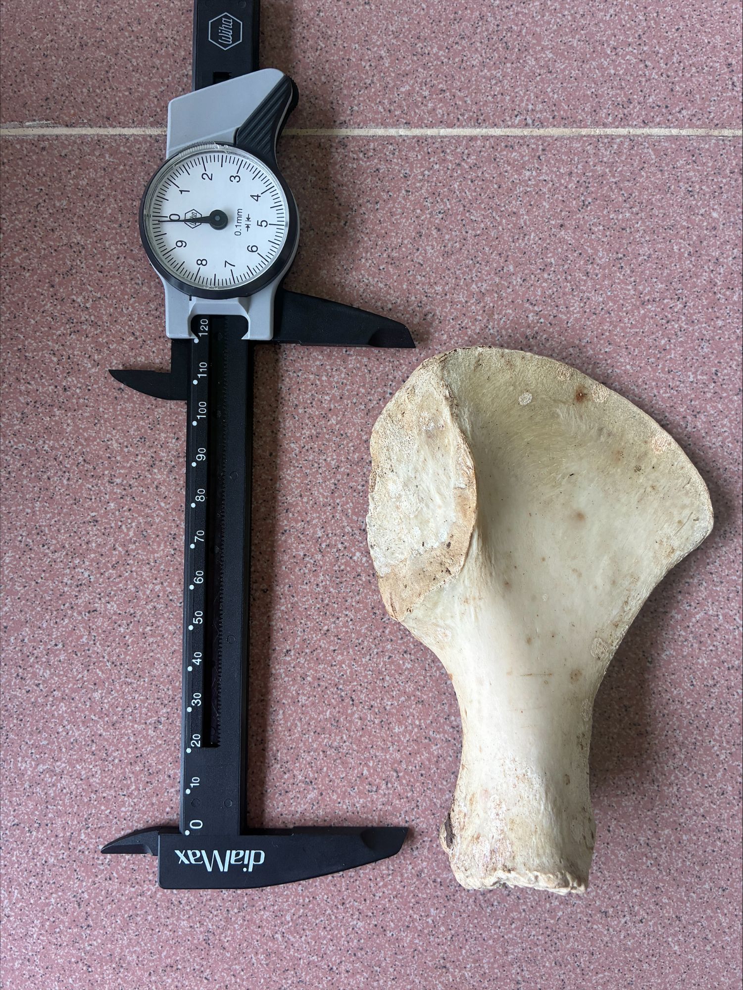
This week I have bony mystery object for you.:
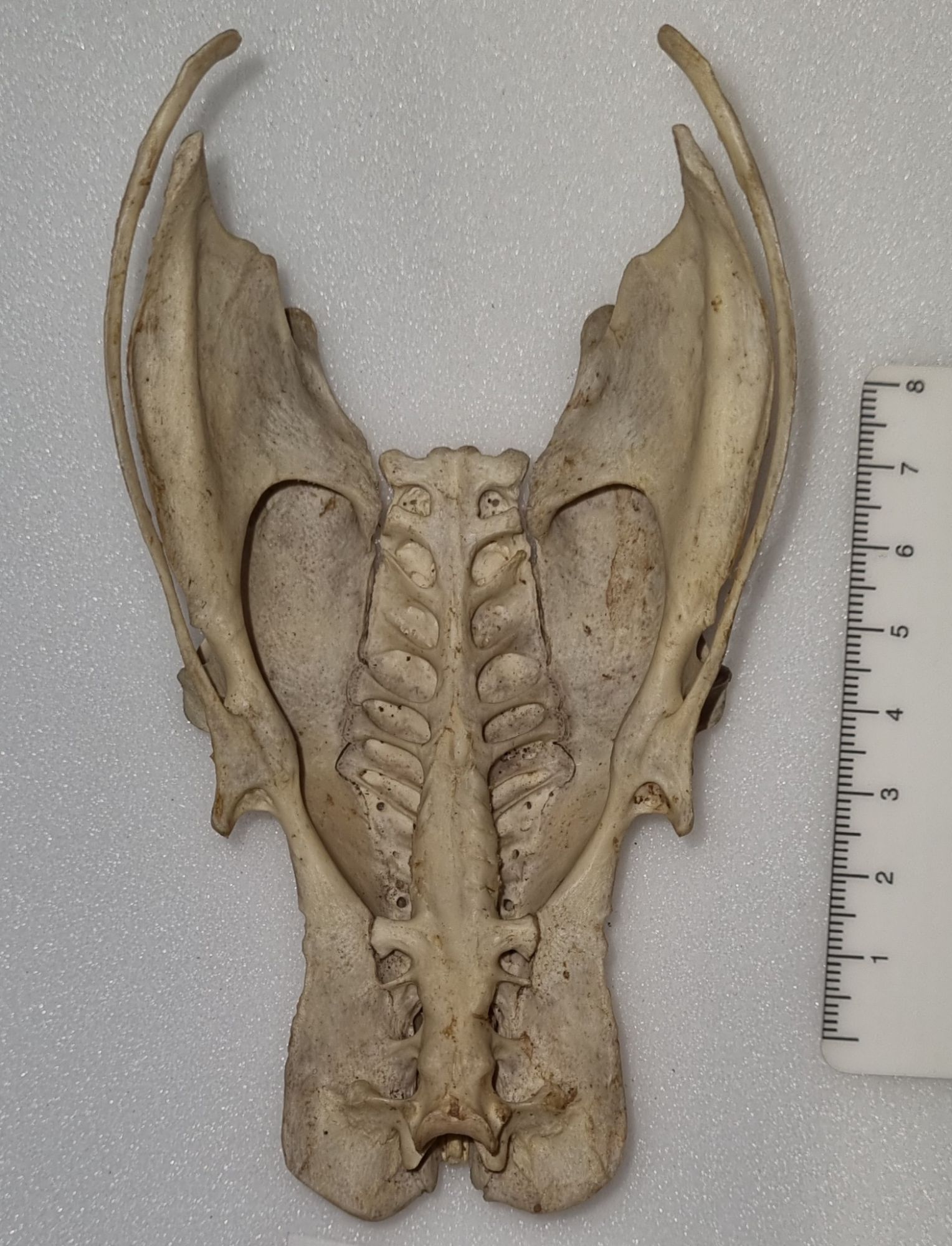
Do you have any idea of the species that this came from?
As ever, you can leave your questions, observations and suggestions in the comments section below. Have fun with it!
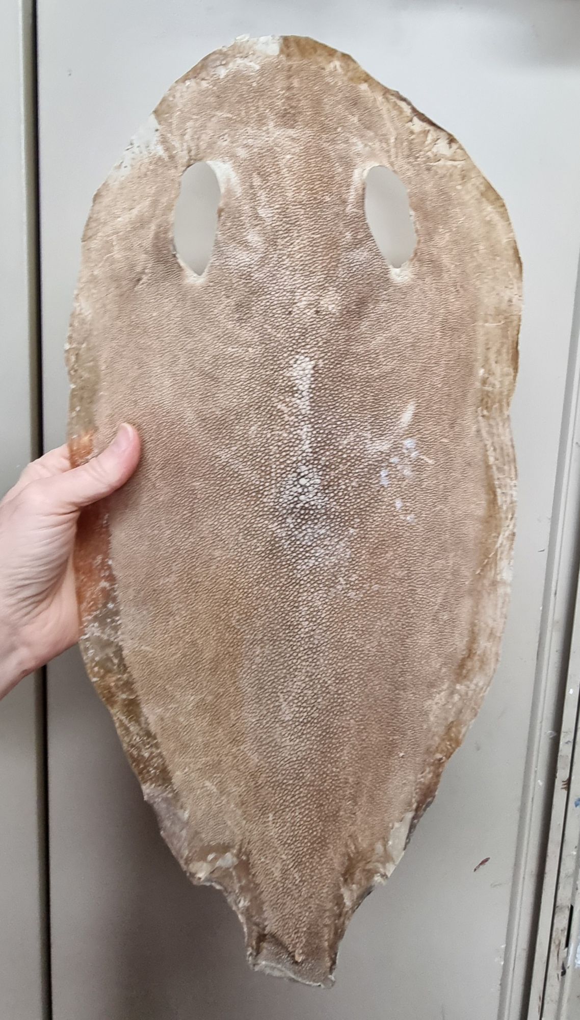
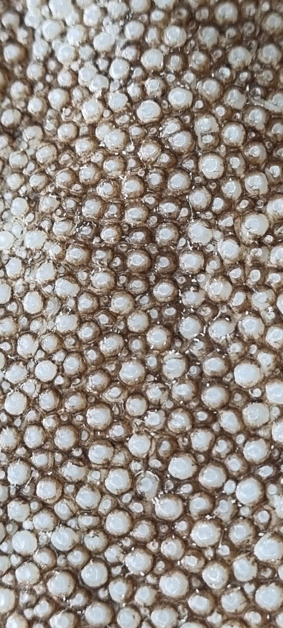
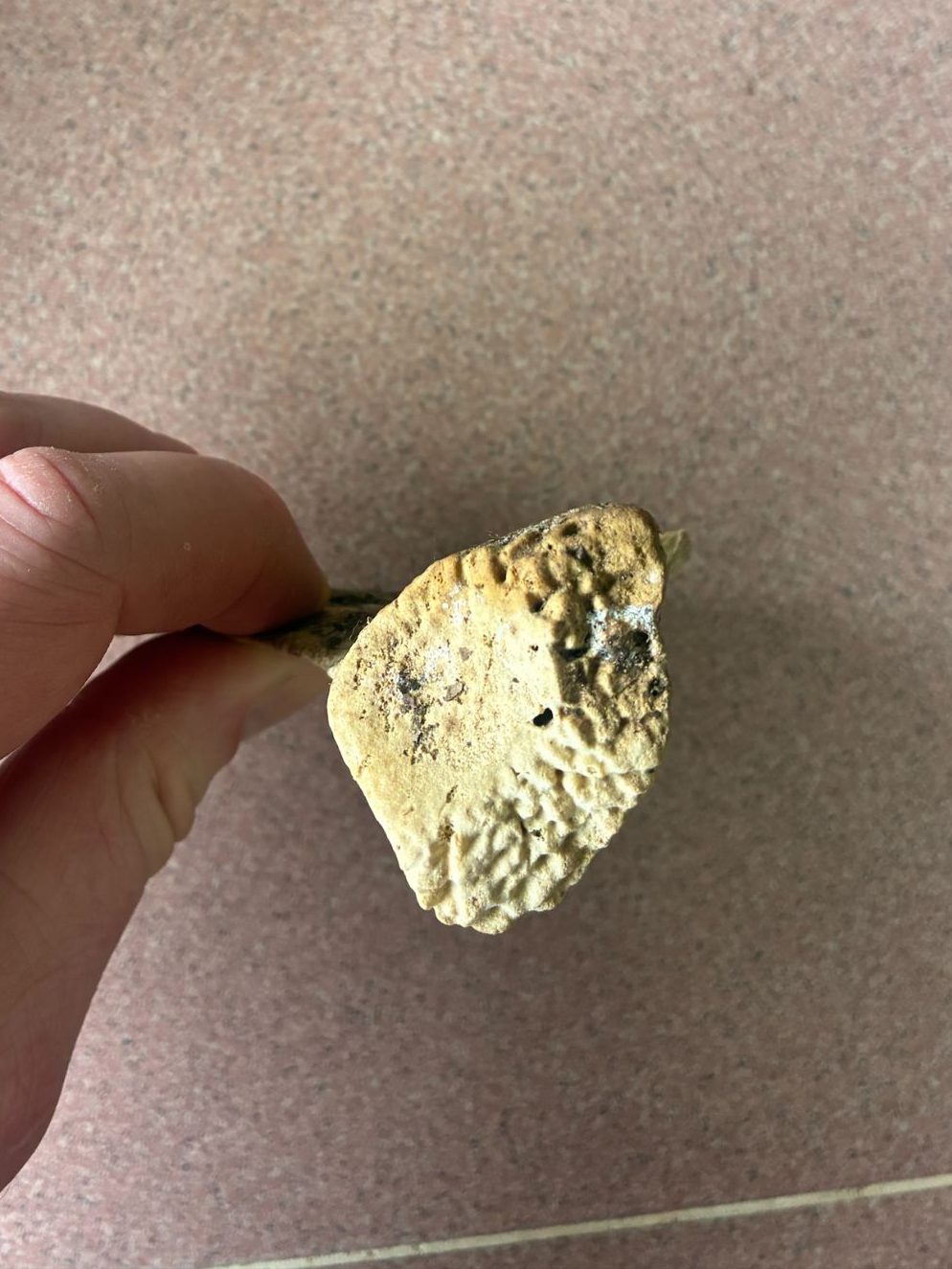
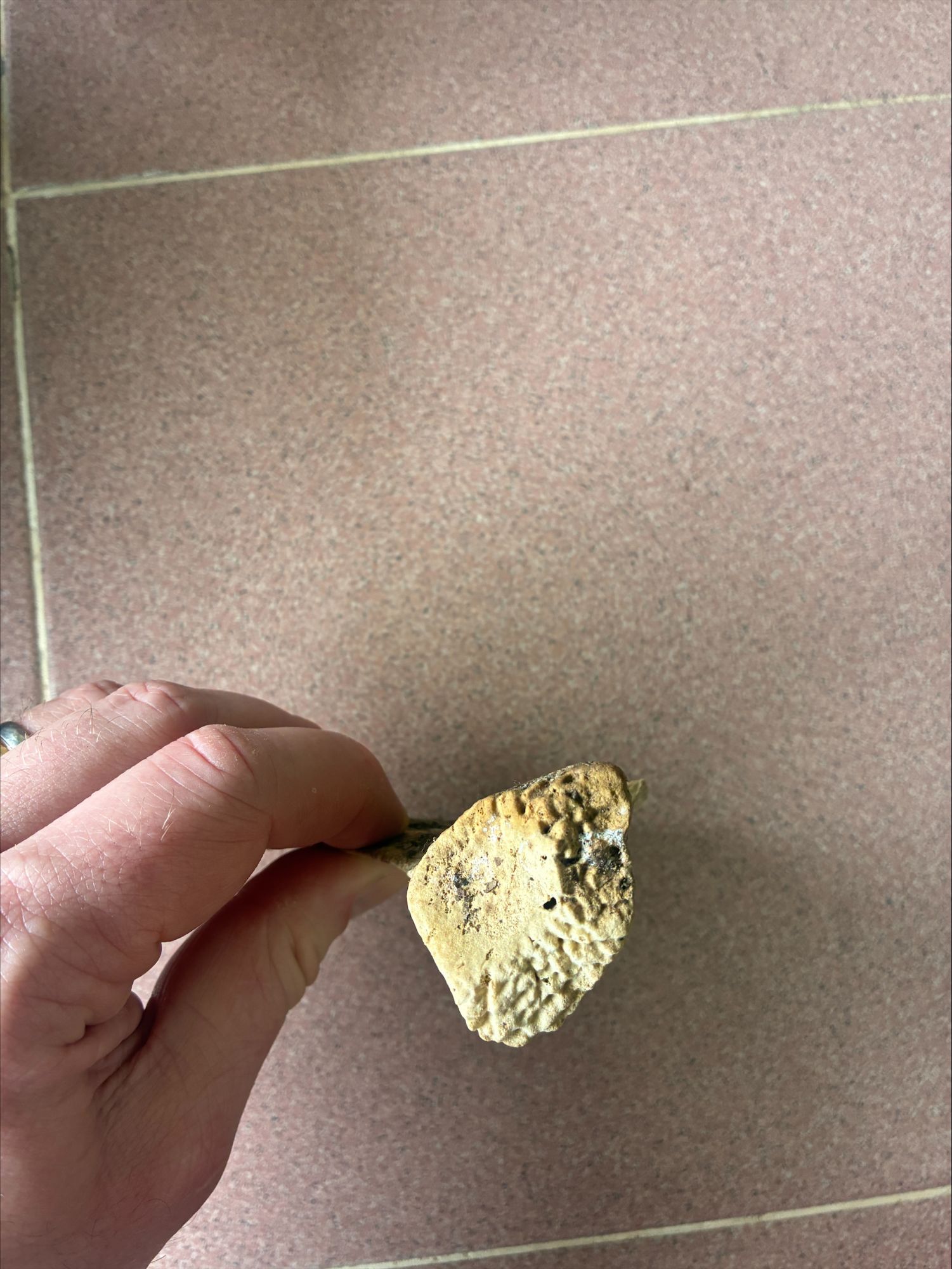
One type of enquiry we get in the museum relates to the identification of natural materials. Often these come from law enforcement or customs officials, who may need an expert eye cast over a material to check whether it’s been imported or sold illegally.
Here’s a worked piece of natural material – I’d be keen to hear your thoughts on what it might be:
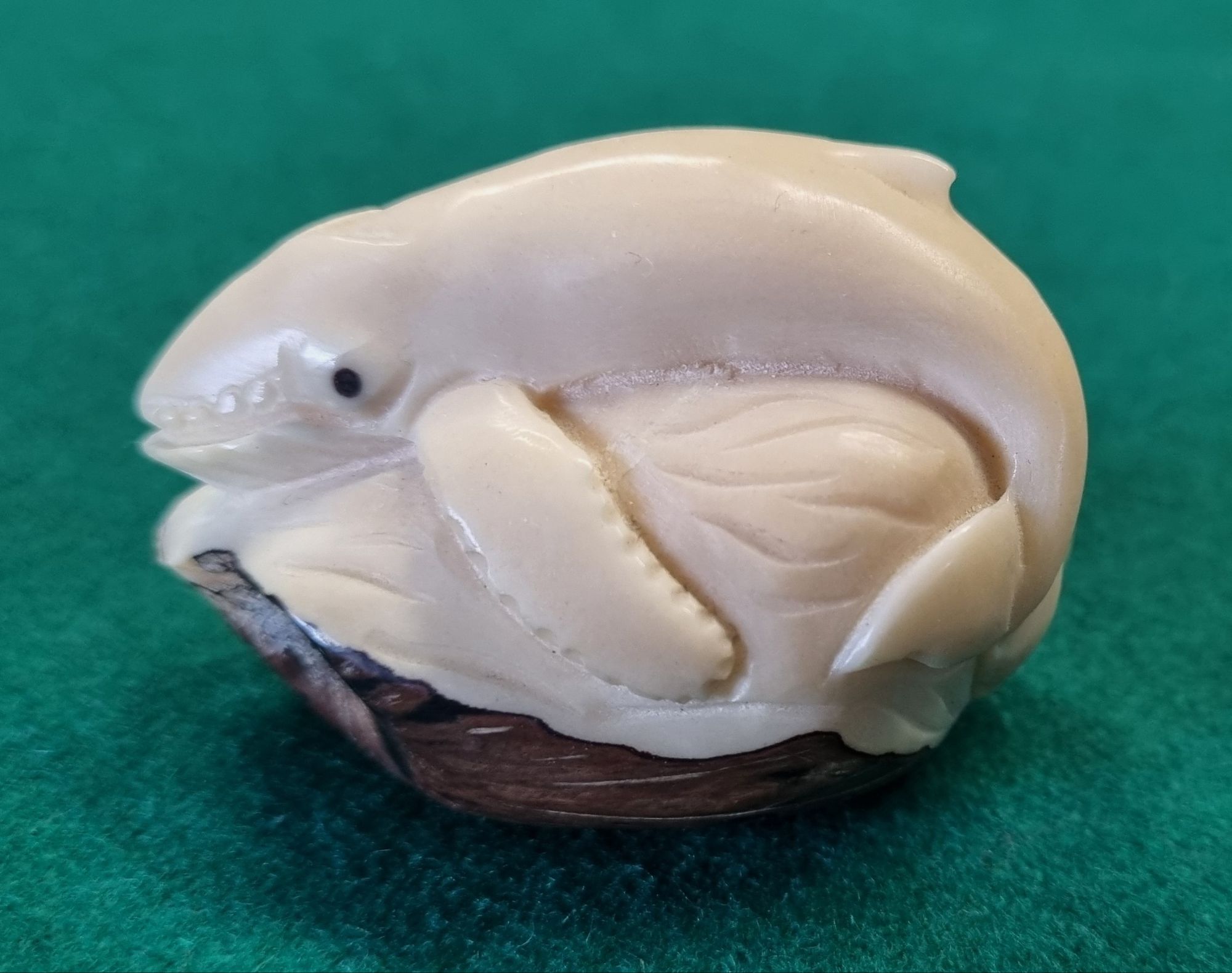
As ever, you can leave your suggestions in the comments section below. I look forward to hearing your thoughts!
Last week I gave you this somewhat faded and oddly shaped fish to have a go at identifying as a New Year challenge:
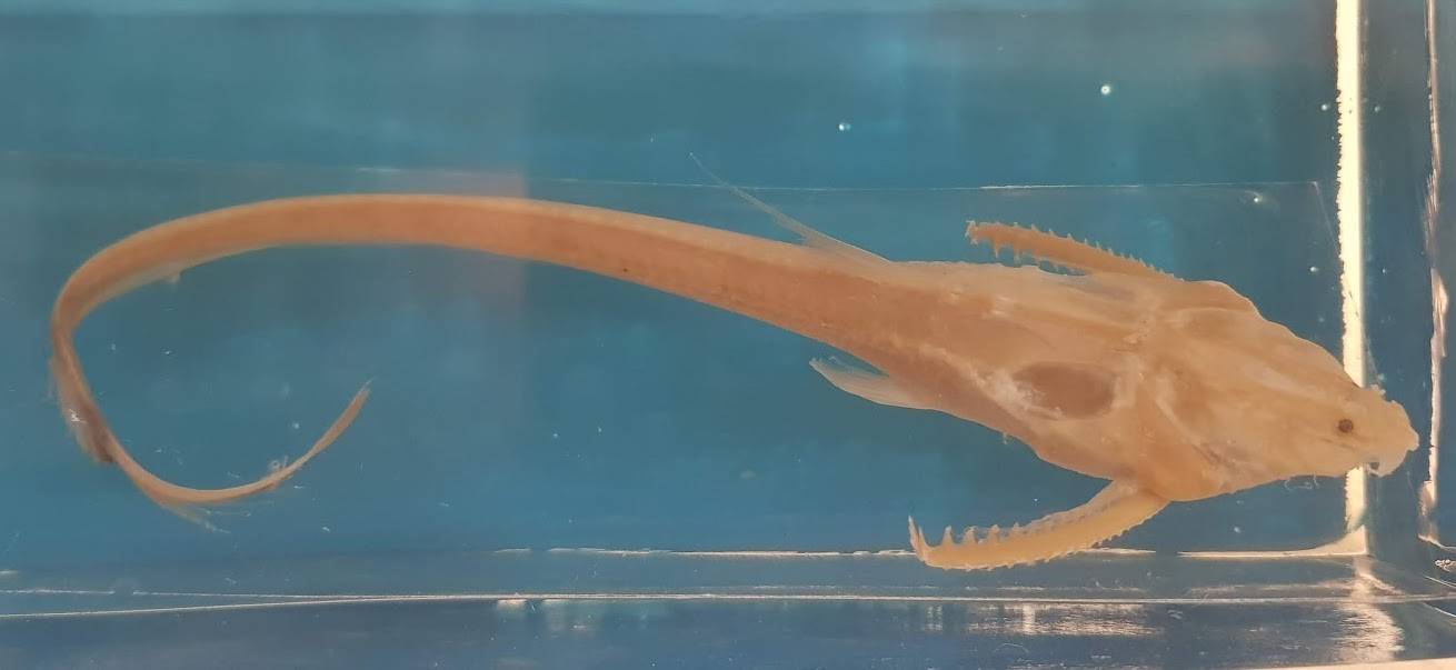
It was a very big ask, as the image isn’t really detailed enough to allow a species identification, but it was great to hear your various thoughts.
Adam Yates got very close with the suggestion it could be one of the Loricariidae – a family containing the armoured suckermouth catfish, but while this is from the same Order (the Siluriformes) this particular catfish is from a different Family.
While it has an armoured appearance, and shares those ornamented pectoral fin spines that are found in many catfish, this one has a filamentous tail and dorsal fin (that you can only just make out). What you can’t really see are the eight pectoral fin rays and the seven barbels present on the head.
This long thin tail is a hint that this is one of the Banjo Catfish, and the details of the barbels and pectoral fin rays I mentioned above let us know that it’s the Sevenbarbed Banjo Aspredinichthys filamentosus (Valenciennes, 1840).
For some reason the name Banjo Catfish always makes me think of this scene from the film Deliverance:
Musical shenanigans aside, these South American fish are bottom feeders in brackish waters, and have the unusual reproductive trait of the female attaching her eggs to her underside, so they can be moved around in the muddy waters in order to keep them oxygenated during their development.
That was certainly a challenging mystery object to start 2026, so I may see if I can find a slightly easier, but hopefully no less interesting specimen for the next mystery object!
Happy New Year! I hope you enjoyed seeing in 2026 and I wish you all a very healthy and enjoyable year ahead.
This Friday’s mystery object is a specimen from the spirit collections in the Dead Zoo:
Do you know what this mystery aquatic beastie might be?
Let me know your thoughts in the comments below – I hope you enjoy the challenge!
