Last week I gave you this detail of a specimen from the Dead Zoo to identify:
I don’t think anyone nailed the identification to species, but several of you get very close, with a trend towards the ursine.
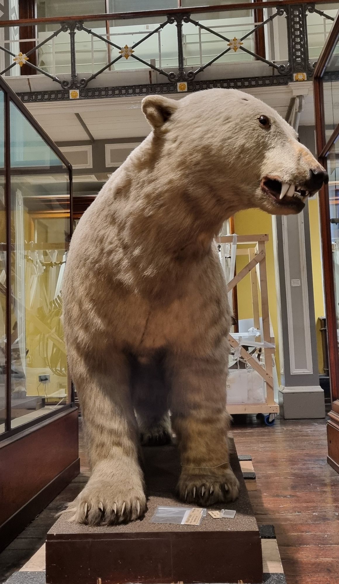
This is the mouth of a Polar Bear Ursus maritimus Phipps, 1774 and even more precisely, it’s the mouth of the Polar Bear shot by Irish explorer Leopold McClintock in April of 1851 on the pack ice between Bathurst Island and Byam Martin Island, in the Canadian Arctic Archipelago.
McClintock’s Polar Bear
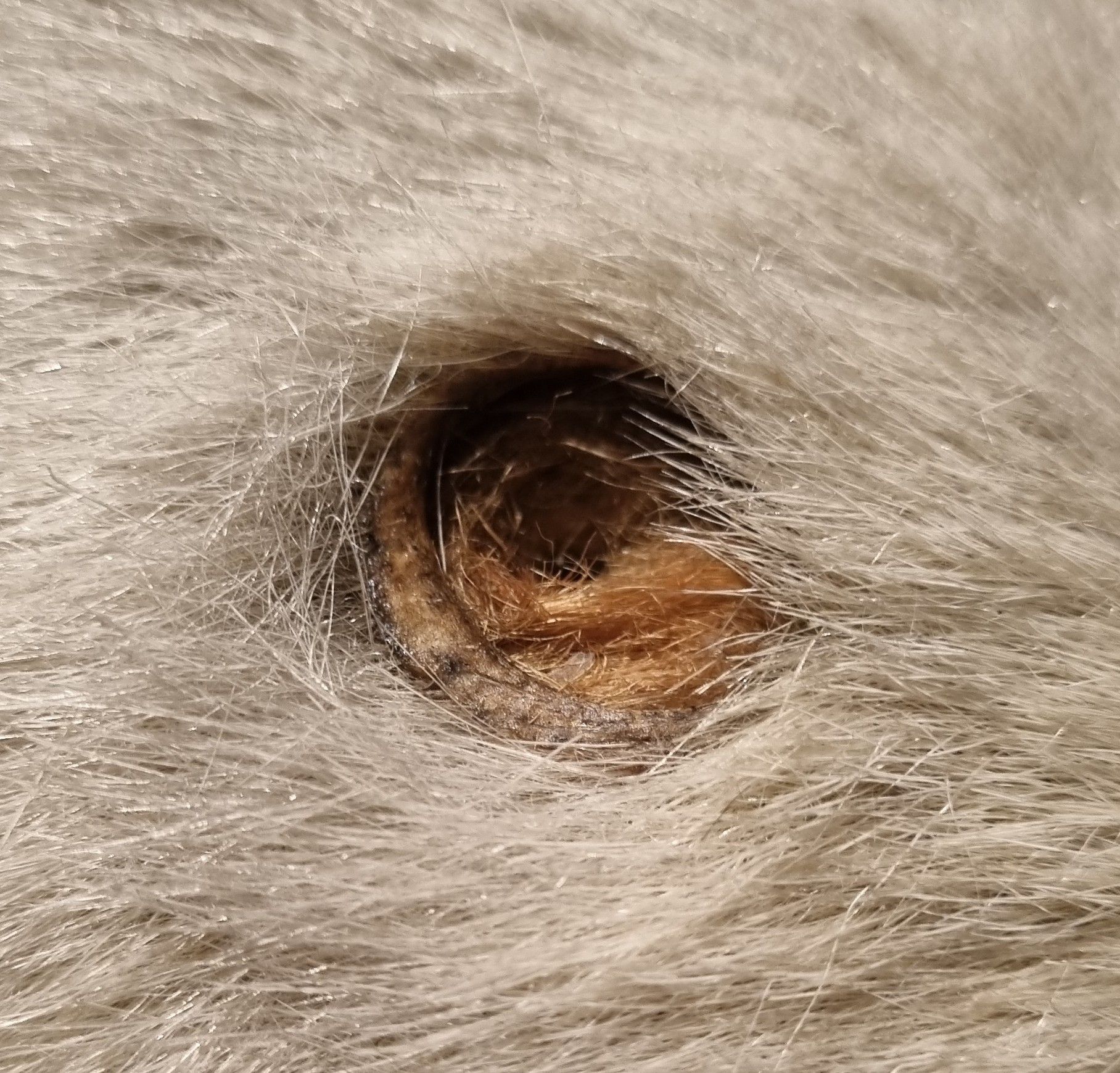
As I understand it, this particular bear was tracking McClintock’s group on the ice, and was shot by McClintock to protect the party (and provide some fresh meat). However, the specimen has several bullet holes, which suggest that more than one member of the group took a pop at the bear.
One of several bullet holes in the Polar Bear
Regardless, McClintock is identified as the collector and the specimen was presented to the Royal Dublin Society by Erasmus Ommaney in December that same year. Ommaney was second in second in command of the HMS Resolute, the ship being used to on an expedition to discover the fate of Sir John Franklin, who was lost while attempting to discover the Northwest Passage.
So well done to everyone who managed to recognise the toothy grin of this enormous predator – the animal reputed to give the Arctic its name (“Arctos” meaning “Bear” in Greek – although that probably refers to the constellation Ursa Major or Ursa Minor, which are key navigable features in the northern sky).
Last week I gave you this specimen from the Dead Zoo to have a go at identifying:
It didn’t take long for Chris Jarvis to drop a great clue to the correct answer, and it seems that overall it was a bit of an easy one for quite a few of you. I admit that I’m not overly surprised by that, since it’s pretty distinctive.
The scapula is long and curved, the humerus is relatively short and robust, the radius and ulna are robust and quite flattened, as are the digits on that wide and splayed-out hand. All of these elements add up to a flipper shape, but unlike the flipper of a cetacean or seal, it has quite short digits.
That’s because this is the left arm of a slow-moving aquatic mammal that plods along under the water (as much as plodding is possible whilst being underwater), rather than a hydrodynamic fast-swimming beastie with a flipper shaped to cut through the water.
There are still a few possible species that fall into this category, but of then all, there is only one with such a curved scapula shape – the Dugong Dugong dugon (Müller, 1776).
Dugong (Dugong dugon) underwater. Image by Gejuni, 2015
I picked this mystery object because it’s from one of several specimens that we’ve been working on recently, in preparation for transport out of the Dead Zoo, as we prepare for a major capital project on the building. The limbs, skull and tail were removed and the vertebrae and ribs stabilised in a structure that we call a “stillage”:
With enough wrapping and packing this specimen will be ready to crane out of the building in the near future, and it’s just one of several thousand specimens that will be making the move. If you’re interested in hearing more about the project, you can listen to an interview I did recently with Sean Moncrieff on NewsTalk.
I hope you enjoyed working out what this limb belonged to, and I suspect that there will be more tales (and possibly tails) from the decant coming up in future posts. Happy Friday!
Last week I gave you this specimen to identify, which came to me as an enquiry, after being found in the sea by a fisherman:
I don’t think it posed too much of a challenge, despite some damage, which has left sections looking a bit different to usual for this skeletal element – which is a section of the lower jaw or mandible.
This piece of the mandible includes the ramus (the rear part of the jaw behind the toothline where it rises up), coronoid process (the section of bone that rises up through the inside of the cheekbones, and where the temporalis muscles attach to power part of the action of the jaw) and the mandibular condyle (the hinging articulation point where the lower jaw meets the rest of the skull).
The scale bar shows that it’s fairly large, and the shape of the coronoid process and articular condyle are what I would consider to be quite distinctive to herbivores, since a long ramus isn’t well suited to resisting forces from struggling prey or meat-cutting bites.
From this point I find it’s useful to check an image I prepared earlier (and by earlier, I mean about 10 years ago):
A quick comparison makes it fairly clear that the mystery object is part of the mandible of a Cow Bos taurus Linnaeus, 1758, based mainly on the shape and orientation of the mandibular condyle.
There are of course species that could possibly turn up in Irish waters, that aren’t on my mandibles photo – in particular, Giant Deer, which Ireland seems to have a lot of. However, I have easy access to those specimens in the Dead Zoo and they have a similar mandibular condyle orientation to a Red Deer.
So well done to everyone who worked it out – I hope my explanation of the anatomy of the rear part of the mandible makes sense and maybe offers some pointers for identifications you might be faced with in the future!
Last week I gave you this skeleton fron the Dead Zoo to test your identification skills:
In retrospect I think I was a little unfair with this one – the photo is not very clear and there is no scale bar, so the identification relied mainly on the context provided by the mount and a lot of deduction. Not an easy task with a rodent, since there are so many different species.
The branch used as a setting for the skeletal mount provided the main and most important clue – it indicates that the species is arboreal. A lot of people picked up on this, with guesses ranging from a flying squirrel to a viscacha. However, the answer is something from a bit closer to home (i.e. Europe).
This is the skeleton of the Edible Dormouse Glis glis (Linnaeus, 1766), a plump (and presumably tasty if you happen to be an ancient Roman), tree-dwelling rodent, with a reputation for somnolence.
Edible dormouse (Glis glis) in an old shed in an abandoned plum orchard in Luc-en-Diois, France. Image by Bouke ten Cate, 2011
This isn’t the first time I’ve featured a dormouse in the blog, although the previous one was a giant extinct example. The Edible Dormouse is the largest species alive today, but it’s still smaller than the fairly diminutive Red Squirrel.
They are fairly well distributed around central Europe, with a small population in Southern England due to escapees from Walter Rothchild’s menagerie in Tring in my home County of Hertfordshire. I’ve heard tell that they can be a bit of a pest in the area, due to their habit of seeking out attics to hibernate in, but then chewing through wires and cables, thus causing fires and broadband outages.
This UK population didn’t arrive until the early 20th Century, so the species that inspired Charles Dodgson (AKA Lewis Carroll) to include his sleepy character was almost certainly the smaller Hazel Dormouse, which occurs in Britain, and which also turned up in Ireland around County Kildare around 14 years ago (and which we have specimens of, thanks to a gift from someone’s pet cat).
I should have either provided a better image or a clue to point you in the right direction for this mystery object, so I feel I’d better apologise for setting this vexatious conundrum and promise to better next time!
Last week I gave you this doe-eyed specimen from the collections of the Dead Zoo to try your identification skills out on:
I didn’t provide a scalebar as I think it would have made it too easy, but even so, it’s clear that the specimen is a very small species of artiodactyl (the group containing pigs, deer, antelope, bovids and a variety of related herbivores).
There were some suggestions that it could be a Dik-dik, but as Adam Yates pointed out, this specimen lacks the large preorbital glands that are very visible in Dik-diks (and makes them look like they got carried away with the eyeliner):
Dik-diks with their distinctive preorbital glands
The mystery object lacking preorbital glands
The other popular suggestion for the identity of the mystery object was a Java Mouse-deer (or Javan Chevrotain), which is the smallest ungulate alive. However, while that’s exactly what it says it is on the label, the location of collection rings alarm bells for me:
There are two species of chevrotain found in Singapore, and the Javan species is not one of them.
Of the two, one is the Greater Mouse-deer and the other is the Lesser Mouse-deer. The Greater, as you probably guessed, is on the large side for a chevrotain, weighing in between 5 and 8kg. This species also has a dark stripe from its nose to its eye, which is missing from the mystery object.
The Lesser Mouse-deer Tragulus kanchil Raffles, 1821 lacks the dark stripe and is almost as tiny as the Javan Mouse-deer, making it the most likely candidate for the mystery object:
Lesser Mouse-deer alongside some rodents
This specimen not only has that likely identification error on the label (easily done considering the complexities of chevrotain taxonomy across Southeast Asia), but it had somehow also had a completely incorrect label associated with it in the past, which said it was a Siberian Musk Deer – a species that’s on the small side, but by no means as tiny as this.
This specimen was of particular interest at the end of last year, when we had a visit by a group of researchers from Singapore, who are undertaking a fantastic project to digitise specimens collected from Singapore that are held in museum collections all around the world. The project is called SIGNIFY and the team were not only absolutely lovely people, but they achieved a huge amount of research and detailed imaging work in a very short time:
The SIGNIFY portable imaging setup in use on a specimen from the Dead Zoo bird skin collection
The SIGNIFY project has huge value for helping to understand the historic baseline biodiversity of Singapore prior to industrialisation, but it also helps foster links between organisations and allows the inextricably linked social and personal histories of collectors to be explored. I loved getting a chance to spend time with the team, learning more about their project and the collections I care for. It also turns out that we have a wealth of spiders from Singapore that still need to be investigated, so I really look forward to welcoming the team back soon!
Last week I shared this fuzzy critter as mystery object for you to identify:
It was probably a bit of a mean one, as I didn’t provide a scalebar. It’s also a species from a group of small carnivores that contains over 30 species that can look quite similar, and (perhaps most importantly) the specimen is old and very faded from being on display in a gallery space with lots of light for the last 100 years or more.
Natural light in the Dead Zoo, image taken in May 2020.
Regardless, Chris Jarvis figured it out (after an initial near miss), while I believe that Joe Vans cheated by checking out the 3D tour of the Dead Zoo. Most other comments on social media came close, with a lot of people working out that this is a Mongoose, but then not quite getting the species.
This specimen is a Common Kusimanse Crossarchus obscurus G. Cuvier, 1825. These are also known as the Long-nosed Kusimanse, as their snout is a bit more elongated than that of most other Mongooses (or should that be Mongeese?):
The Common Kusimanse is one of the African Mongooses in the Subfamily Mungotinae. They are a good bit shorter than most of the other species of Mongoose – for comparison here’s this specimen next to a Small Indian Mongoose (well, that’s what the label says, although I have some doubts):
They are normally a dark brown colour, like this example:
Common Kusimanse by LA Dawson, 2006.
However, the Dead Zoo specimen is now bleached blonde, so I’m not surprised that this identification was tricky. Fading leads to all sorts of issues for the accurate representation and identification of species, to the point where the Giant Panda on display in the building had to be dyed black in places a few years ago, because it had ended up looking like a Polar Bear cub due to the sun damage.
At the moment that’s no longer an ongoing issue in the Museum, as a temporary floor has been installed just beneath the old glass ceiling, to allow investigations on the roof space to take place – this blocks almost all of the natural light. This has been great for conditions in the building, as it no longer heats up like a greenhouse on sunny days, and the bleaching of the specimens has been put on hold.
At some point the tempoprary floor will be removed, but I sincerely hope that a more permanent solution to the light issue will have been put in place by then. Still plenty of work to be done to get to that point though!
Dead Zoo with temporary floor installed to allow roof access. This blocks the natural light and keeps the environment more stable.
Last week, I gave you this shiny blob to have a go at identifying:
There wasn’t much to go on, since it is just a blob that looks like a chunk of hardened tar, but it is in fact a rare and valuable natural material. It’s actually a small piece of ambergris.
Ambergris has always maintained an air of mystery, since it’s formed deep within the bile duct of a Sperm Whale and its function in the animal is still only suggested rather than fully understood. General agreement seems to be that this tarry substance provides protection for the intestines from the sharp beaks of the squid that Sperm Whales prefer to eat.
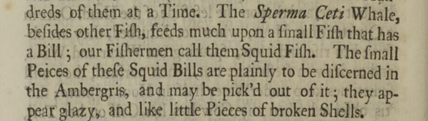
Evidence for this comes from the fact that squid beaks are often found embedded in ambergris – an observation recorded from as early as 1725:
An essay upon the natural history of Whales, with a particular account of the ambergris found in the Sperma Ceti Whale. In a letter to the publisher, from the Honourable Paul Dudley, Esq; F. R. S Published:30 April 1725https://doi.org/10.1098/rstl.1724.0053
This letter goes on in some detail about various whale species, offering details of their economic yield in terms of barrels of oil, quantity of whalebone (baleen) and medicinal uses of teeth, as well as some aspects of their biology. When the discussion gets onto ambergris, much of the focus is on its location in the whale and the method of extraction (N.B. it’s not pretty.)
Of course, the suggested function of ambergris as a mechanism to aid the passing of sharp objects through the gastrointestinal tract would indicate that ambergris also might emerge naturally, and doesn’t necessarily need to be ripped from an unwilling victim.
However, evidence for this is quite hard to find, since it’s remarkably difficult to follow a Sperm Whale and keep track of its bowel movements or regurgitations (which have also been suggested as an exit route for these masses of indigestible items). What is known is that ambergris can be found floating at sea and washed up on beaches, sometimes persisting for years (there has even been fossilised ambergris discovered in Italy).
This non-invasive method of harvesting ambergris by beachcombing may not be hugely efficient, but it supplies the majority of the ambergris now used in perfume manufacture. Yes, you heard me right.
Human interest in ambergris may seem surprising given its somewhat revolting source, but as we all know, humans are pretty weird when it comes to making use of the fruits of nature, especially when searching for ingredients for perfume – just think of the African Civet and its anal excretions.
The natural complex aromatic compounds found in waxy substances like ambergris and civet musk provide long-lasting base odours that have played an important role in creating perfumes for centuries. Modern chemical synthesis of similar products has taken over to a large extent, but the naturally occurring compounds are still in use today.
This particular blob of ambergris was photographed when we were getting it out for sampling to inform a rather different line of scientific questioning, since there is still a lot to learn about this very unusual natural material.
Last week I gave you some festive-looking specimens to have a go at identifying:
Image by Charles Fisher, published in Microfauna–Macrofauna Interaction in the Seafloor: Lessons from the Tubeworm. Boetius A PLoS Biology Vol. 3/3/2005, e102 doi:10.1371/journal.pbio.0030102
I thought these were some specimens in the care of Andy Taylor, FLS, but this was my error – Andy sent me the images to suggest the species as mystery objects, but I didn’t realise that he hadn’t photographed his specimens to use at that point. These images are actually from a paper (referenced above) that discusses the species and the blue-green colour is a stain added to allow the growth rate of the tubeworms to be calculated (spoiler alert – it’s very slow).
Here are Andy’s specimens:
Image by Andy Taylor FLS, 2023.
A bit less colourful, but the tubes retain the same structure, with those clearly defined rings.
As Adam Yates said in the comments, these are specimens of Lamellibrachia luymesi van der Land & Nørrevang, 1975. They have similarities to other genera, such as Hilary Blagbrough’s suggestion of Ridgeia and katedmonson’s suggestion of Riftia.
Image of Ridgeia specimens by Andy Taylor FLS, 2023.
Image of Riftiapachyptila specimen by Andy Taylor FLS, 2023.
Species like Riftiapachyptila are from hydrothermal vents and that nutrient rich and high temperature environment gives their symbiotic bacteria a boost that allows Riftia to be the fastest growing invertebrate, reaching around 1.5m long in just a couple of years. This is useful as it allows rapid colonisation of these ephemeral volcanic environments that occur at mid-ocean ridges.
On the flip side, Lamellibrachia luymesi tubeworms live in cold seeps of hydrocarbons in the deep ocean, where their symbiotic bacteria have to work at temperatures of 4°C or less, making their energy production a slow process. Consequently, L.luymesi are one of the slowest growing invertabrates, taking around 125 years to reach 1.5m long. Cold seeps are much more stable than the hydrothermal vents however, so L. luymesi have been found to continue growing up to 3m, taking around 250 years, and therefore being among the longest lived invertebrates (and indeed animals) on the planet.
Some might suggest that there’s a lesson to be learned here about “slow and steady winning the race”, but slow growth would be disastrous for a species that relies on a rapidly changing environment. Both species are remarkably adapted to their environment and neither would do well in the other’s place.
It’s worth noting that both of these remarkable organisms are only as successful as their symbionts allow them to be, so if there’s any lesson to be shared, it’s probably that the value of teamwork should never be underestimated.
On that (somewhat cheesy) note, I would like to thank Andy once again for sharing his collections. I’ll be back in the New Year with another Mystery Object – I hope you enjoy the celebrations!
Last week I gave you this mystery object to have a go at identifying:
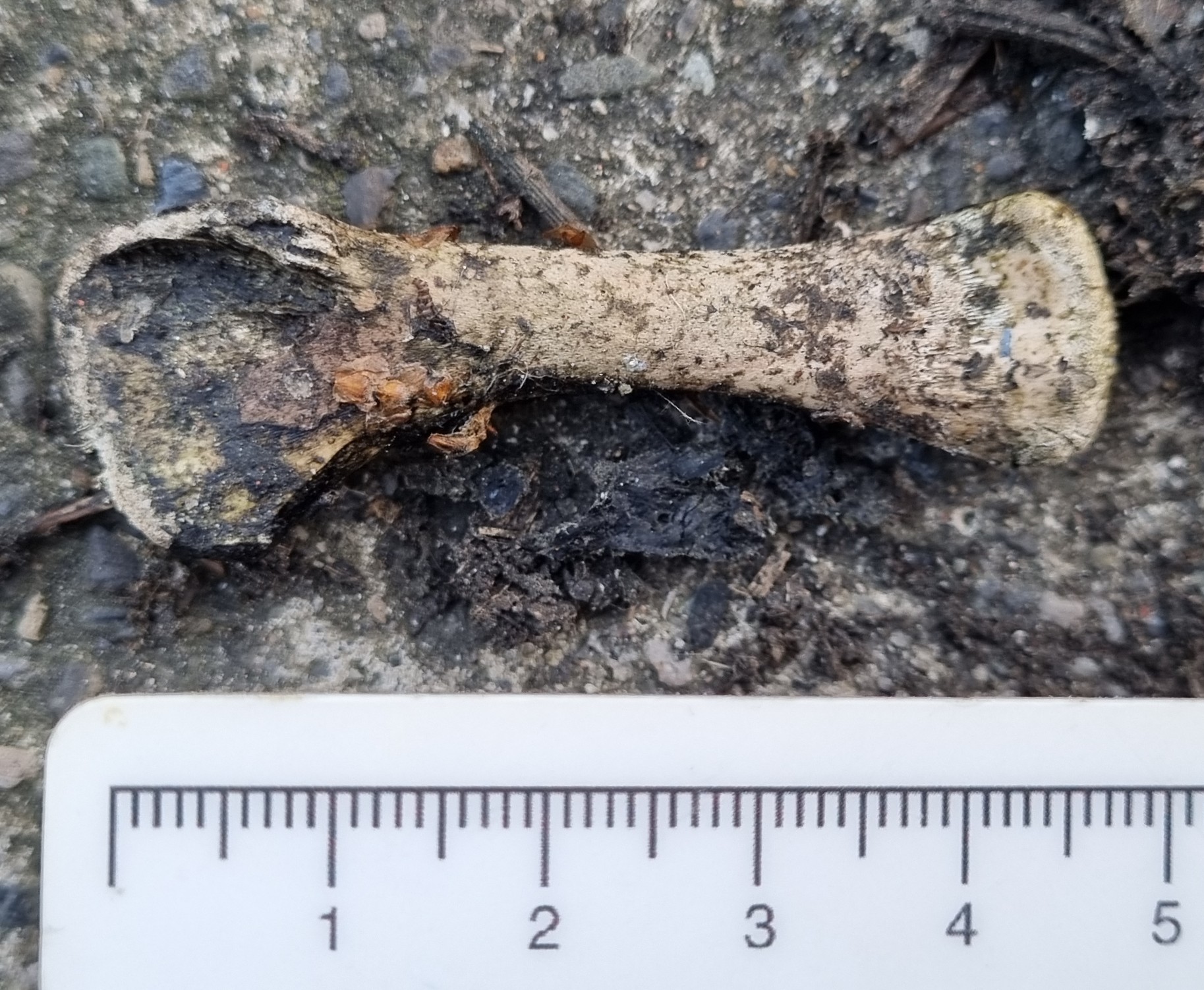
Not the prettiest object perhaps, but I did find it in the gutter on my street, so I think that’s excusable.
This isn’t the most difficult specimen to identify – in fact, I think pretty much everyone should be familiar with it, since it’s probably one of the most commonly found bones in the world.
Chris was the first reply, within 23 minutes of the blog being posted. I was lucky enough to see Chris in Oxford this week, and he confirmed that most of that time was spent coming up with a suitable cryptic clue. And it was a spot on:
Foul! You should of cleaned it first, Paulo (although it is quite funny!)
Chris says: September 1, 2023 at 8:23am
As Chris hinted, this is of course the humerus of a Chicken Gallus domesticus (Linnaeus, 1758).
As I mentioned, this is probably the single most commonly encountered bone you’ll find. There are an estimated 34 billion Chickens alive at any given time, with around 74 billion being slaughtered for food each year, so it’s no surprise that their leg and wing bones accumulate wherever you find people.
In fact, the presence of a high density of Chicken bones in sediments is considered to be one of the features that will help to define the Anthropocene period.
The high density bit is important, since the Red Jungle Fowl has been around for 4-6 million years in Asia at low densities, but with the domestication taking place over 3,500 years ago, Chickens have travelled the globe with Humans, providing eggs and meat for a huge range of cultures.
But it’s not until huge numbers started being reared commercially in the 20th Century that landfills started containing vast numbers of bones from these birds.
Image from the George F. Landegger Collection of Alabama Photographs in Carol M. Highsmith’s America, Library of Congress, Prints and Photographs Division. 2010.
Alongside a variety of other materials generated by human activity, from soot to radiactive isotopes dispersed around the globe by nuclear testing, chicken bones are providing a diagnostic features for geologists of the future to recognise the start of the Anthropocene.
So, bravo to Chris, and be sure to remember what this bone looks like, as I’m sure you’ll see plenty of them in future!
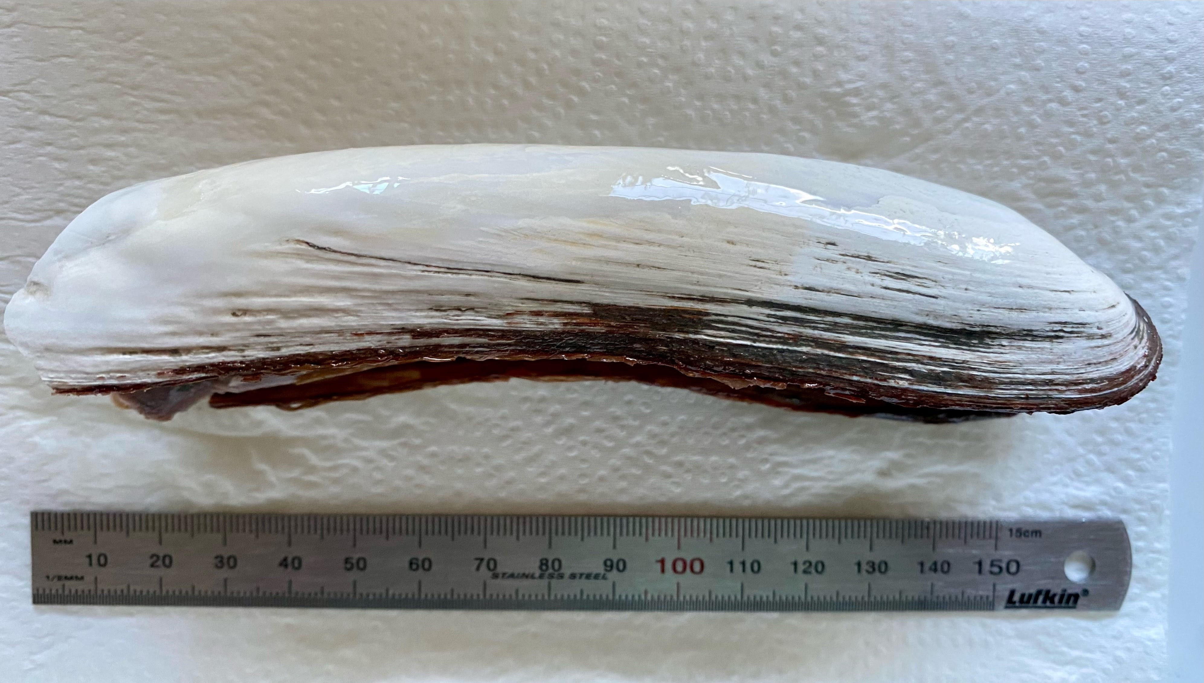
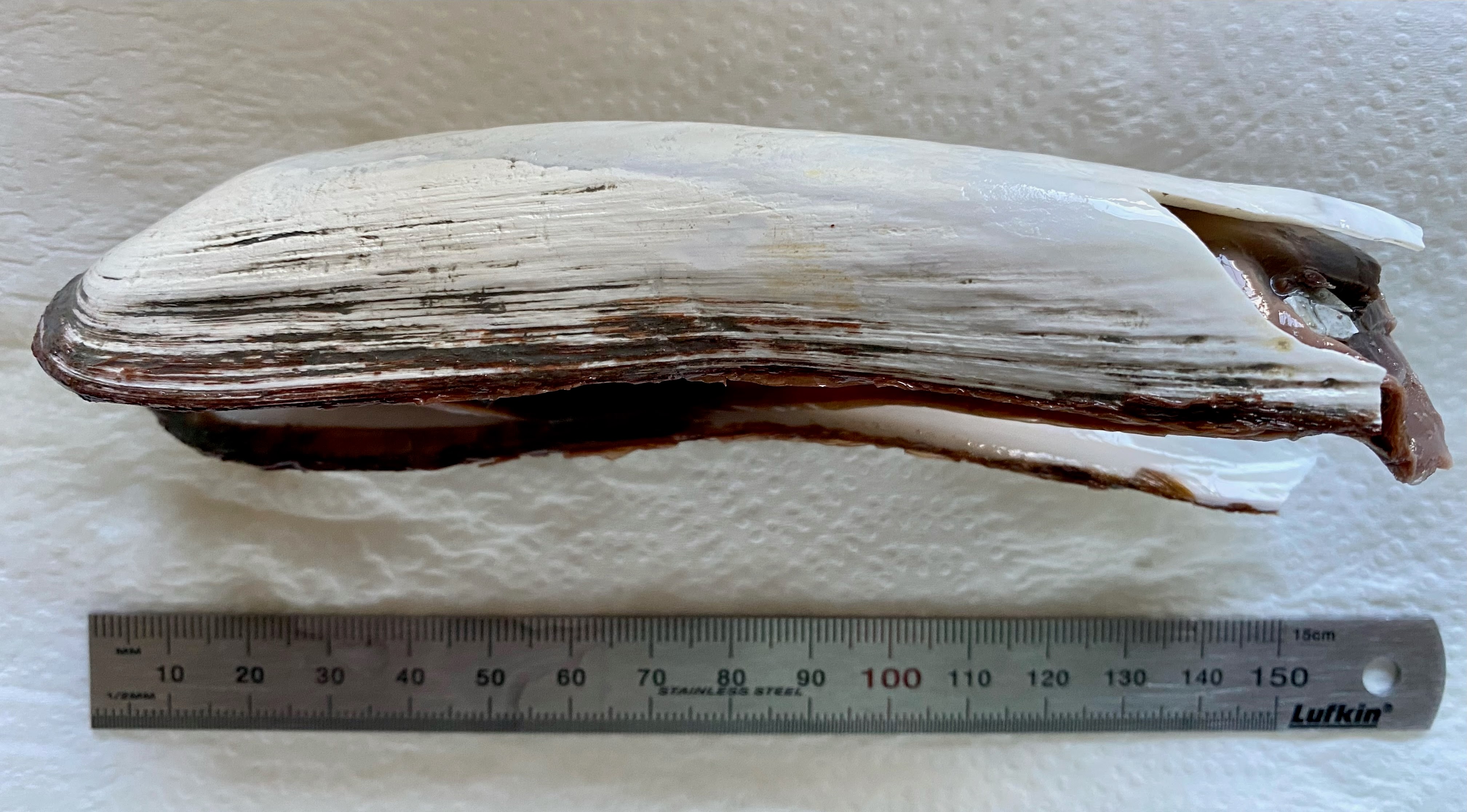
Last week we had a very difficult guest mystery object (or objects, as there were two specimens). These are from the collections of Andy Taylor, FLS:
Image by Andy Taylor, 2023
The general consensus in the comments was that they are a type of mollusc, and due to the elongated nature there were a few suggestions of something in the Razor Clam area of the crunchy-yet-squishy zone of the tree of life.
But these are a bit more unusual than that, and unfortunately nobody seems to have picked up on my ever so cryptic clue:
You may need to delve into the depths of the internet to work it out
This refers to the fact that this species is one of the denizens of the deepest parts of the world’s oceans.
This combined with the characteristically elongated shell shape does help to narrow it down, although it takes a lot of work – or a degree of familiarity to work it out.
Remarkably, Dennis C. Nieweg on LinkedIn did manage to figure it out to the previous generic name of Calyptogena, which is hugely impressive for such an unusual and generally unfamiliar specimen.
These are specimens of Abyssogena (was Calyptogena) phaseoliformis (Métivier, Okutani & Ohta, 1986). They are very deep living bivalves in the Order Venerida, that survive around deep-water vents and seeps in the Abyssal zone and which were first described when submersibles were developed that could sample at great depths – opening up a whole new realm of discovery.
The details provided by Andy are as follows:
First specimen is from the Japan Trench and was collected at a depth of 6347m in 1997 by ’Shinkai 6500’ DSV (Deep Submergence Vehicle) operated by JASTEC (Japan Agency for Marine and Earth Science).
This second specimen was collected from the Aluetian Trench at a depth of 4776m – 44949m in 1994. This specimen was collected by the ‘RV Sonne’ with a remote submersible and TVG (TV guided grab).
Andy Taylor, FLS on 17 Aug 2023
So these specimens represent some of the deepest living organsims on Earth, which we’ve only known about the existence of for about 40 years. That’s pretty cool in my book!
Last week I gave you this genuine mystery object from Rohan Long, curator of the comparative anatomy collection of the Harry Brookes Allen Museum of Anatomy and Pathology at the University of Melbourne:
Image by Rohan Long, 2023
This specimen may have been collected by Frederic Wood Jones, a British comparative anatomist who headed up the Anatomy Department at the University of Melbourne in the 1930s. This may mean it could have come from almost anywhere, given Wood Jones’ links with other anatomists.
So all we really have to go on is the morphology of the object.
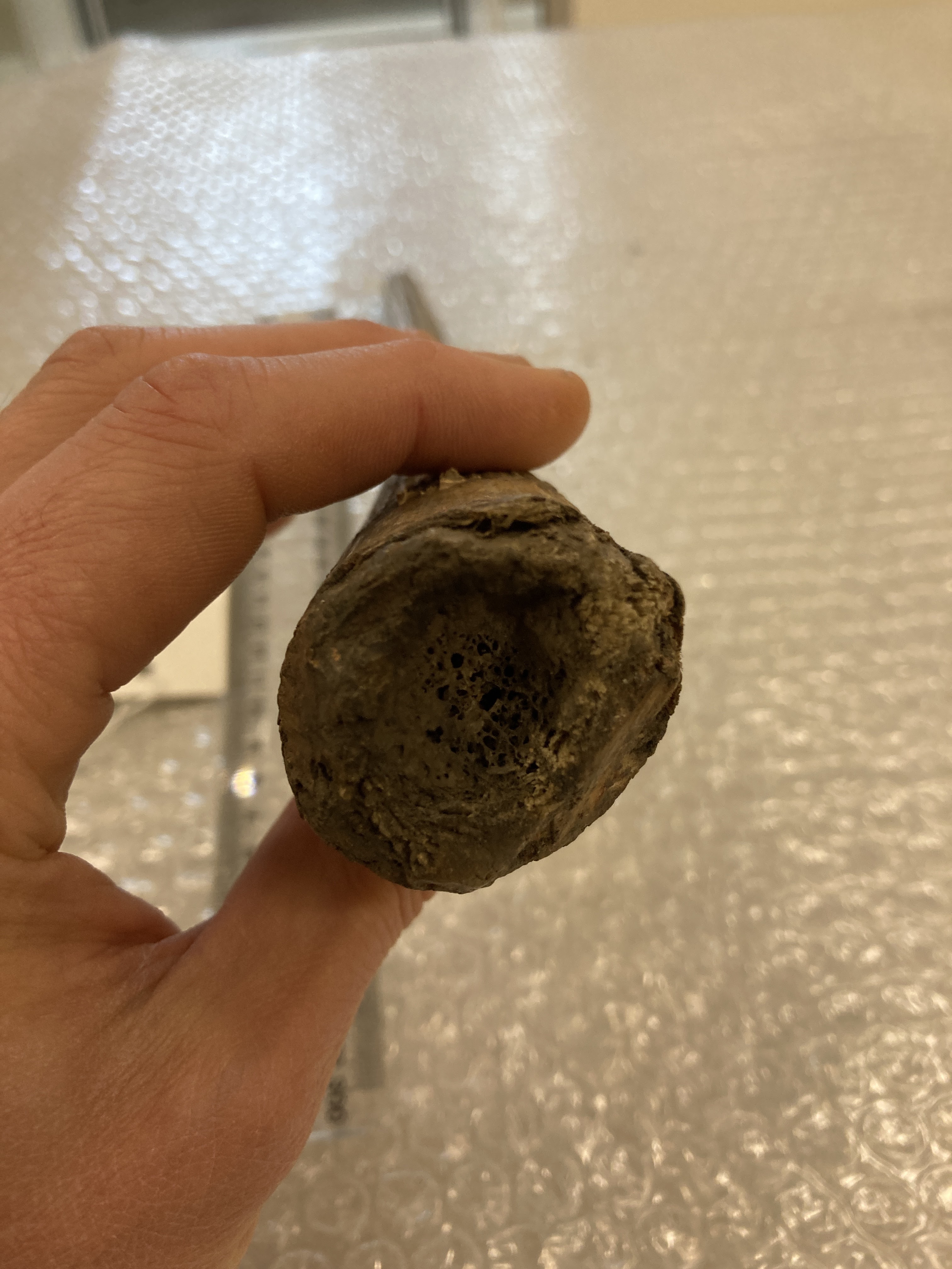
It’s clearly made from long section of quite highly vascularised bone:
It seems to be missing the smooth surface normally seen on bone, but that can be caused by a variety of factors, from disease and infection in the live animal to weathering after it’s been dead for a while.
The ends of the bone don’t have any indications of an articular surface:
The larger end has a bit of a hollow, but the smaller end appears to be broken and you can see a hollow core to the bone.
Overall, the shape is not really reminiscent of any long bone I can think of. It lacks a normal articular surface at the unbroken end and it has no crests or ridges that I would normally expect muscles to attach to. It tapers quite consistently and has a slight curve.
My first thought was shared by others in the comments, with Chris Jarvis getting in first with the simple but effective pun:
This is of course a reference to Oosik, which is the name in Native Alaska Languages for the baculum or os penis of a Walrus Odobenus rosmarus (Linnaeus, 1758).
There are some differences between this specimen and some of the other Walrus bacula I’ve seen and which show up in an image search, but there are a variety of possible explanations for that.
One is simply that Walrus bacula are quite variable. They can vary significantly through the life of the animal as it develops, but it can also vary quite a lot between individuals. If you spend as long looking at Walrus penis bones on the internet as I have (what on Earth happened to my life?!) then you’ll notice that some have a strong double curve while others can be almost straight, others are thick and some are quite thin.
This variability is also seen in other pinnipeds with a high degree of sexual dimorphism, like Sealions and Elephant Seals.
So while it’s hard to be 100% certain of the identification, I think it is the most likely solution to this mystery.
Last week I gave you an entomological mystery to solve, in the triangular(ish) shape of this moth:
For the real insect aficionados out there, this probably wasn’t too much of a challenge (I’m looking at you Tim), but for the rest of us it wasn’t quite so easy.
The overall appearance of this moth, with its size, shape (especially wing position), and fuzzy wing fringes is what you expect from a member of the family Noctuidae – the Owlet Moths. However, it’s a big family, with almost 12,000 species. A lot of the species also look similar to each other and some have a wide variety of different colour morphs – just to make things more complicated.
Even with helpful visual resources, with good photos of the different Irish species, it can be hard to work out what the diagnostic features might be. For some species of moths you have to get into the fine detail of the genitals, but thankfully there are wing patterns that are distinctive in this instance.
In this case, the wing pattern of interest is the small triangular black mark on the lower portion of the leading edge of the upper wing.
That’s it.
The rest of the colour and pattern of the upper wing is very variable in this species, so you can’t rely on any of those features as a reliable indicator. Of course, if the specimen was alive and had its wings open, it would be a much easier identification:
This is, of course, a Large Yellow Underwing Noctua pronuba (Linnaeus, 1758), which is pretty obvious once the underwing is visible. So well done to everyone who worked it out from just the upper wings.
Last week I gave you this really difficult, but incredibly cool mystery object to identify:
Definitely not a simple one for the uninitiated, but most of you got impressively close.
It looks a bit like ancient chewed gum at first glance (hey, it’s a thing!):
A nearly 6,000 year old piece of chewing gum, found in Scandinavia, perfectly preserved the DNA of its chewer. She was a young, hunter-gatherer girl, with dark skin, brown hair & blue eyes, who had recently eaten a meal of hazelnuts & duck. Archaeology is amazing. pic.twitter.com/kfav9PkonE
However, on closer inspection, some of the features start to emerge – including teeth:
Obviously, this is the fossilised skull or some critter, but what kind of critter is harder to determine.
The length suggests it’s something about the size of a rabbit:
And if you’re looking for a good fossil rabbit, you can’t beat Palaeolagus:
Palaeolagus skull. Image by Smithsonian Institution, 2019
NotPalaeolagus skull.
As you can see, the mystery object has a few differences, but due to the various missing parts, it’s a little hard to be confident exactly how different they are – although the shape of the orbital margin (the front of the eyesocket) gives a bit of a hint.
But, even more useful, is the curve in the maxilla (the upper jaw bone) that traces the root of the first incisor. In lagomorphs (rabbits, hares and even pikas), the incisor roots terminate with quite a big gap before the orbital margin, often with a triangular fenestrated region of cancellous bone (a sort of window of bony struts) in between.
The mystery specimen doesn’t have that – in fact the end of the incisor root is very close to the orbital margin. This is something you see in rodents.
I would have been impressed if you got that far, since the overall shape and size of this specimen definitely gives off a rabbity vibe, but believe it or not, this a dormouse. More specifically, it’s the Gigantic Dormouse Leithia miletensis (Adams, 1863) or if you want to go with the commonly used and more technically accurate, but nomenculatorily incorrect, L. melitensis, since Adams made a spelling error in his original description.
In fact, this is one of the specimens collected and figured by Adams in that original work describing the species, making this part of the type series for the species (although the holotype is more likely to be a very well preserved half mandible from the same site).
The fact that this is a fairly large and intact part of the type series means that it is of great interest to researchers. The reason I had this specimen to hand for the mystery object, is because I was preparing it for a research loan to some of my old colleagues in UCL, where it’s being MicroCT scanned.
This research will help refine an understanding of the morphology of the Gigantic Dormouse and offer some clues to what happens on islands that leads to the development of giants, building on work that they’ve been doing on this fascinating species, which is an interesting read that you can find here (you may even recognise Fig. 1B).
Virtual Cranial Reconstruction of the Endemic Gigantic Dormouse Leithia melitensis (Rodentia, Gliridae) from Poggio Schinaldo, Sicily, By Jesse J. Hennekam , Victoria L. Herridge, Loïc Costeur, Carolina Di Patti, Philip G. Cox – CC BY 4.0, https://commons.wikimedia.org/w/index.php?curid=92037373
Last week I gave you a somewhat tricky object to identify:
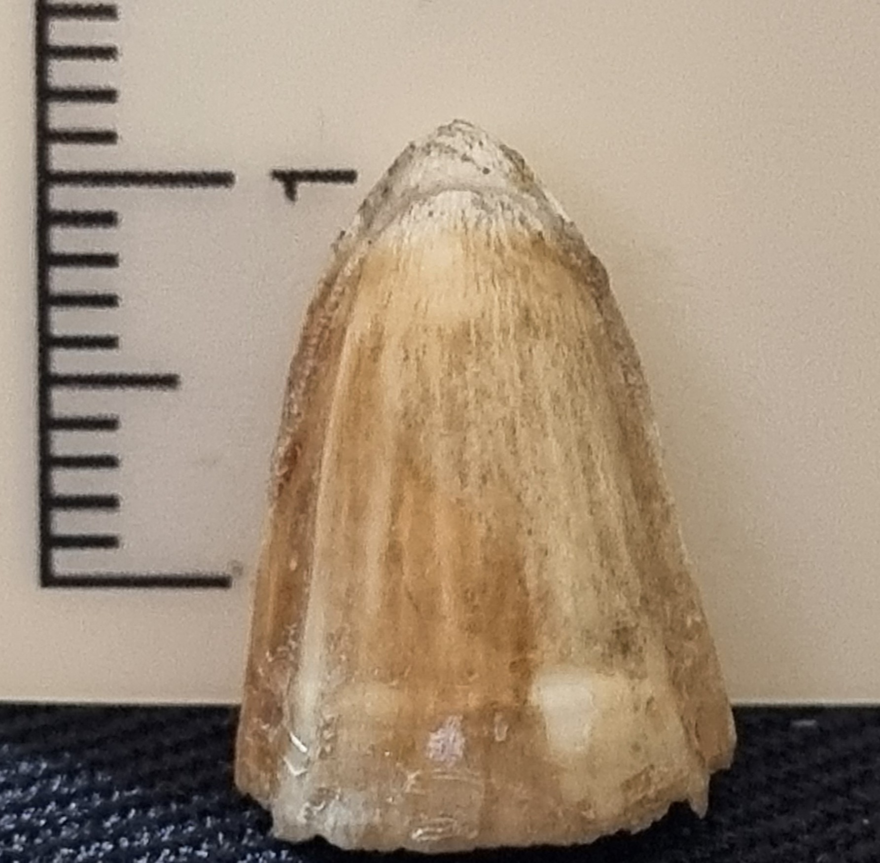
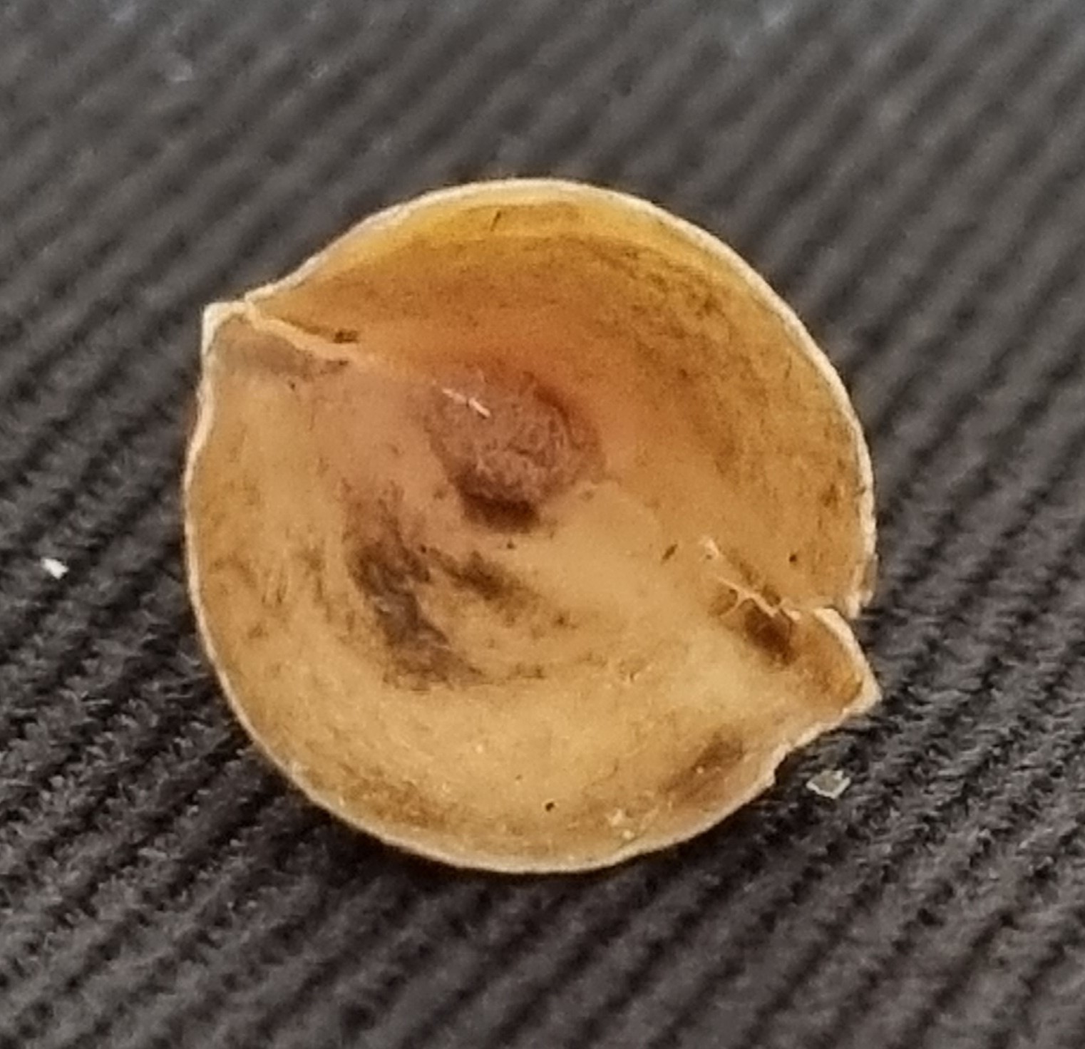
It’s only about 14mm long, it’s hollow and it has a seam down either side:
These features seemed to raise more questions than answers for many of you – and I’m not surprised, since this really does require some experience with the object to identify it.
Fortunately, some of you clearly had the necessary experience. Chris Jarvis and Adam Yates and Kat Edmonson all chimed in with some very useful suggestions, observations and discussion.
It’s a tooth from a Mugger Crocodile Crocodylus palustris (Lesson, 1831), in fact, if you want to be specific, it’s a newly emerging tooth from the 3rd socket from the rear in the left maxillary row.
I really wasn’t expecting anyone to figure out the exact species, but I was impressed that several of you figured out it was from the genus Crocodylus rather than Alligator.
Last week I gave you a guest mystery object from Catherine McCarney, the manager of the Dissection Room at the UCD School of Veterinary Medicine:
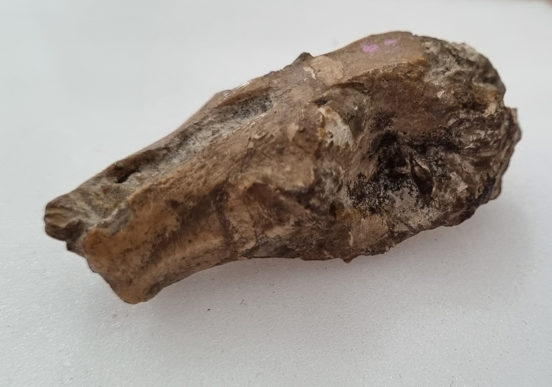
The first question I normally try to answer when undertaking an identification is “what kind of bone is this?”, but in this instance it’s not immediately obvious.
There is a broad section with articulation points, a foramen (or at least something that looks like a hole, which might be a foramen) and a flattish section that looks like it probably butts up against something with a similar flat section. This would normally put me in mind of the ischium of a pelvis.
But it’s not a pelvis as the articulations are all wrong and the shape of the skinny piece of bone that projects off doesn’t fit any functional ilium shape that I’m aware of.
The pectoral girdle has a similar set of structural features and this object starts to make more sense with that in mind. Things like turtles and whales may have a structure like this, but there’s something to keep in mind: despite being fairly large, this object only weighed in at 26g.
Turtles and whales have dense bone that helps reduce buoyancy, to make remaining submerged less energetically demanding, but this bone must be full of air spaces – which offers a clue as to likely type of animal it came from. A bird – as Joe Vans noted in the comments.
Considering the size of this object there are very few possible candidates. Most birds are pretty small and this object is pretty big, so we just need to look at some of the Ratites.
The comparisons I managed to find have led me to the conclusion that this is most likely part of the pectoral girdle of an Ostrich Struthio camelus Linnaeus, 1758.
More specifically, I think it’s the coracoid (#2 on image), clavicle (#3 on image), and scapula (#4 on image) from the left hand side of the pectoral girdle of an Ostrich.
I was delighted to see that Wouter van Gestel agreed with this assessment in the comments, since he knows more about bird bones than I could ever hope to learn!
Finally I’d like to thank the fatastic Catherine McCarney for sharing this mystery object from the depths of the Vet School’s collections. I hope you all enjoyed this challenge!
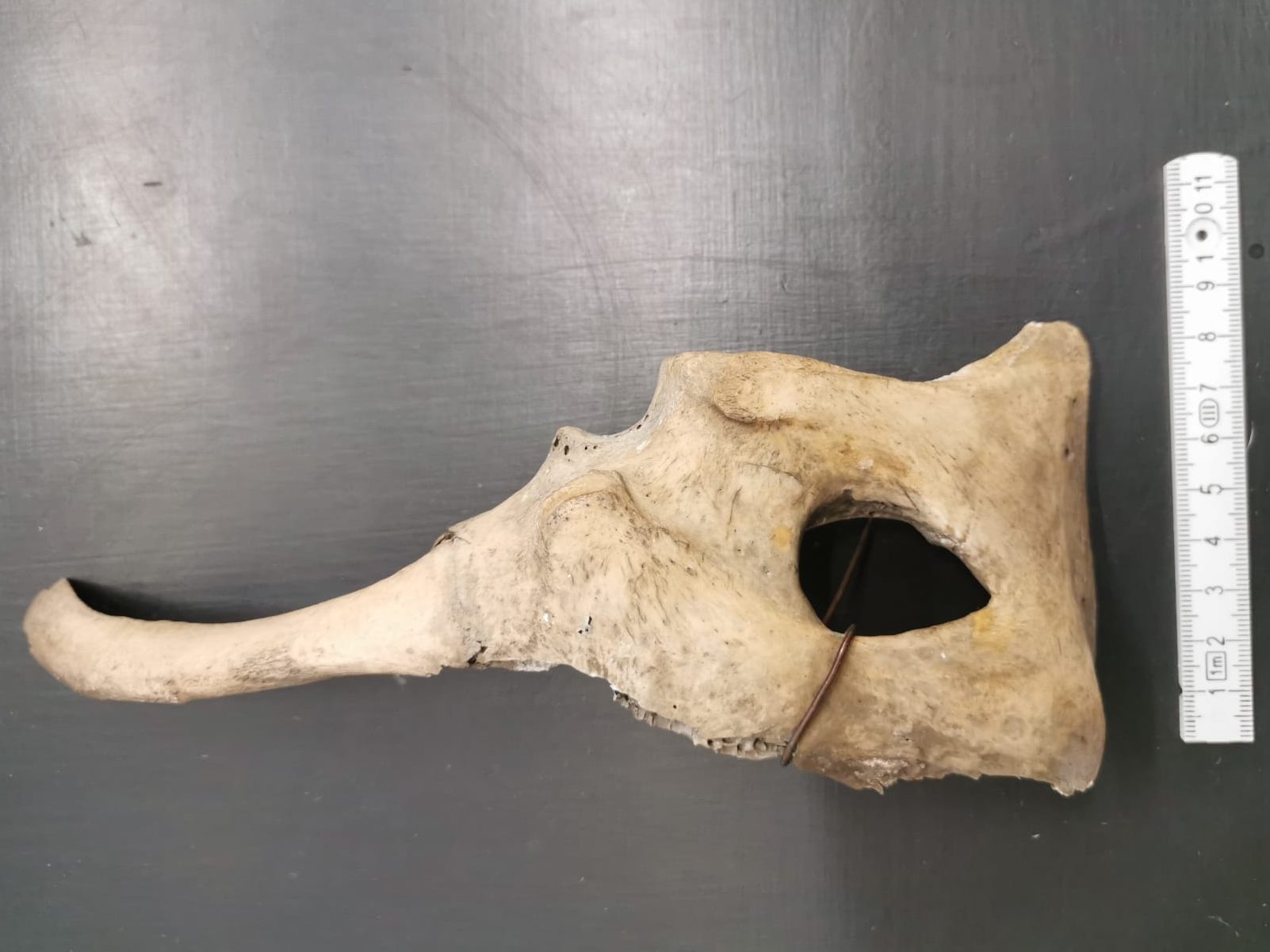
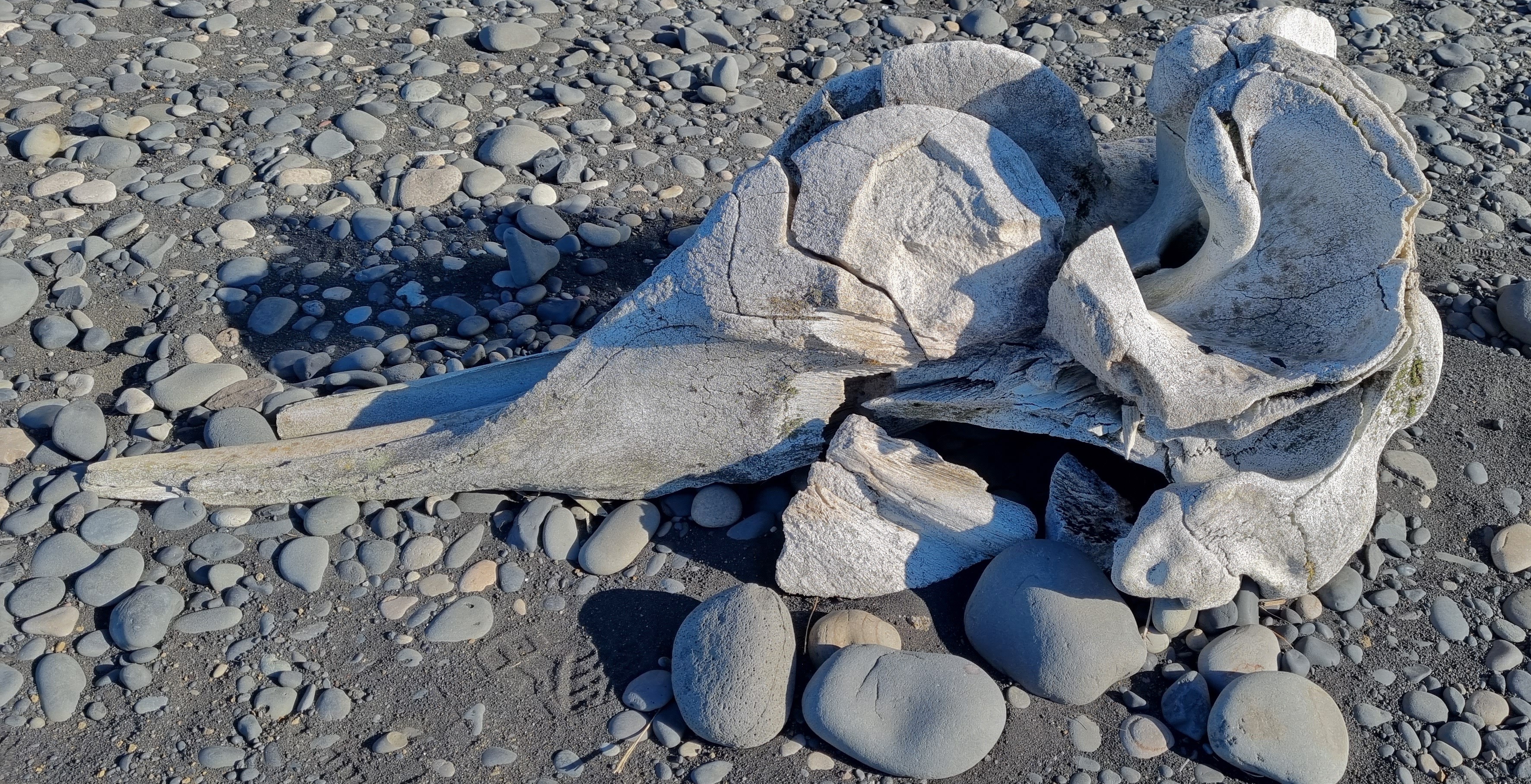
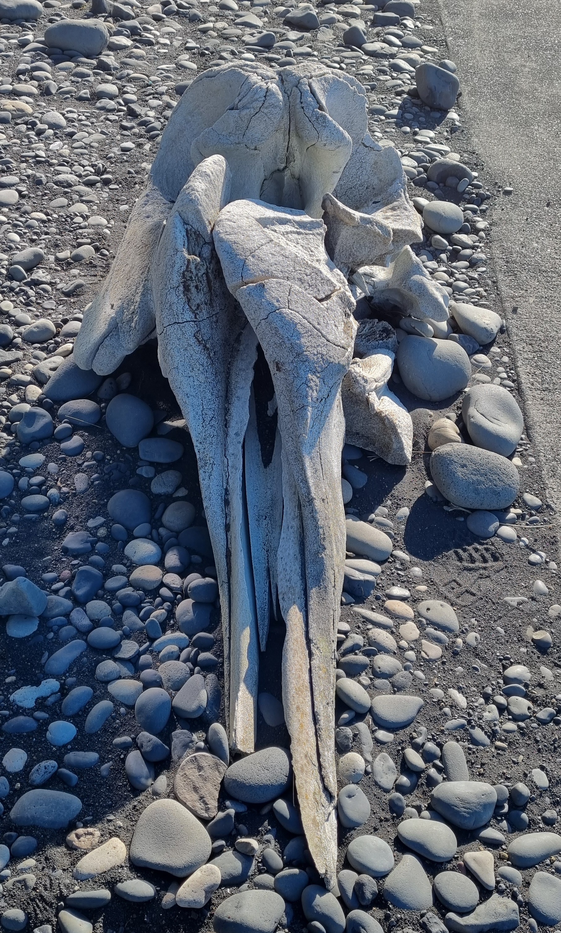
Last week I gave you this rugged skull, from a rugged place, to have a go at identifying:
As everyone spotted, this is a whale of some sort (what else has a skull that weird-looking?), but the question is, which species?
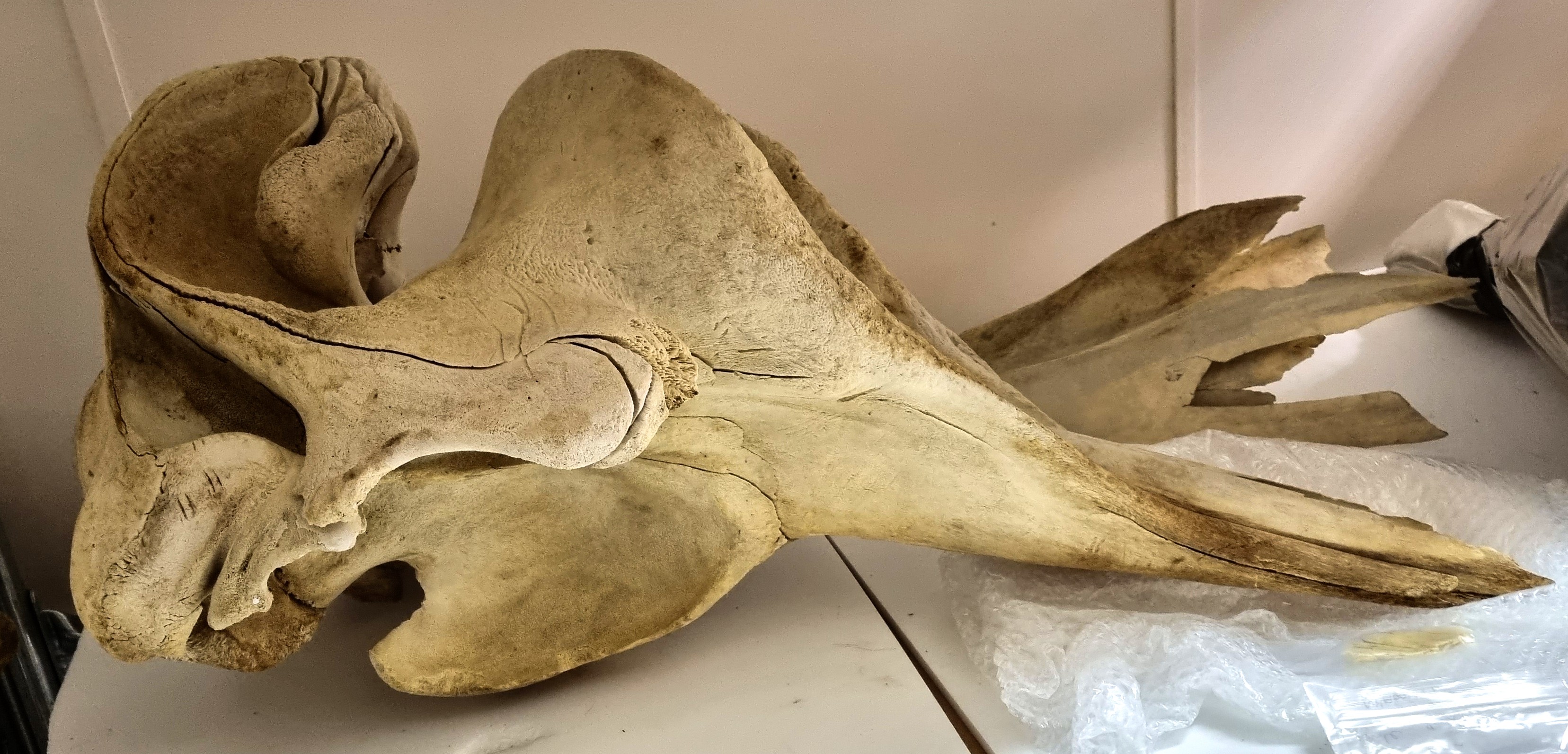
The location led to a few suggestions of Arctic / sub-Arctic species like Narwhal or Beluga, but they have a much flatter top section of the skull. In fact, those huge vertical lobes of the maxillae seen here is pretty unusual and quite distinctive (even if it is a ittle weathered and broken):
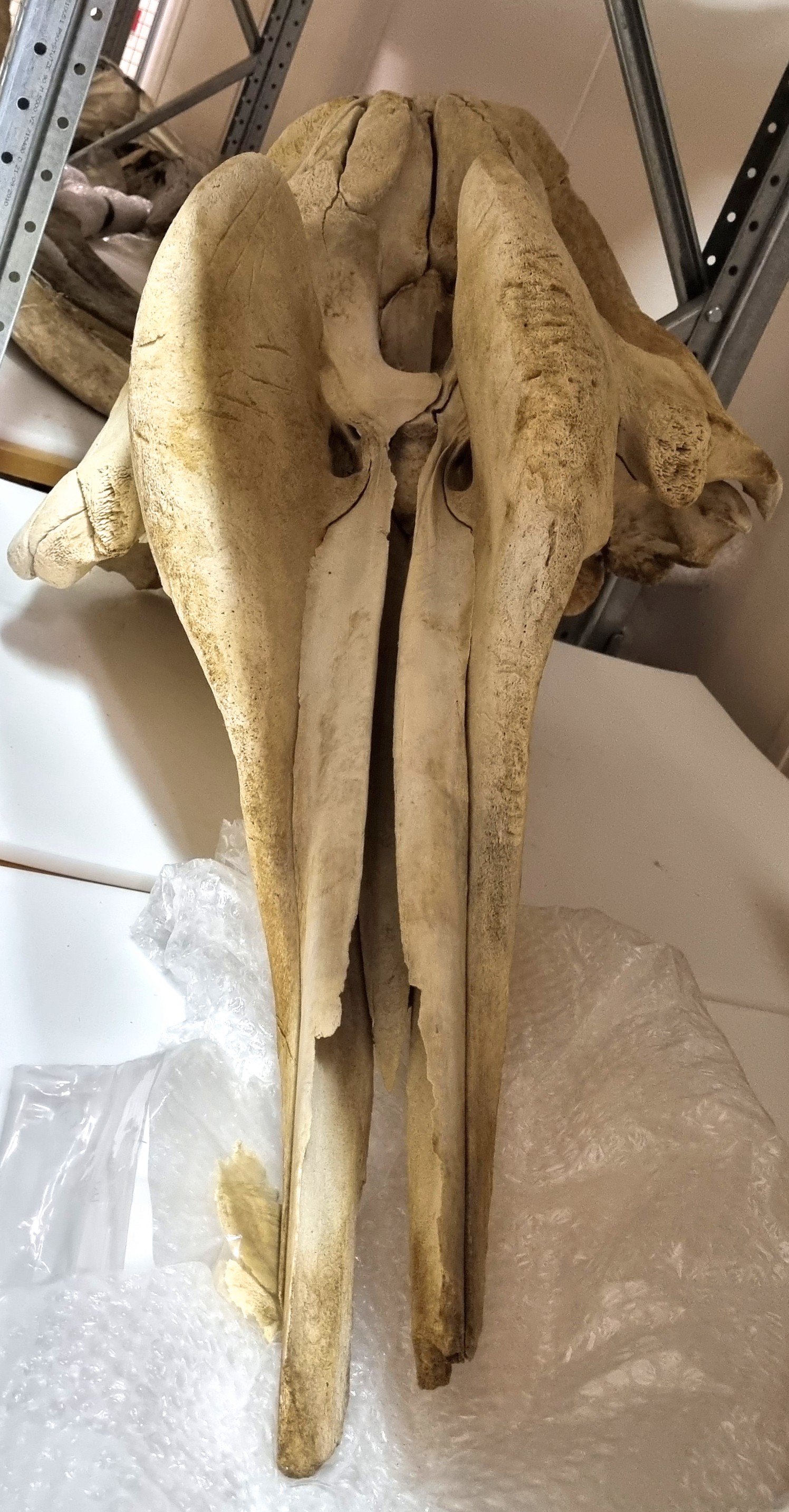
This reminded me of a specimen in the collections of the Dead Zoo and which I had to check, just to be sure of my identification:
As spotted immediately by Chris and not too long afterwards by Adam Yates and Wouter van Gestel, this is the skull of a Northern Bottlenose Whale Hyperoodon ampullatus (Forster, 1770).
This sub-Arctic species has a distribution across much of the North Atlantic. They tend to stick to quite deep water, which makes sense in the case of the specimen I shared from Iceland, since the Reynisfjara beach is infamously dangerous because it shelves off very steeply into very deep water, making the waves that break along the beach behave in an unusual (and frankly terrifying) way.
Occasionally this species will come into shallower waters, in one (somewhat tragic) case a female Bottlenose Whale swam up the Thames (and is now in NHM, London). Our specimen came from an animal stranded on the Irish coast and there are theories that maritime sound pollution is connected to them being driven into shallower waters.
Well done to everyone who worked out which species this skull is from – hope you’re ready for another mystery next week!
Last week I gave you a challenge to get your teeth into:
As I suspected, everyone managed to figure out what type of animal this is, since these teeth are quite distinctive (as mammal teeth often are).
To start with, there are canines and incisors in the premaxilla (the top jaw). These are missing from things like cows, sheep and deer. So it’s not one of them. The premolars are adapted to grinding rather than cutting, so it’s not some kind of pig or carnivore.
The molar teeth are low-crowned, unlike the teeth of grazers like horses which are high crowned, to cope with the wear and tear of silica-toughened grasses. This suggests an animal that browses on softer vegetation. Also, the lophs (those ridges of enamel that join the tooth cusps) are well defined and quite distinctive in their shape. That rules out most other herbivores, including the camels and their relatives.
I think it’s understandable that nobody got the correct species, since the specimen is a subadult (check out the molar in the jaw that’s still developing) which will somewhat alter the proportions compared to an an adult – especially considering the photos I gave you were restricted to the teeth and missed all the useful features of the rest of the skull.
So well done to everyone who worked out that the teeth belonged to a tapir!
Last week I gave you this nice robust skull to have a go at identifying:
It proved a little bit more of a challenge than I originally expected, at least in terms of getting a species level identification.
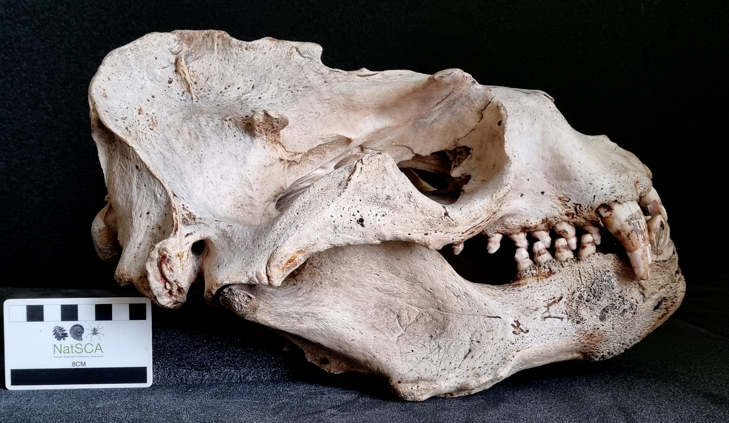
So despite a somewhat ursine (bear-like) overall appearance, that may have confused a few people at first, this has all the features you’d expect to see in a male sea lion. In that it’s big, craggy, has huge open sinuses opening into the orbital region (nobody wants their eyes to be overly pressurised when they’re diving) and the teeth are relatively undifferentiated in the back part of the mouth, but they’re well adapted for fighting up front.
However, it turns out that there aren’t a huge number of resources online to see and compare the skulls of these beasties (and the ones that do exist aren’t necessarily the easiest to navigate). So while almost everyone figured out the sea lion bit, the species choice went a bit off track.
Most people plumped for the Steller’s Sea Lion, which (it must be admitted) looks very similar. But this is actually the skull of a Southern Sea Lion Otaria flavescens (Shaw, 1800).
I talked about this species before on Zygoma (many years ago now), with a specimen from the Horniman Museum, where I provided some links to the Marine Species Identification Portal. Sadly, that resource has been retired, but fortunately Naturalis Biodiversity Centre rescued the content and has kept it available online. It provides drawings of the skulls of both Steller’s and Southern Sea Lions and if you take a look at few key features you’ll spot the differences.
One major indication is the length and shape of the palate. The Southern Sea Lion has a very long palate, which terminates almost in line with the mandibular articulation, whereas the Steller’s terminates further forward. There are a few other features, but that one is the most immediately obvious.
So, a hearty congratulations to a variety of folks on Twitter who spotted that this was the Southern Sea Lion, but there’s no shame in not getting the correct species if you picked Steller’s, given how few resources there are that allow a really good comparison. I hope you enjoyed the challenge!
Last week I gave you this guest mystery object from the comparative anatomy collection of the Harry Brookes Allen Museum of Anatomy and Pathology at the University of Melbourne, courtesy of Rohan Long:
Image by Yijie Cheng, 2023
This is one of those specimens that it can take a while to get your head around, as most of the key features are entirely missing. From the top, the skull almost looks mammalian. Perhaps a little like a large rodent missing part of its zygomatic arches:
Skull of a Striped Ground Squirrel
Even from the side there are some similarities, although it looks a bit more like a turtle:
Image by Yijie Cheng, 2023
Skull of a Loggerhead Turtle
If you look closely at the underside of the skull, you’ll notice that it has a single occipital condyle, which is something you see in reptiles and birds, but that view of the underside also becomes clear that the front section of the mystery object doesn’t taper to create a bill, like you’d see in a turtle:
Image by Yijie Cheng, 2023
In fact, a bill is the most diagnostic feature that’s missing, and that’s because it’s fallen off.
Those cervical vertebrae are quite distinctively avian – and from a long-necked avian at that. Once you realise that this is the braincase of a fairly large long-necked bird, the next task thing is to look at birds with a bulbous and cleft region on the head, just at the base of the bill (most bird skulls taper down to the bill).
For me that indicates one species above all others – the Mute Swan Cygnus olor (Gmelin, JF, 1789).
Mute swan skulls “Cygnus olor”. Technique of bone maceration on display at the Museum of Veterinary Anatomy, FMVZ USP.
I’d like to offer a hearty ‘bravo’ to Adam Yates, who was the first to comment and correctly identify this with a great cryptic clue:
It is an anseriform for sure the large oval basipterygoid articulations are a give away. With that profile, i’d lose my voice while trying to say the name of a certain Western Australian River.
Last week I gave you this bird specimen to have a go at identifying:
In the ornithological community, birds like this are sometimes referred to as an LBB / LBJ (Little Brown Bird / Job), because they are small, brown and hard to identify (especially in the field) due to the large number of similar looking species.
This specimen has a robust, conical bill and grey-streaked breast, which led some of you to think it could be a juvenile Crossbill or perhaps a female Grosbeak. However, as indicated by Wouter van Gestel in an excellent cryptic clue (and by Tim Dixon in a rather rude one), this is a Corn Bunting Emberiza calandra Linnaeus, 1758. It was collected in County Dublin and donated to the Dead Zoo in 1880.
These seed-eating passerines were widespread in arable farmland across Ireland in the 19th and early 20th centuries. However, changing land use practices reduced the available habitat until they became locally extinct as a breeding species in the late 1990s – well within living memory for many people.