This week I have a mystery object for you that came in as an enquiry from a regular donor to the Dead Zoo’s collections. It was found by a fisherman in the Irish Sea, just off Howth, which is a lovely seaside village on a peninsula that marks the northern tip of Dublin Bay :
Any thoughts on what it might be? I suspect some of you will have a pretty good idea, so keep your suggestions cryptic if you can, so everyone has a chance to figure it out for themselves. I hope you have fun figuring it out!
Last week I gave you this skeleton fron the Dead Zoo to test your identification skills:
In retrospect I think I was a little unfair with this one – the photo is not very clear and there is no scale bar, so the identification relied mainly on the context provided by the mount and a lot of deduction. Not an easy task with a rodent, since there are so many different species.
The branch used as a setting for the skeletal mount provided the main and most important clue – it indicates that the species is arboreal. A lot of people picked up on this, with guesses ranging from a flying squirrel to a viscacha. However, the answer is something from a bit closer to home (i.e. Europe).
This is the skeleton of the Edible Dormouse Glis glis (Linnaeus, 1766), a plump (and presumably tasty if you happen to be an ancient Roman), tree-dwelling rodent, with a reputation for somnolence.
Edible dormouse (Glis glis) in an old shed in an abandoned plum orchard in Luc-en-Diois, France. Image by Bouke ten Cate, 2011
This isn’t the first time I’ve featured a dormouse in the blog, although the previous one was a giant extinct example. The Edible Dormouse is the largest species alive today, but it’s still smaller than the fairly diminutive Red Squirrel.
They are fairly well distributed around central Europe, with a small population in Southern England due to escapees from Walter Rothchild’s menagerie in Tring in my home County of Hertfordshire. I’ve heard tell that they can be a bit of a pest in the area, due to their habit of seeking out attics to hibernate in, but then chewing through wires and cables, thus causing fires and broadband outages.
This UK population didn’t arrive until the early 20th Century, so the species that inspired Charles Dodgson (AKA Lewis Carroll) to include his sleepy character was almost certainly the smaller Hazel Dormouse, which occurs in Britain, and which also turned up in Ireland around County Kildare around 14 years ago (and which we have specimens of, thanks to a gift from someone’s pet cat).
I should have either provided a better image or a clue to point you in the right direction for this mystery object, so I feel I’d better apologise for setting this vexatious conundrum and promise to better next time!
I have a potentially perplexing mystery object for you this week from the Dead Zoo. Any idea who this bony character might be?
If you think this is an easy one, then I would be both surprised and impressed, so let me know your thoughts through a crytic clue or two below. Enjoy the challenge!
Last week I gave you this doe-eyed specimen from the collections of the Dead Zoo to try your identification skills out on:
I didn’t provide a scalebar as I think it would have made it too easy, but even so, it’s clear that the specimen is a very small species of artiodactyl (the group containing pigs, deer, antelope, bovids and a variety of related herbivores).
There were some suggestions that it could be a Dik-dik, but as Adam Yates pointed out, this specimen lacks the large preorbital glands that are very visible in Dik-diks (and makes them look like they got carried away with the eyeliner):
Dik-diks with their distinctive preorbital glands
The mystery object lacking preorbital glands
The other popular suggestion for the identity of the mystery object was a Java Mouse-deer (or Javan Chevrotain), which is the smallest ungulate alive. However, while that’s exactly what it says it is on the label, the location of collection rings alarm bells for me:
There are two species of chevrotain found in Singapore, and the Javan species is not one of them.
Of the two, one is the Greater Mouse-deer and the other is the Lesser Mouse-deer. The Greater, as you probably guessed, is on the large side for a chevrotain, weighing in between 5 and 8kg. This species also has a dark stripe from its nose to its eye, which is missing from the mystery object.
The Lesser Mouse-deer Tragulus kanchil Raffles, 1821 lacks the dark stripe and is almost as tiny as the Javan Mouse-deer, making it the most likely candidate for the mystery object:
Lesser Mouse-deer alongside some rodents
This specimen not only has that likely identification error on the label (easily done considering the complexities of chevrotain taxonomy across Southeast Asia), but it had somehow also had a completely incorrect label associated with it in the past, which said it was a Siberian Musk Deer – a species that’s on the small side, but by no means as tiny as this.
This specimen was of particular interest at the end of last year, when we had a visit by a group of researchers from Singapore, who are undertaking a fantastic project to digitise specimens collected from Singapore that are held in museum collections all around the world. The project is called SIGNIFY and the team were not only absolutely lovely people, but they achieved a huge amount of research and detailed imaging work in a very short time:
The SIGNIFY portable imaging setup in use on a specimen from the Dead Zoo bird skin collection
The SIGNIFY project has huge value for helping to understand the historic baseline biodiversity of Singapore prior to industrialisation, but it also helps foster links between organisations and allows the inextricably linked social and personal histories of collectors to be explored. I loved getting a chance to spend time with the team, learning more about their project and the collections I care for. It also turns out that we have a wealth of spiders from Singapore that still need to be investigated, so I really look forward to welcoming the team back soon!
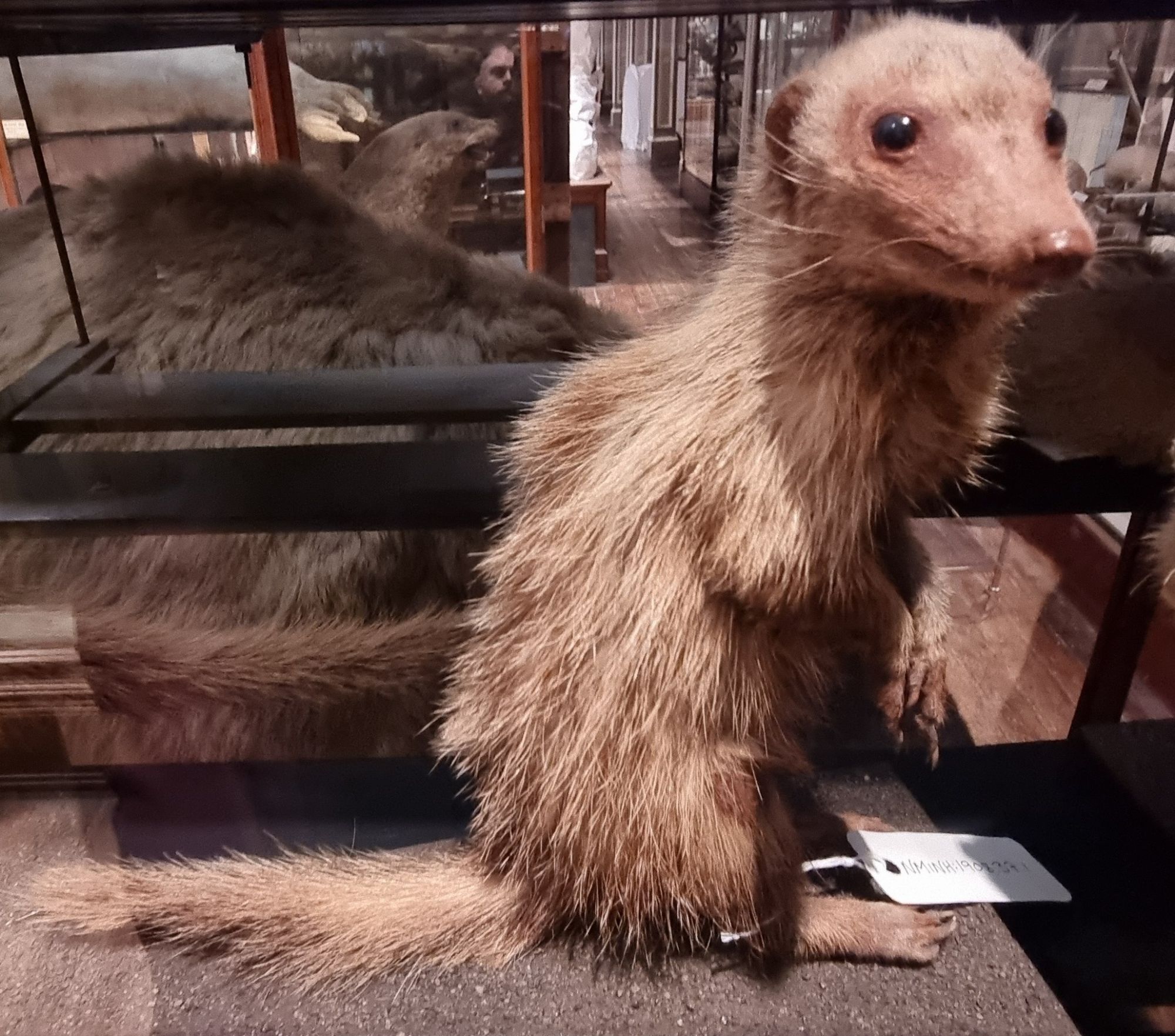
Last week I shared this fuzzy critter as mystery object for you to identify:
It was probably a bit of a mean one, as I didn’t provide a scalebar. It’s also a species from a group of small carnivores that contains over 30 species that can look quite similar, and (perhaps most importantly) the specimen is old and very faded from being on display in a gallery space with lots of light for the last 100 years or more.
Natural light in the Dead Zoo, image taken in May 2020.
Regardless, Chris Jarvis figured it out (after an initial near miss), while I believe that Joe Vans cheated by checking out the 3D tour of the Dead Zoo. Most other comments on social media came close, with a lot of people working out that this is a Mongoose, but then not quite getting the species.
This specimen is a Common Kusimanse Crossarchus obscurus G. Cuvier, 1825. These are also known as the Long-nosed Kusimanse, as their snout is a bit more elongated than that of most other Mongooses (or should that be Mongeese?):
The Common Kusimanse is one of the African Mongooses in the Subfamily Mungotinae. They are a good bit shorter than most of the other species of Mongoose – for comparison here’s this specimen next to a Small Indian Mongoose (well, that’s what the label says, although I have some doubts):
They are normally a dark brown colour, like this example:
Common Kusimanse by LA Dawson, 2006.
However, the Dead Zoo specimen is now bleached blonde, so I’m not surprised that this identification was tricky. Fading leads to all sorts of issues for the accurate representation and identification of species, to the point where the Giant Panda on display in the building had to be dyed black in places a few years ago, because it had ended up looking like a Polar Bear cub due to the sun damage.
At the moment that’s no longer an ongoing issue in the Museum, as a temporary floor has been installed just beneath the old glass ceiling, to allow investigations on the roof space to take place – this blocks almost all of the natural light. This has been great for conditions in the building, as it no longer heats up like a greenhouse on sunny days, and the bleaching of the specimens has been put on hold.
At some point the tempoprary floor will be removed, but I sincerely hope that a more permanent solution to the light issue will have been put in place by then. Still plenty of work to be done to get to that point though!
Dead Zoo with temporary floor installed to allow roof access. This blocks the natural light and keeps the environment more stable.
Last week, I gave you this shiny blob to have a go at identifying:
There wasn’t much to go on, since it is just a blob that looks like a chunk of hardened tar, but it is in fact a rare and valuable natural material. It’s actually a small piece of ambergris.
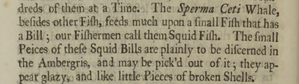
Ambergris has always maintained an air of mystery, since it’s formed deep within the bile duct of a Sperm Whale and its function in the animal is still only suggested rather than fully understood. General agreement seems to be that this tarry substance provides protection for the intestines from the sharp beaks of the squid that Sperm Whales prefer to eat.
Evidence for this comes from the fact that squid beaks are often found embedded in ambergris – an observation recorded from as early as 1725:
An essay upon the natural history of Whales, with a particular account of the ambergris found in the Sperma Ceti Whale. In a letter to the publisher, from the Honourable Paul Dudley, Esq; F. R. S Published:30 April 1725https://doi.org/10.1098/rstl.1724.0053
This letter goes on in some detail about various whale species, offering details of their economic yield in terms of barrels of oil, quantity of whalebone (baleen) and medicinal uses of teeth, as well as some aspects of their biology. When the discussion gets onto ambergris, much of the focus is on its location in the whale and the method of extraction (N.B. it’s not pretty.)
Of course, the suggested function of ambergris as a mechanism to aid the passing of sharp objects through the gastrointestinal tract would indicate that ambergris also might emerge naturally, and doesn’t necessarily need to be ripped from an unwilling victim.
However, evidence for this is quite hard to find, since it’s remarkably difficult to follow a Sperm Whale and keep track of its bowel movements or regurgitations (which have also been suggested as an exit route for these masses of indigestible items). What is known is that ambergris can be found floating at sea and washed up on beaches, sometimes persisting for years (there has even been fossilised ambergris discovered in Italy).
This non-invasive method of harvesting ambergris by beachcombing may not be hugely efficient, but it supplies the majority of the ambergris now used in perfume manufacture. Yes, you heard me right.
Human interest in ambergris may seem surprising given its somewhat revolting source, but as we all know, humans are pretty weird when it comes to making use of the fruits of nature, especially when searching for ingredients for perfume – just think of the African Civet and its anal excretions.
The natural complex aromatic compounds found in waxy substances like ambergris and civet musk provide long-lasting base odours that have played an important role in creating perfumes for centuries. Modern chemical synthesis of similar products has taken over to a large extent, but the naturally occurring compounds are still in use today.
This particular blob of ambergris was photographed when we were getting it out for sampling to inform a rather different line of scientific questioning, since there is still a lot to learn about this very unusual natural material.
This week I have a specimen that we recently had an enquiry relating to at the Dead Zoo:
Do you have any ideas what this shiny blob might be? If you’re confident in your blob identification skills, maybe try to keep your answer cryptic, so everyone gets a chance to work it out for themselves. Have fun!
Last week I gave you this mystery object from the Dead Zoo as a way to kickstart 2024:
While it does indeed look a bit like an old Roman shoe (thanks Adam Yates – I will never unsee that now), these are in fact the gill rakers from a Basking Shark Cetorhinus maximus (Gunnerus, 1765).
This was spotted early on by Dennis Nieweg and several other people worked it out, both here and on social media (Mastodon, Bluesky and LinkedIn). However, there were also a lot of suggestions of whale baleen, which is not surprising, since they perform the same function in filter feeding on plankton.
Basking Sharks are the second largest fish on the planet reaching around 8m in length, although they trail behind another far bigger filter feeding cartilaginous fish, the Whale Shark, by quite a margin (the Whale Shark can be twice as long). These two species have little overlap in their ranges – with the Whale Sharks in tropical waters and Basking Sharks in the temperate marine zones:
Range of basking shark (Cetorhinus maximus). Map by TheEmirr, 2012
Range of the whale shark (Rhincodon typus). Map by TheEmirr, 2011
Basking Sharks occur in the waters around Ireland, where this specimen was collected. The Dead Zoo also has a full taxidermy specimen collected off the coast near Galway in 1870, which has pride of place hanging from the ceiling in the Ground Floor Irish Room:
At the moment this specimen appears to be wearing a nappy – and this isn’t intended to capitalise on the popularity of the “baby shark” earworm, it’s an emergency stabilisation of the specimen. This became necessary after the rudimentary taxidermy (the skin is basically nailed to a barrel-built internal frame along the top of the specimen) failed in last summer’s unusually humid conditions and the skin started to fall off.
A proper repair will eventually be undertaken, but that will require lowering the specimen from the ceiling and transporting it offsite so it can be fully assessed and conserved. This is a big job, and this year we will be kicking off the next stage in a major capital project in the Museum, to deal with lack of physical access and problems with the environment – including the high humidity issue, so it’s just one big job alongside many, many others.
As the project gains momentum I hope to be able to share some of the work that takes place here on Zygoma, as well as through social media channels – so be sure to watch this space for updates. Here’s looking forward to an exciting 2024!
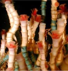
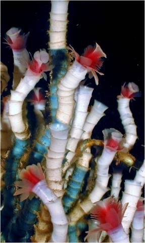
Last week I gave you some festive-looking specimens to have a go at identifying:
Image by Charles Fisher, published in Microfauna–Macrofauna Interaction in the Seafloor: Lessons from the Tubeworm. Boetius A PLoS Biology Vol. 3/3/2005, e102 doi:10.1371/journal.pbio.0030102
I thought these were some specimens in the care of Andy Taylor, FLS, but this was my error – Andy sent me the images to suggest the species as mystery objects, but I didn’t realise that he hadn’t photographed his specimens to use at that point. These images are actually from a paper (referenced above) that discusses the species and the blue-green colour is a stain added to allow the growth rate of the tubeworms to be calculated (spoiler alert – it’s very slow).
Here are Andy’s specimens:
Image by Andy Taylor FLS, 2023.
A bit less colourful, but the tubes retain the same structure, with those clearly defined rings.
As Adam Yates said in the comments, these are specimens of Lamellibrachia luymesi van der Land & Nørrevang, 1975. They have similarities to other genera, such as Hilary Blagbrough’s suggestion of Ridgeia and katedmonson’s suggestion of Riftia.
Image of Ridgeia specimens by Andy Taylor FLS, 2023.
Image of Riftiapachyptila specimen by Andy Taylor FLS, 2023.
Species like Riftiapachyptila are from hydrothermal vents and that nutrient rich and high temperature environment gives their symbiotic bacteria a boost that allows Riftia to be the fastest growing invertebrate, reaching around 1.5m long in just a couple of years. This is useful as it allows rapid colonisation of these ephemeral volcanic environments that occur at mid-ocean ridges.
On the flip side, Lamellibrachia luymesi tubeworms live in cold seeps of hydrocarbons in the deep ocean, where their symbiotic bacteria have to work at temperatures of 4°C or less, making their energy production a slow process. Consequently, L.luymesi are one of the slowest growing invertabrates, taking around 125 years to reach 1.5m long. Cold seeps are much more stable than the hydrothermal vents however, so L. luymesi have been found to continue growing up to 3m, taking around 250 years, and therefore being among the longest lived invertebrates (and indeed animals) on the planet.
Some might suggest that there’s a lesson to be learned here about “slow and steady winning the race”, but slow growth would be disastrous for a species that relies on a rapidly changing environment. Both species are remarkably adapted to their environment and neither would do well in the other’s place.
It’s worth noting that both of these remarkable organisms are only as successful as their symbionts allow them to be, so if there’s any lesson to be shared, it’s probably that the value of teamwork should never be underestimated.
On that (somewhat cheesy) note, I would like to thank Andy once again for sharing his collections. I’ll be back in the New Year with another Mystery Object – I hope you enjoy the celebrations!
This week’s mystery object is a guest specimen from regular contributor Andy Taylor, FLS, chosen for its seasonal colours:
Any ideas what these living party decorations might be?
As ever, you can pop your questions, thoughts, and suggestions in the comments below – no need for cryptic clues this time around as it’s a tricky one!
With that seasonal conundrum to consider, I wish you all a wonderful holiday weekend!
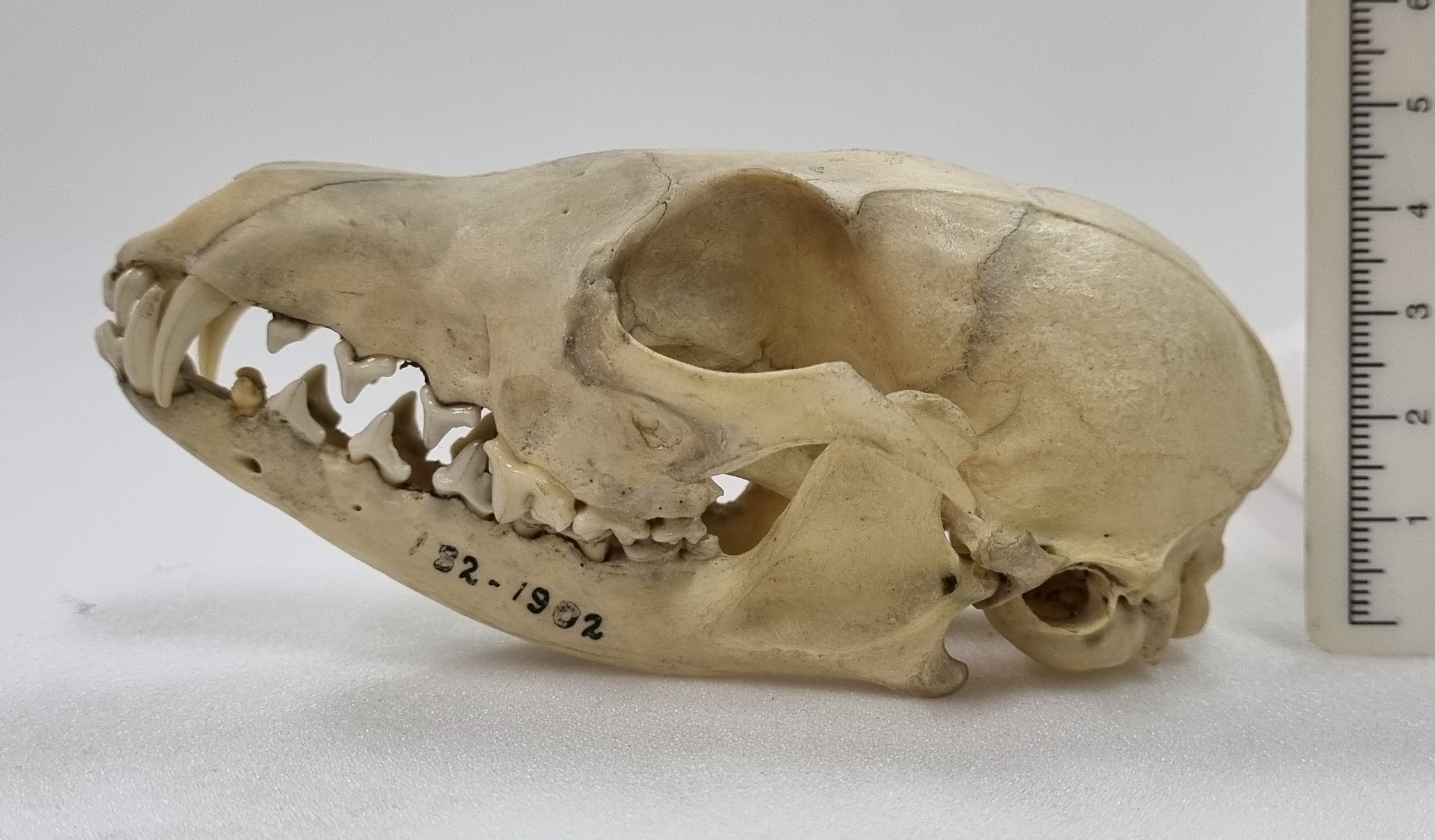
Last week I gave you this unidentified skull from the collections of the Dead Zoo:
I say it was unidentified, but in a strange quirk of coincidence I actually did identify this skull three years ago, just a month before the start of the Covid Pandemic (which might explain why I never had a chance to update the record).
I think the specimen is most likely to be the skull of a White-nosed Coati Nasua narica (Linnaeus, 1766), for the reasons I outlined back in 2020. So well done to Leon and Chris for recognising this cousin of the Raccoon from South and Central America and the southern parts of some North American states .
Coatis have quite distinctive upper canine teeth, that look almost like short tusks. These are useful for defence from other Coatis, but they are not very well adapted for subduing larger prey. This isn’t really a problem for Coatis, since they mainly feed on invertebrates, fruit and small vertebrates that they undcover during their energetic foraging.
So I apologise to everyone for repeating a specimen – this is the first time this has happened (and hopefully the last)!
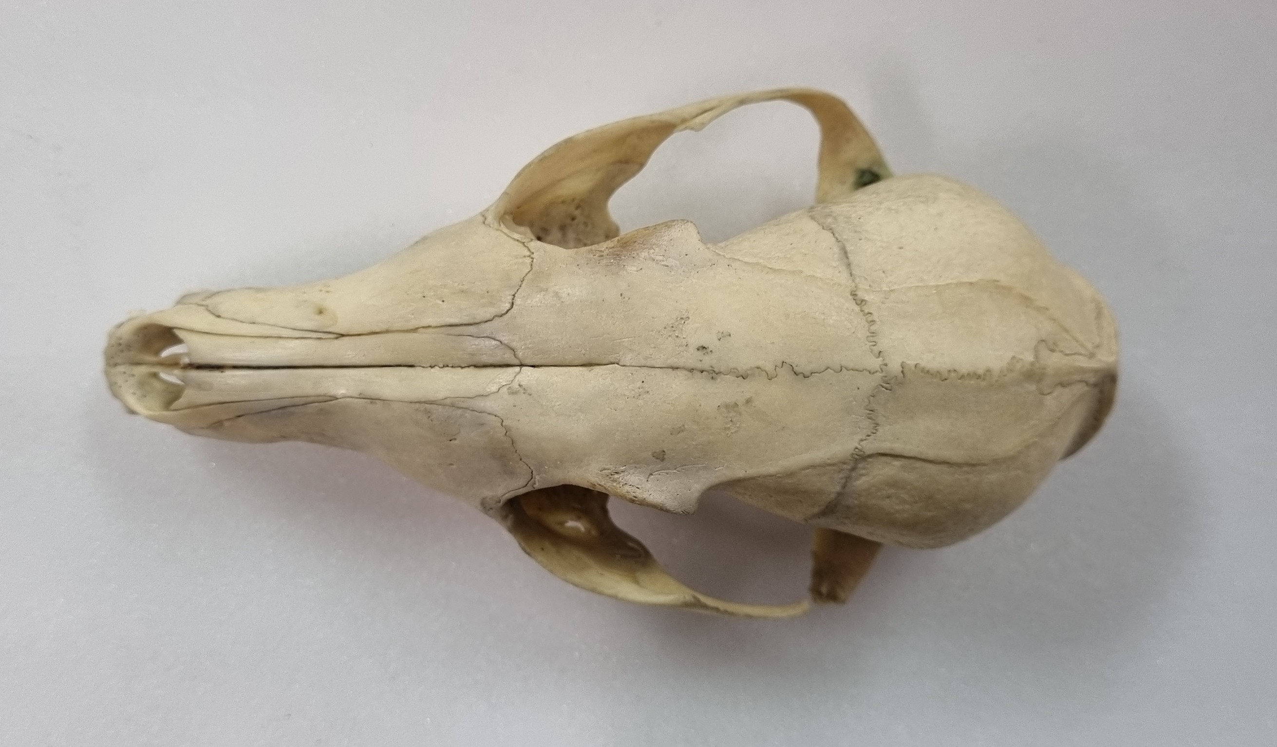
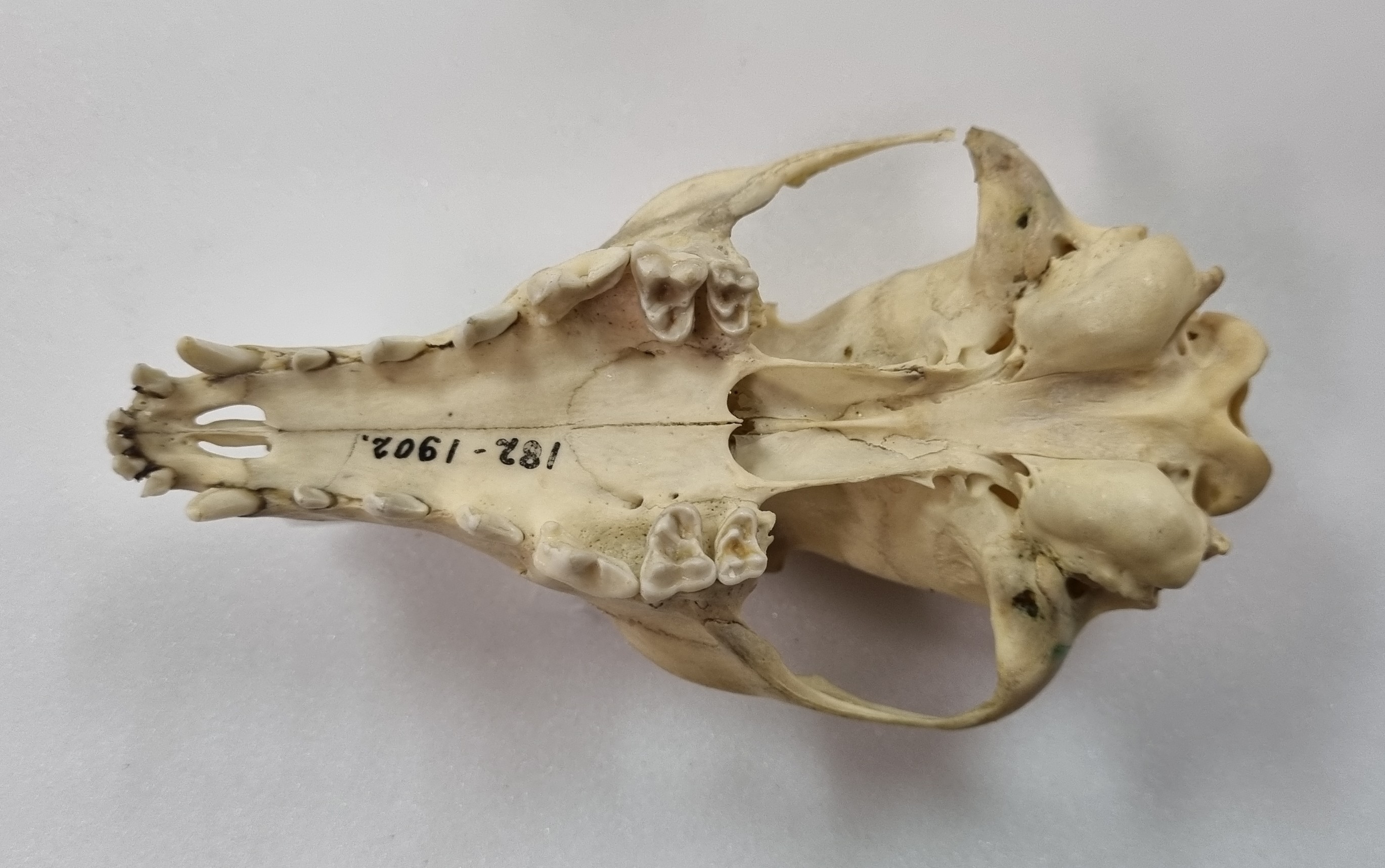
This week I’m continuing with skulls from the collections of the Dead Zoo, and this one was sitting in the “Unidentified” drawer:
Do you have any ideas what this might be? I suspect that some of you will be familiar with this, so perhaps it’s time for some cryptic suggestions in the comments. Have fun!
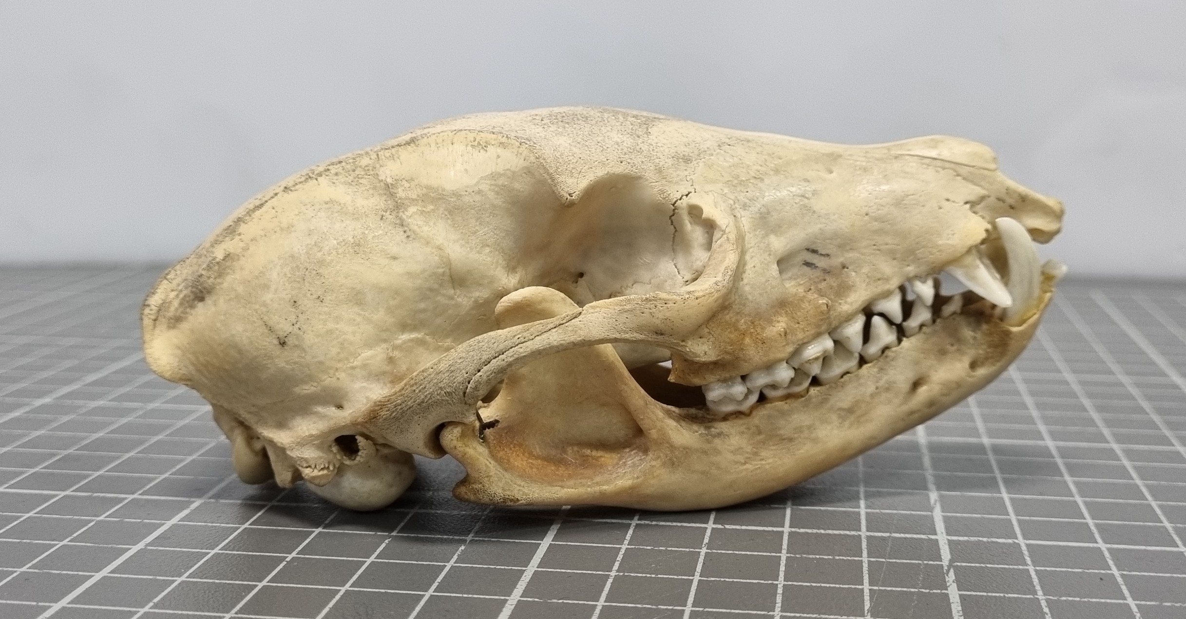
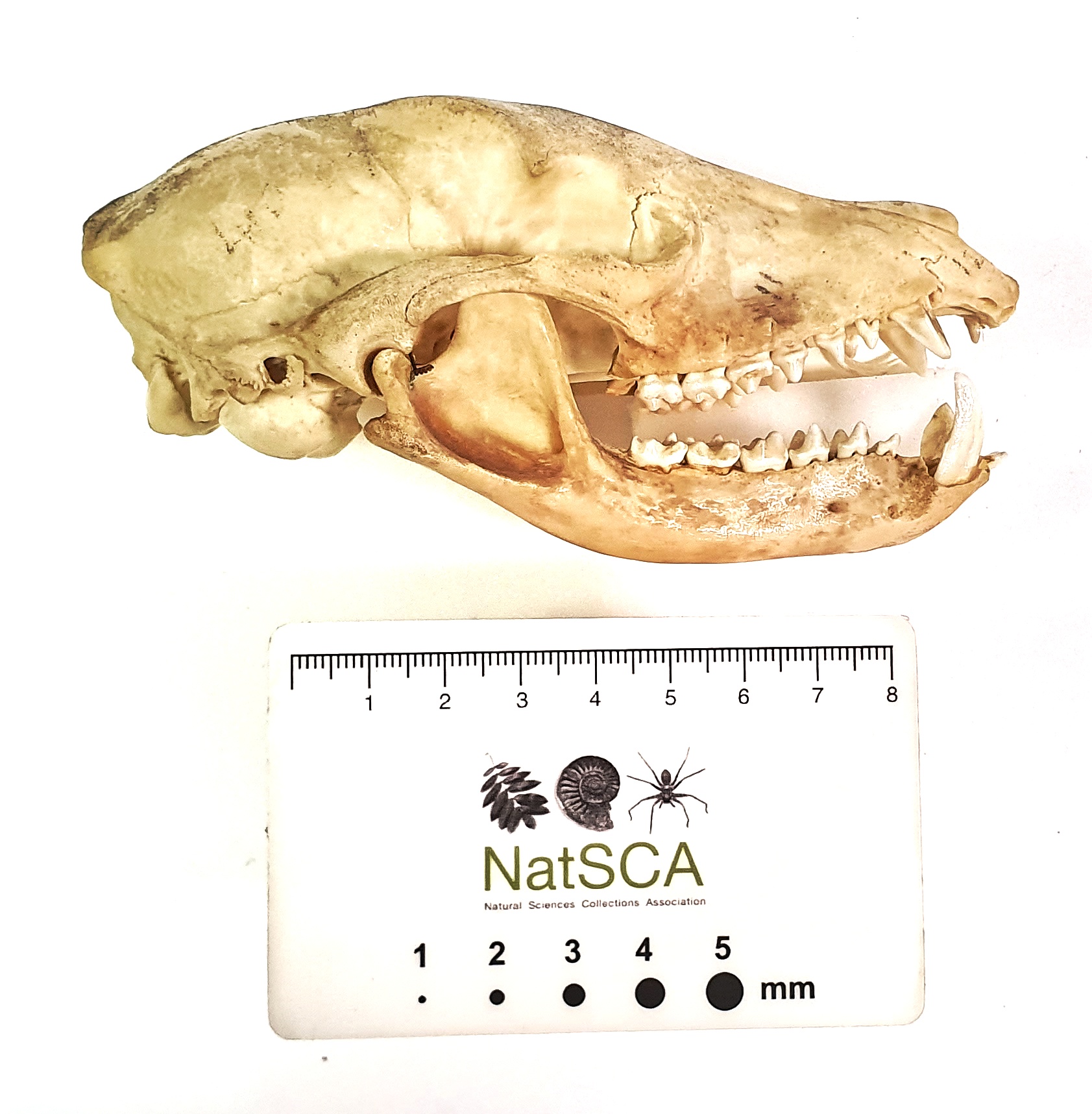
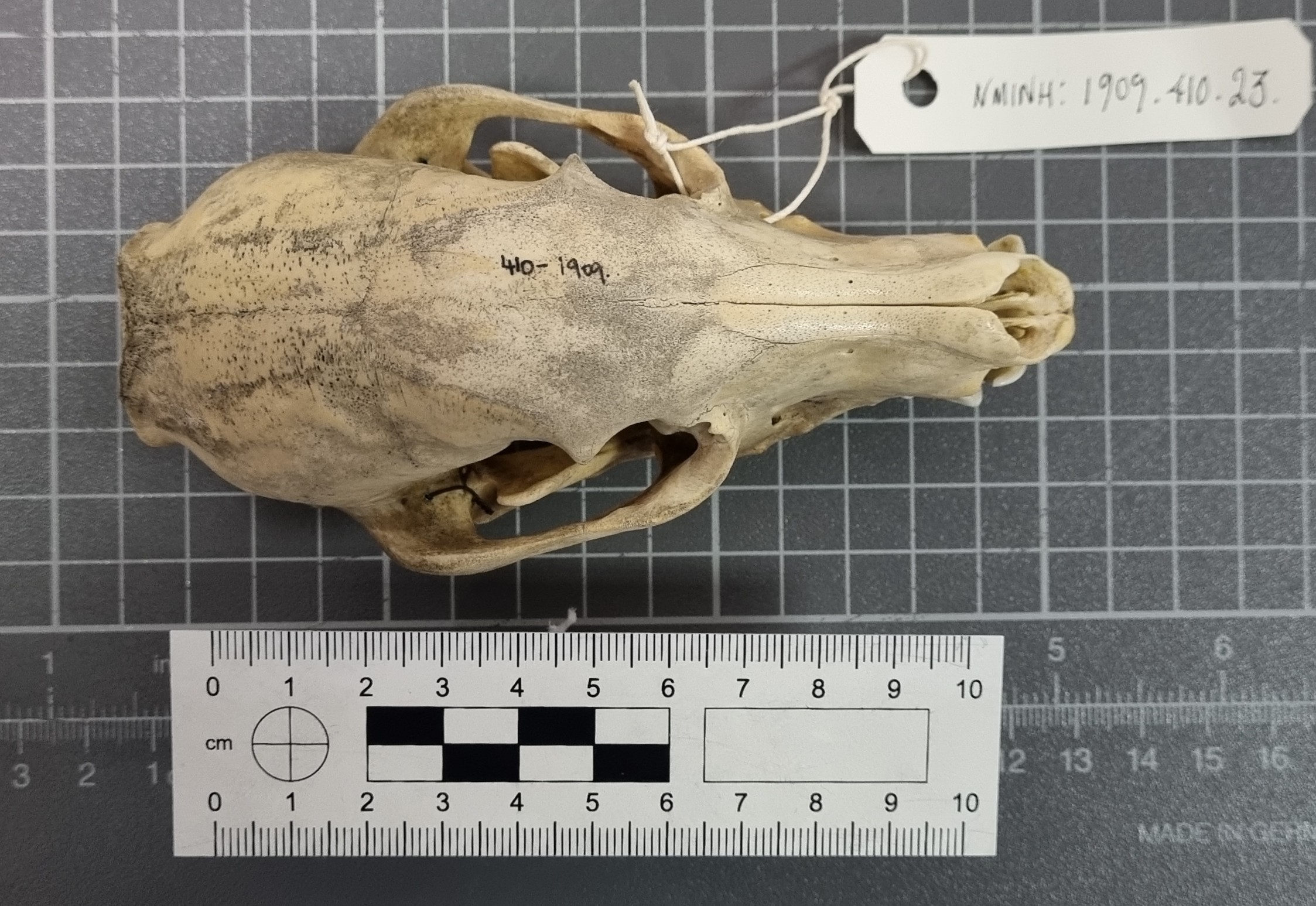
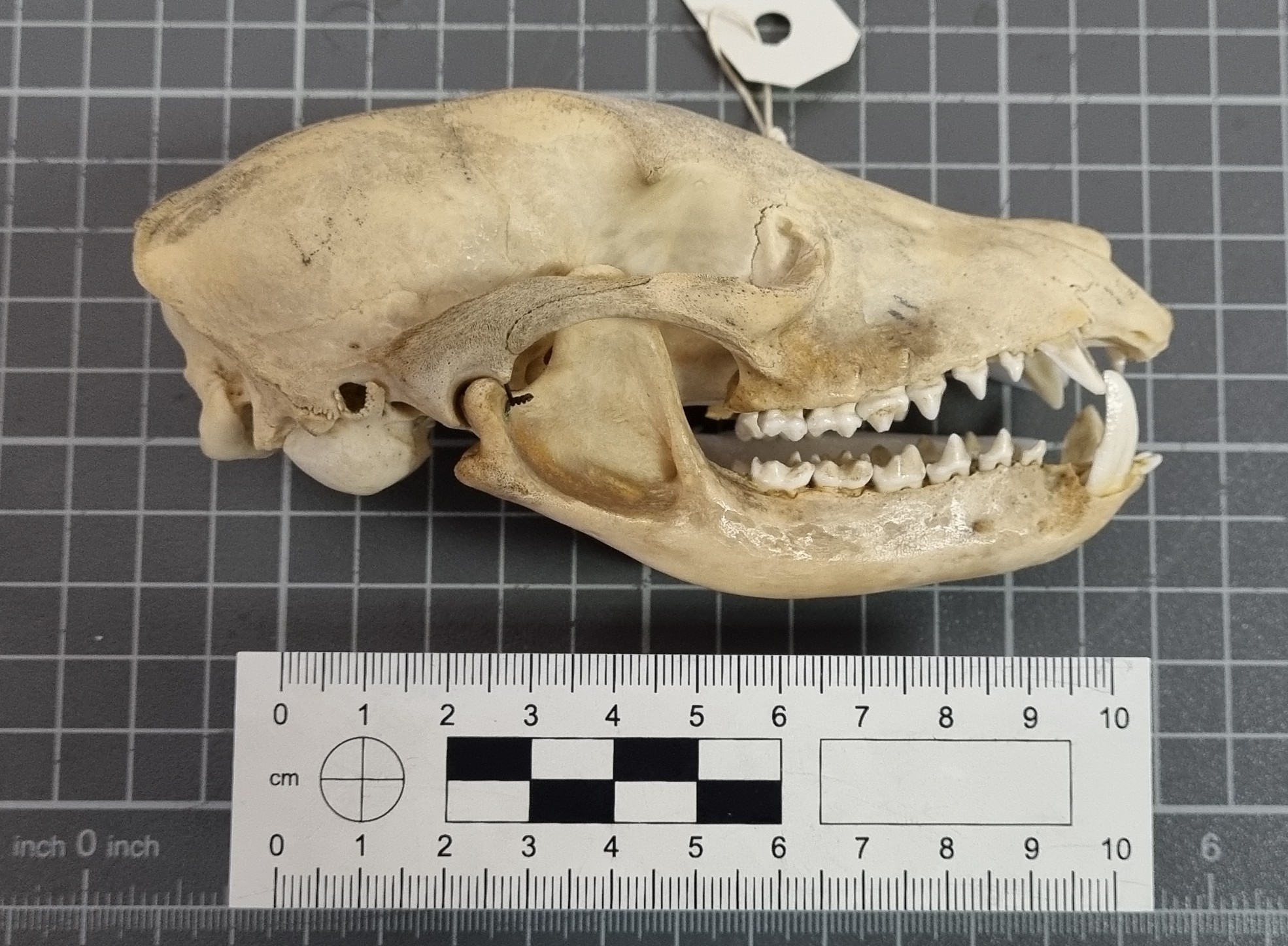
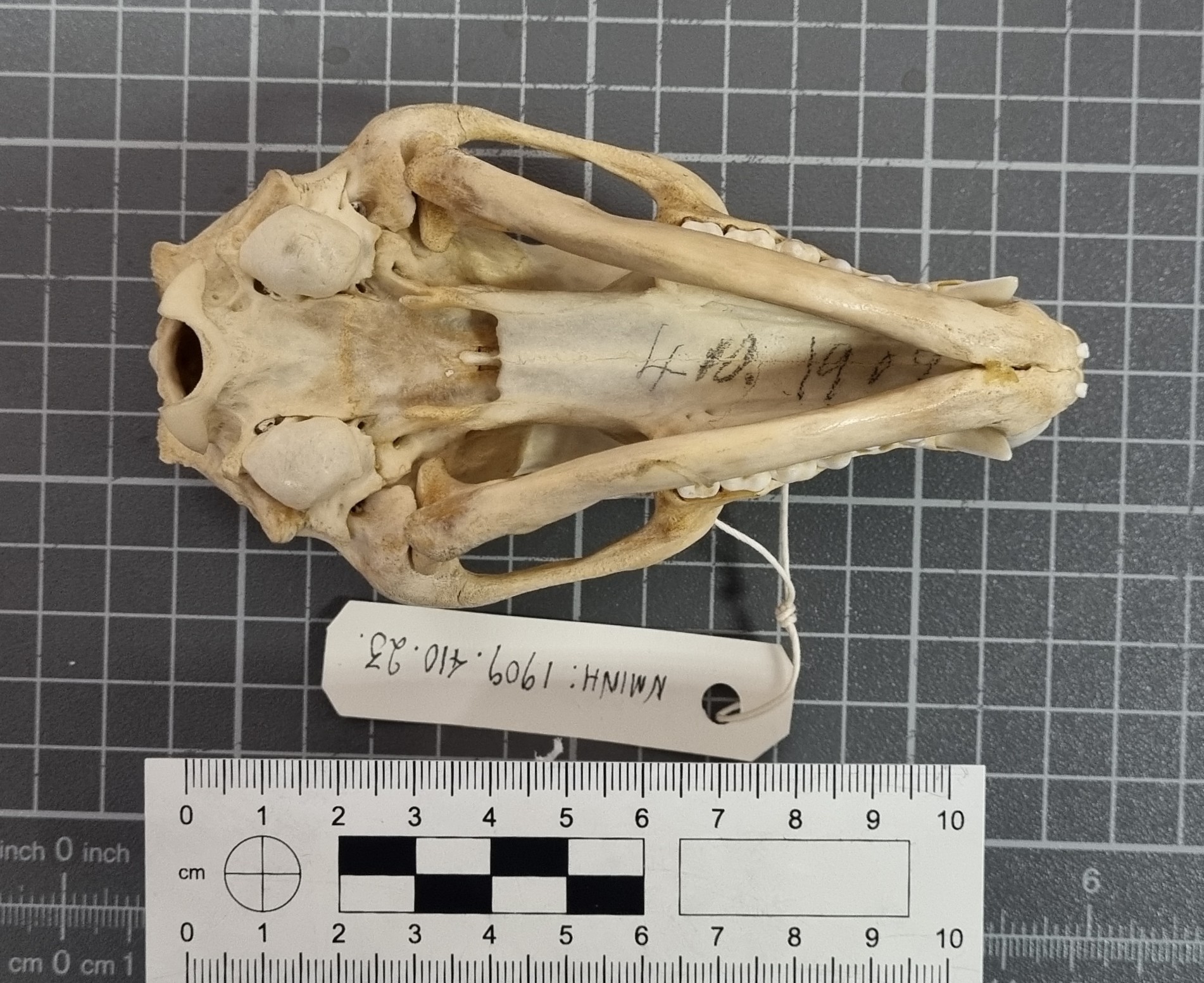
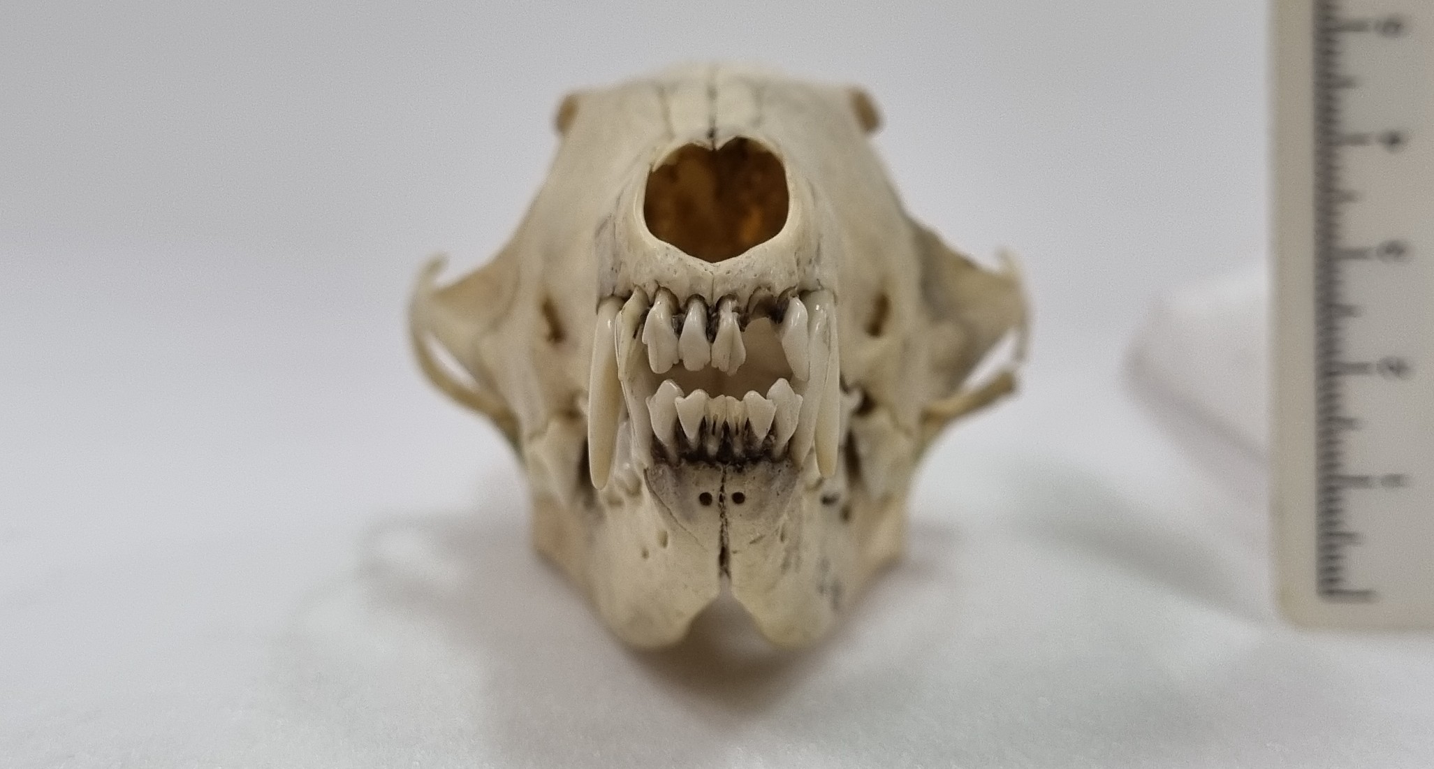
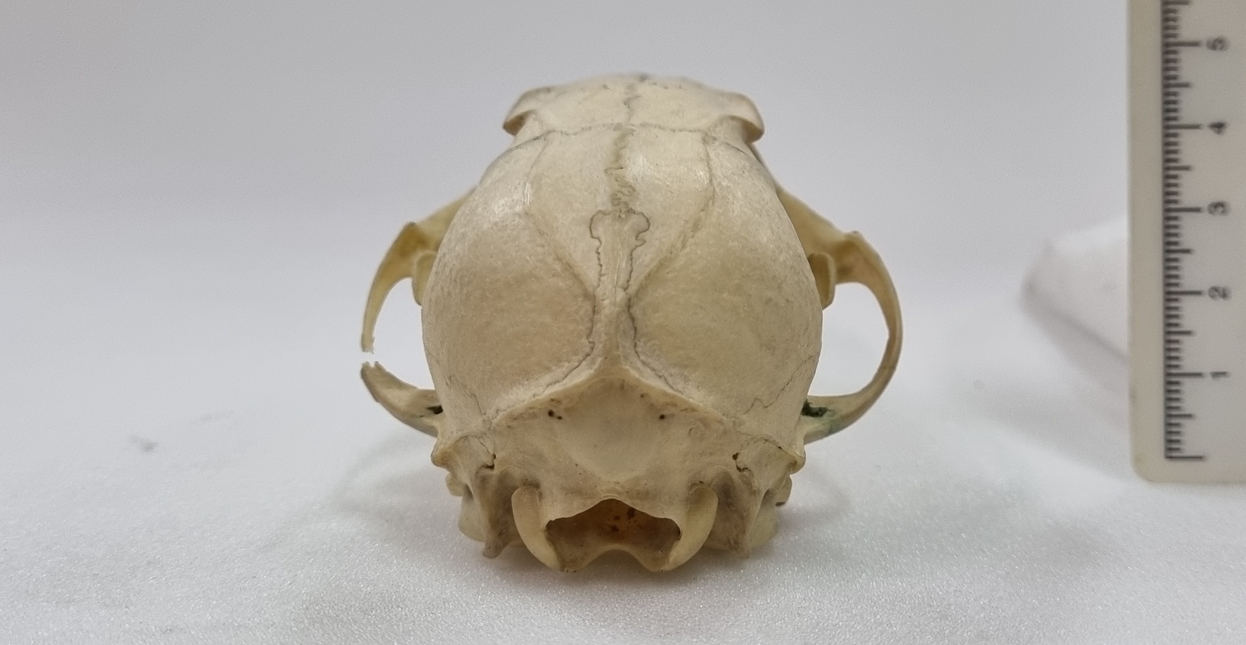
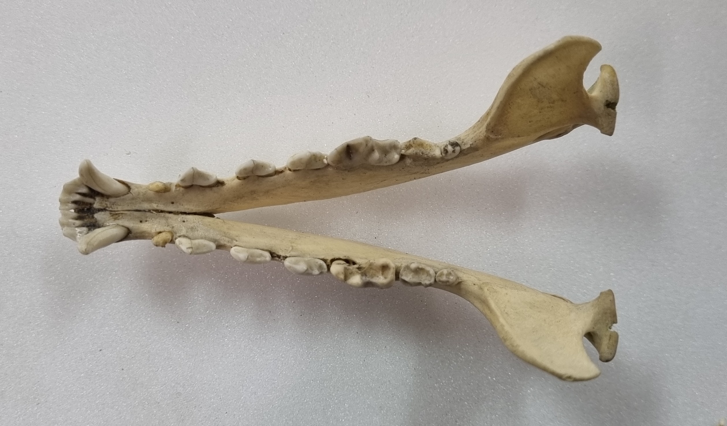
Last week I gave you this neat little skull to have a go at identifying, from research collections of the Dead Zoo in Dublin:
I wasn’t surprised that everyone in the comments worked out that this is the skull of some sort of fox, but I was equally unsurprised that nobody worked out the species. Generally speaking, most people immediately think of the near ubiquitous Red Fox or perhaps Grey Fox (or Gray Fox to our American friends), but there are plenty of others – 24 species commonly referred to as “fox” and 12 species of “true fox” in the genus Vulpes.
This particular specimen has a sagittal crest that forms a lyre-shape – normally something associated with the Grey Fox:
However, this feature can occur in other species, often in females or subadults, where the surface of the bone has not finished remodelling at the margin of the attachment of the temporalis muscles (those are the ones that connect to the lower jaw from the sides of the cranium and are responsible for the operation of the lower jaw during powerful biting).
However, in this specimen the muzzle is more tapered and the postorbital constriction is relatively broad. All of these point away from the Grey Fox.
With foxes there can be a lot of similarities between the skulls of species, with all the usual compounding issues of sexual dimorphism, age and regional variation. However, size can give some clues, and things like the relative size of the external auditory meatus (also known as the ear-hole), and the shape of the auditory bulla, are useful for differentiating between species.
With a bit of patience, a bit of pattern recognition, and a resource with good images of specimens, like the Animal Diversity Web, it is possible to work out what you’re looking at.
In this case, the mystery object is a Swift Fox Vulpes velox (Say, 1823).
These North American foxes are smaller than the Red or Grey Fox, but a bit bigger than their close cousin, the Kit Fox. They live in grasslands and praries, where they prey on rodents, birds, reptiles and pretty much anything they can find – including insects, fruit and grasses.
As with many species, the Swift Fox has declined due to changing land use and the systematic persecution of predators in the first half of the 20thC. In fact, it was wiped out in Canada around this time, although it was subsequently successfully reintroduced and numbers have increased.
So watch out for those foxy skulls – there are more species to consider than you might think and they can be tricky to identify without reference resources. I hope you enjoyed this little detour down the fox hole!
For the first time in quite a while, I managed to escape from my desk and spend a little time in the collections of the Dead Zoo. The main reason was to facilitate access for researchers doing some really cool projects, but it also gave me a chance to spend a little time exploring the collections I’m responsible for.
In one of the cabinets I spotted this skull, and I thought it might make a good mystery object:
So, do you have any thoughts on what this might be? As ever, you can leave your questions, observations and suggestions in the comments section below. I hope you enjoy this specimen as much as I did!
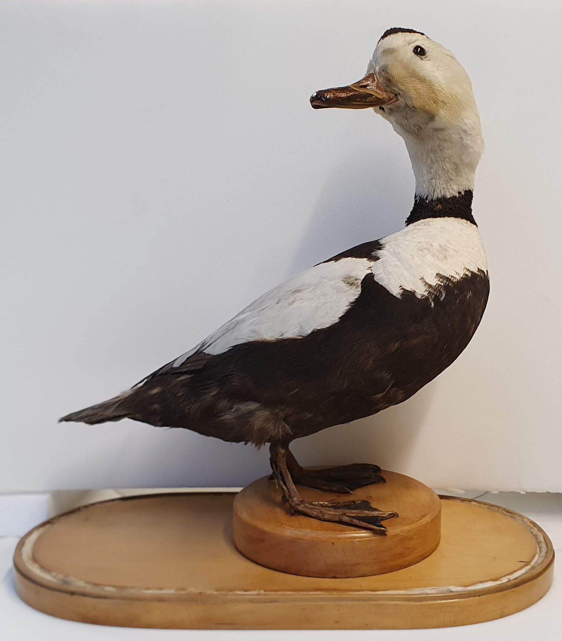
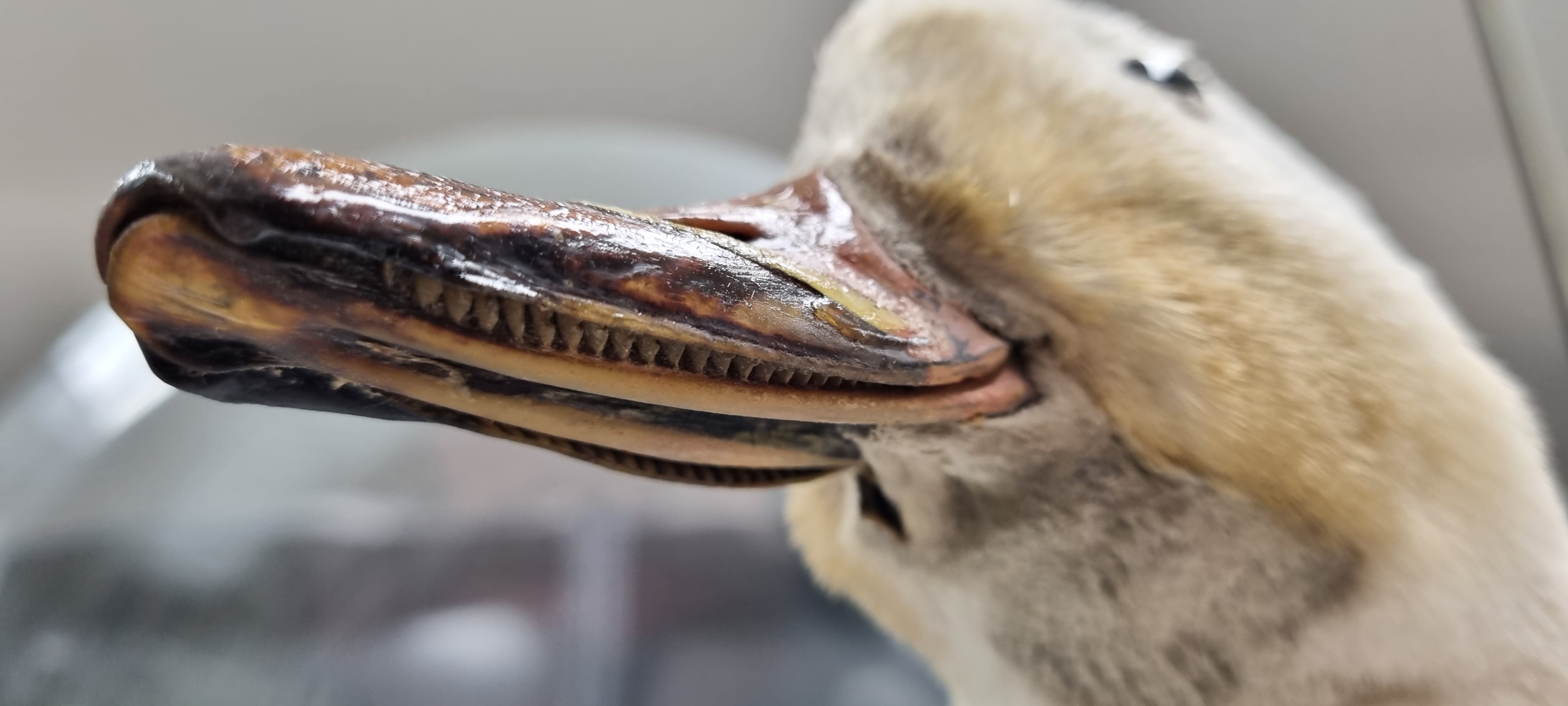
Last week I gave you this specimen from the Dead Zoo to have a go at identifying:
Evidently it was a bit too easy, since everyone who commented not only worked it out, but took the time to come up with clever cryptic clues to reveal the identity. The first was Tony Irwin with:
I suppose that a pencil sketch of this would qualify as a labradoodle?
This is a specimen of Labrador Duck Camptorhynchus labradorius (Gmelin, JF, 1789), an endemic North American duck that has the dubious honour of being the first American species to be pushed into extinction following the European invasion of the continent. The exact reasons for the extinction are unsure, but between the adult birds being shot or caught on fishing lines, their eggs being over-exploited as a food source by settlers, and competition with humans for mussels and other marine molluscs, the already small populations were quickly reduced to nothing.
Lamellae in the bill of the Labrador Duck.
The last sighting was in 1878, but the museum bought this specimen in 1892 for the princely sum of £30-0-0 (that’s the equivalent of about €5,376 in today’s money). It had passed through a few pairs of hands before the Museum got hold of it, but we’re fortunate in knowing that the specimen was brought to Ireland in August 1838 from New York by Lt. W. Swainson R.N. who was in command of The Royal William, a paddle steamer in the service of the City of Dublin Steam Packet Co.
Only 55 specimens of Labrador Duck are known from museums around the world, so all information about them is valuable, especially when you consider how scarce and irreplacable they are. Specimens like this really hammer home the responsibility we have as museum professionals who are responsible for their care.
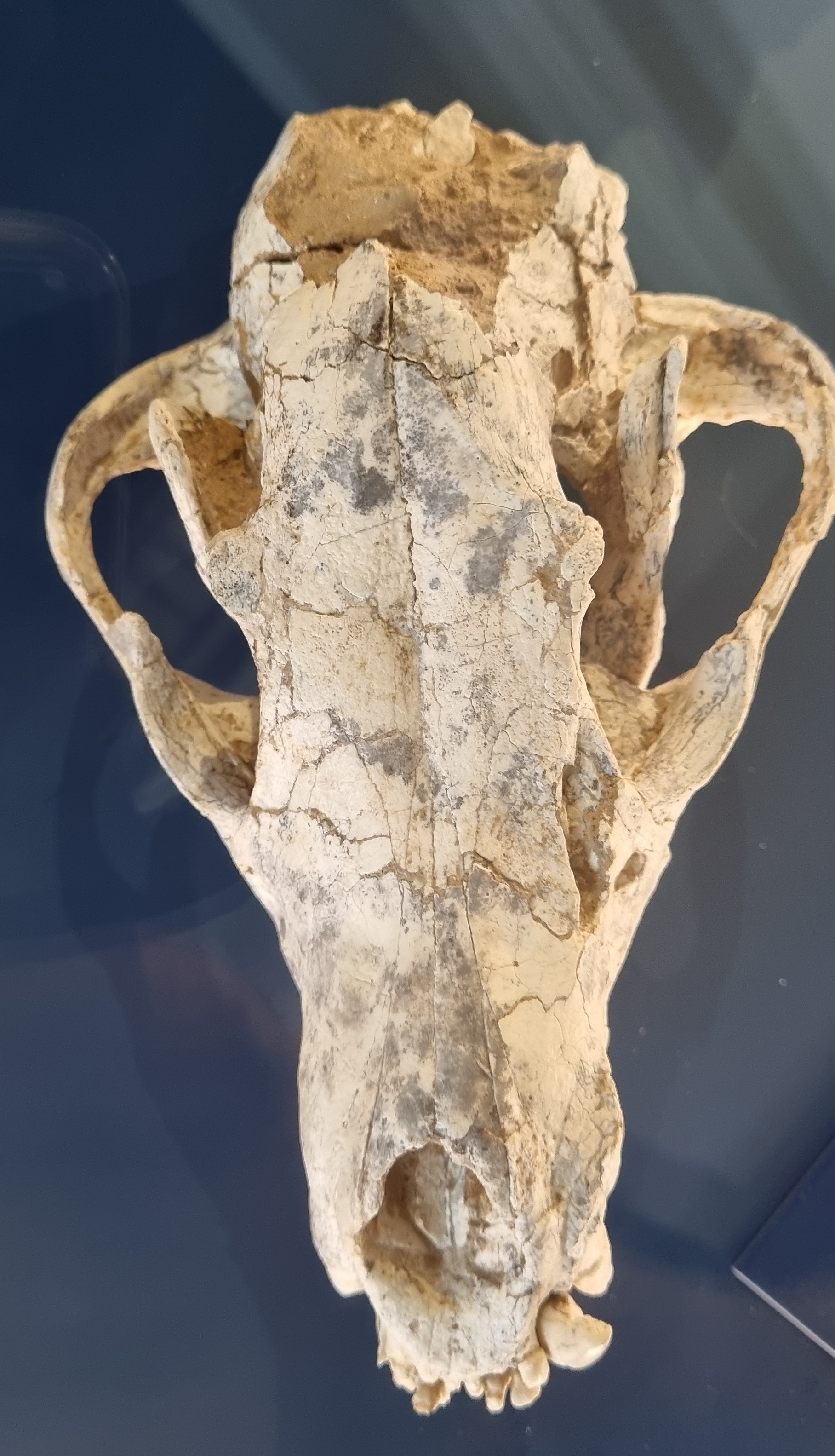
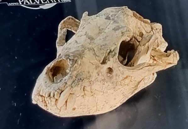
Last week I gave you this 6 million year old fossil skull to have a go at identifying:
The specimen is on display in the geology galleries of the Natural History Museum at the University of Oslo (which is well worth a vist). However, this did mean the photos provided weren’t quite as good as I’d like, particularly notable being the lack of a scale bar (sorry!)
Even without a scale, consensus shifted towards this being some kind of hyena, thanks to the curved mandible and (hint of) robust molars and shorter toothrow than you might expect to see in a canid. The broad and flat profile of the frontals between the eye sockets probably helped too:
Hyenas have an interesting evoloutionary history, branching off from the basal feliforms around 22 million years ago and adapting to fill a terrestrial carviore niche in Eurasia and becoming quite diverse. In America the canids were filling that same niche, which led to some competition when the canids made it to Eurasia (spoiler alert – the hyenas lost that competition, leaving us with just three highly specialised bone crushers and the decidedly weird Aardwolf living today).
Four live specimens of hyenas (clockwise from top left): spotted hyena (Crocuta crocuta), brown hyena (Parahyaena brunnea), aardwolf (Proteles cristata), striped hyena (Hyaena hyaena). Image by Termininja, 2020CC BY-SA 4.0
The mystery specimen was labelled as Thalassictis wongii (Zdansky, 1924), a species described from China and originally placed in the genus Icititherium, but reassessed by Kurtén (1985). A cladistic treatment of the Hyaenidae by Werdelin & Solunias (1991) later placed it in the genus Hyaenotherium, but that may not have been accepted by the curatorial team in Oslo without an accompanying formal taxonomic treatment.
These are the sorts of decisions that need to be made when considering something as simple as a label stating a species name, so you can imagine my sense of trepidation as we are about to embark on a major project at the Dead Zoo, which will allow us to reassess the information with our 10,000 or so display specimens. Fun times ahead!