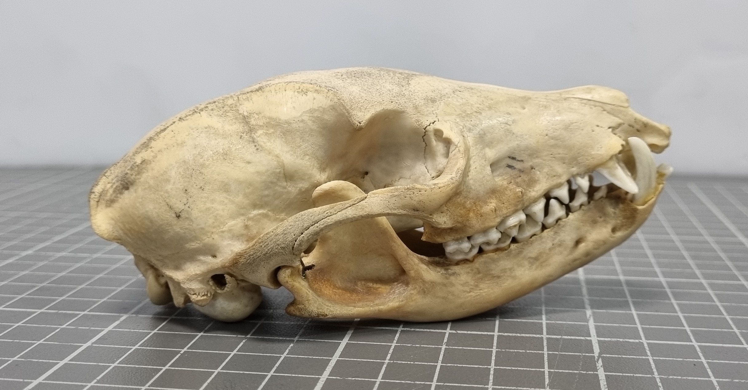
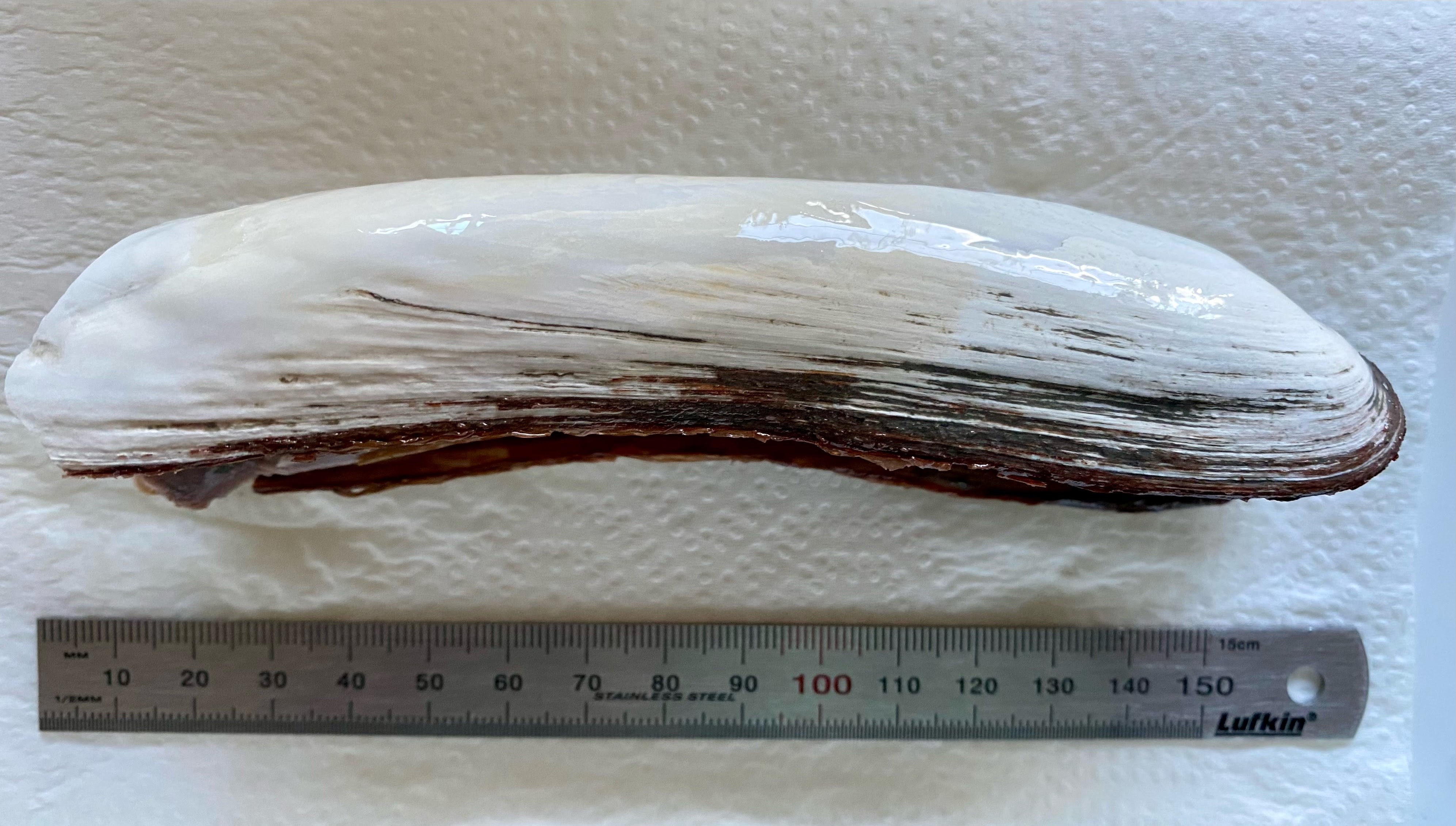
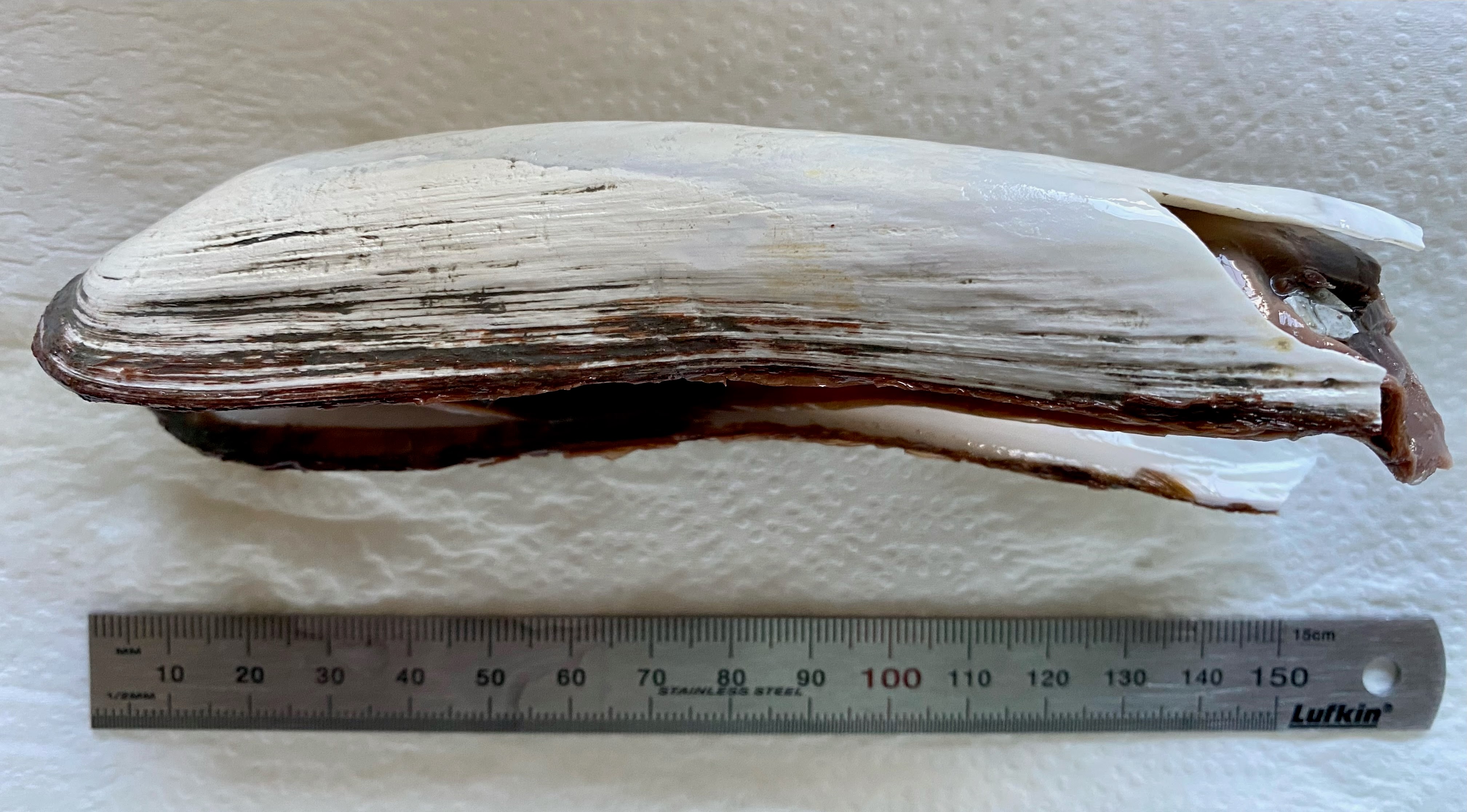
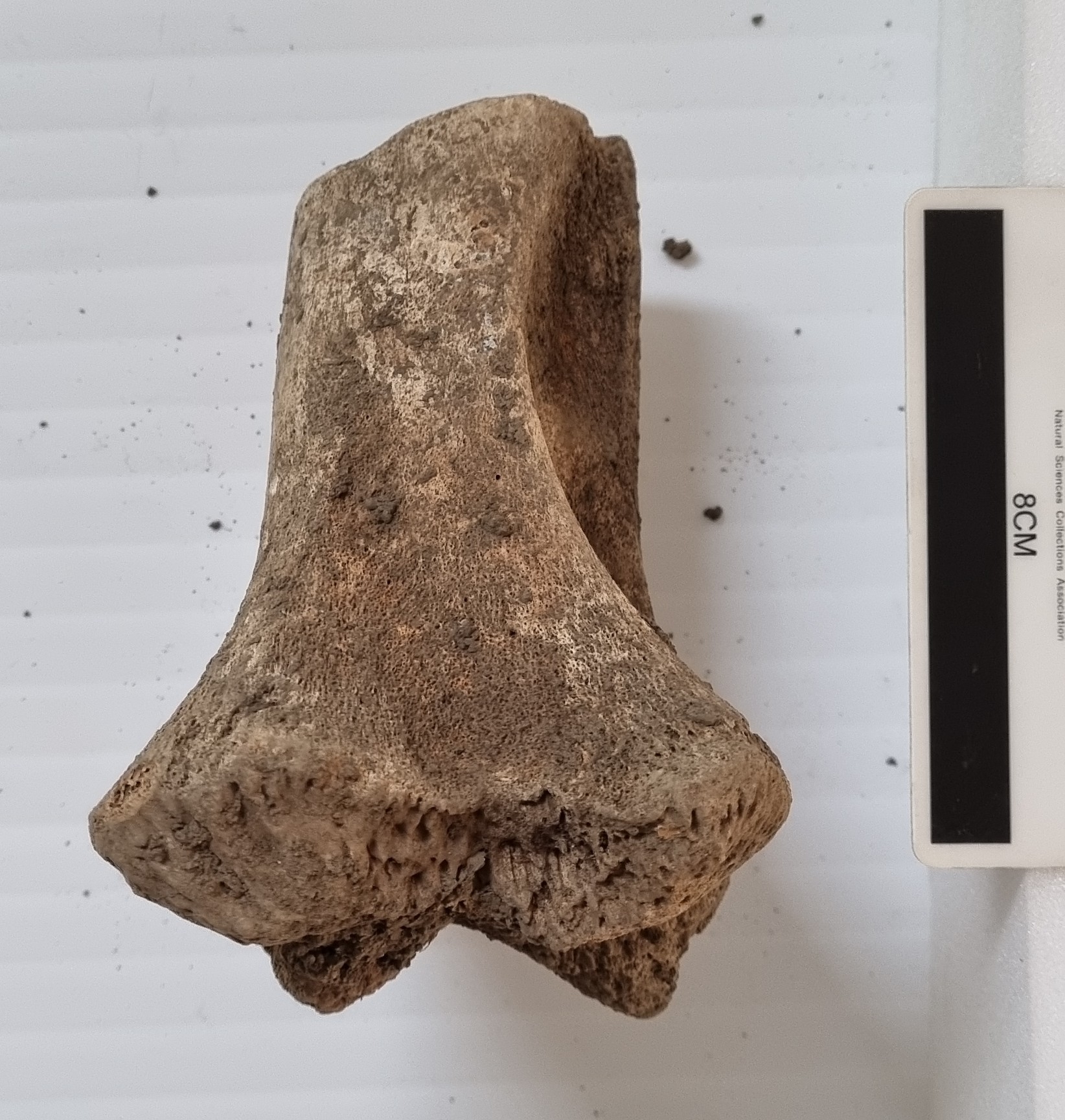
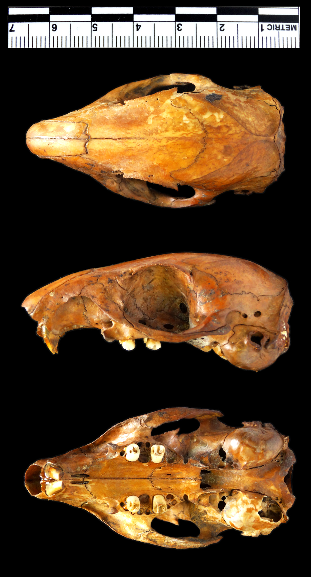
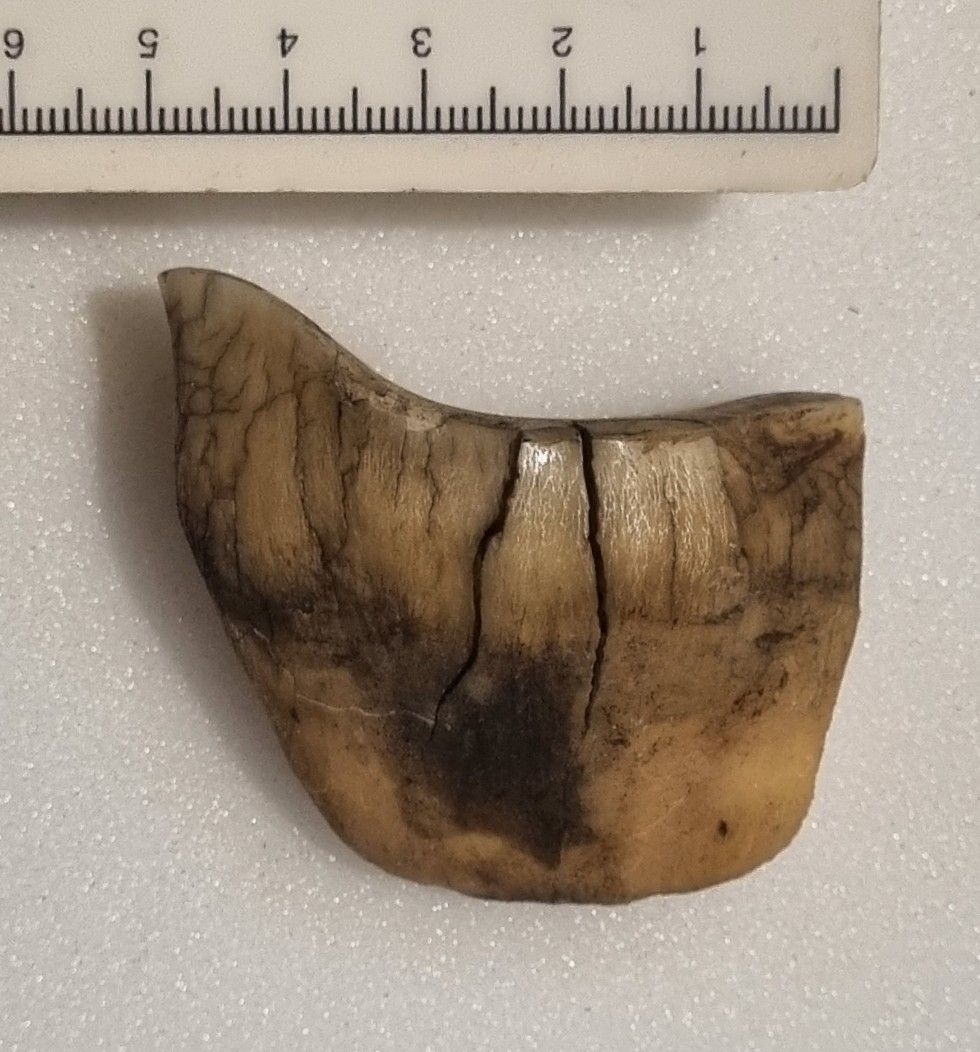
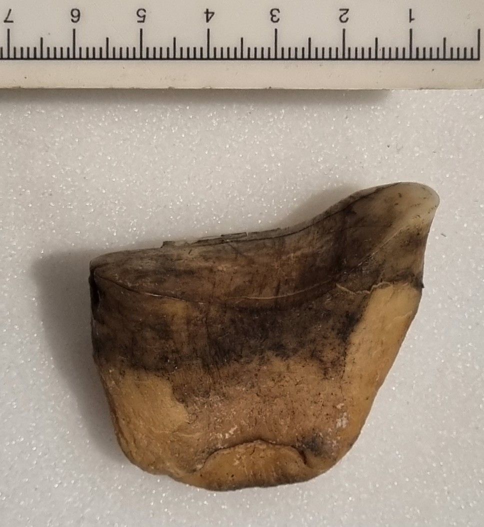
Last week I gave you this specimen to identify, from the collections of the Dead Zoo:
I thought it might prove a little challenging, and I wasn’t mistaken, although some people did manage to get very close. Chris Jervis was the first, but Adam Yates also provided a nice analysis of the specimen:
It is blade shaped without cusps, so the incisor of a large animal seems possible (I’m ruling out a Thylacoleo premolar on the basis of the broad wear facet on the occlusal surface and single root).
The root is really short so I’m guessing its a deciduous incisor.
Horses are large mammals with incisors of this sort of shape. If it were a horse it would be a third incisor because of its less square crown. As the high point (presumed mesial side) is on the left side it is probably a right incisor.
So my guess is a right deciduous incisor three of a horse.
Adam Yates June 7, 2024 at 10:20 am
Adam then followed up with a note about the size being too big for a horse, and leaned in to Chris Jervis’ suggestion – a good decision.
So what is it?
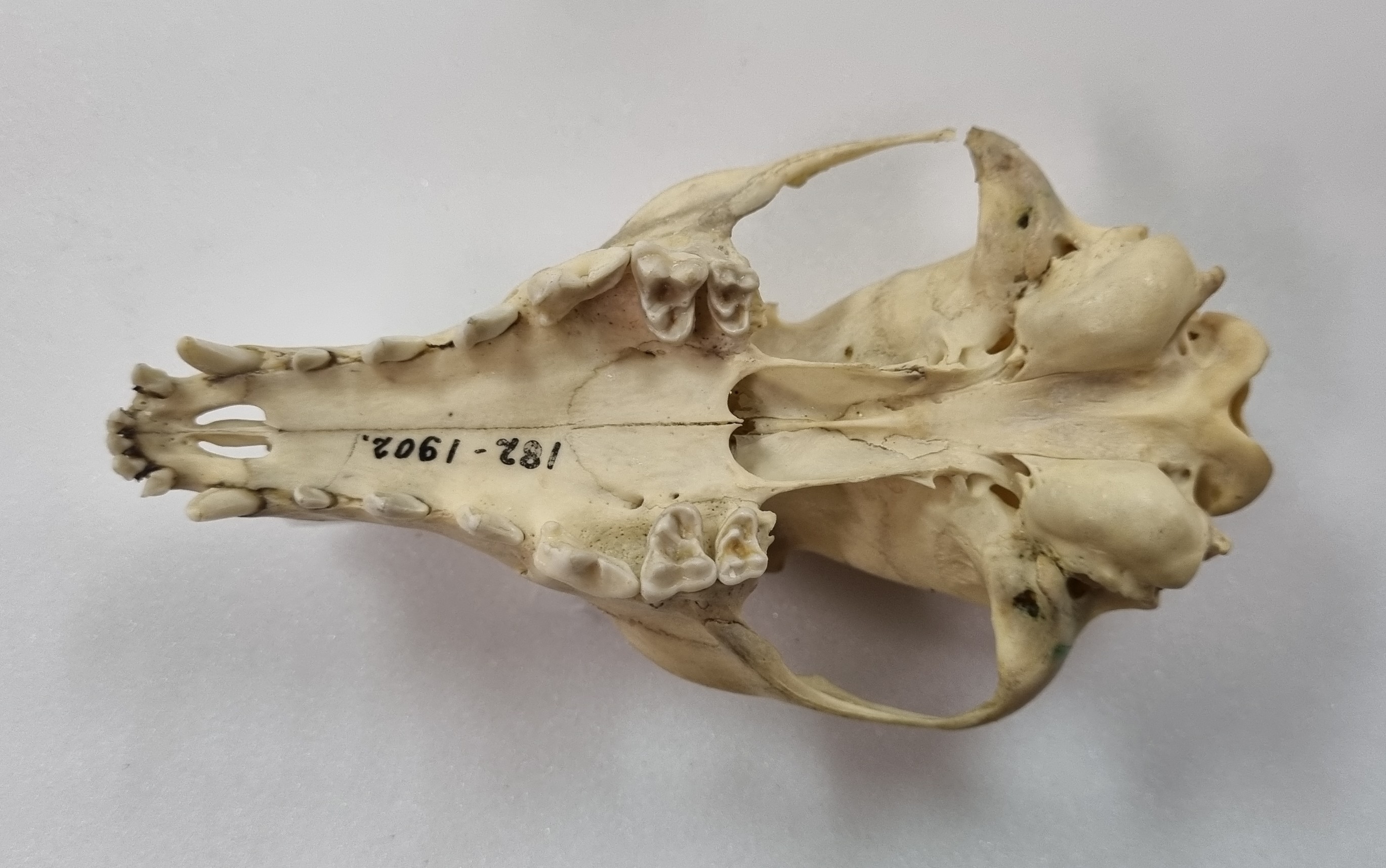
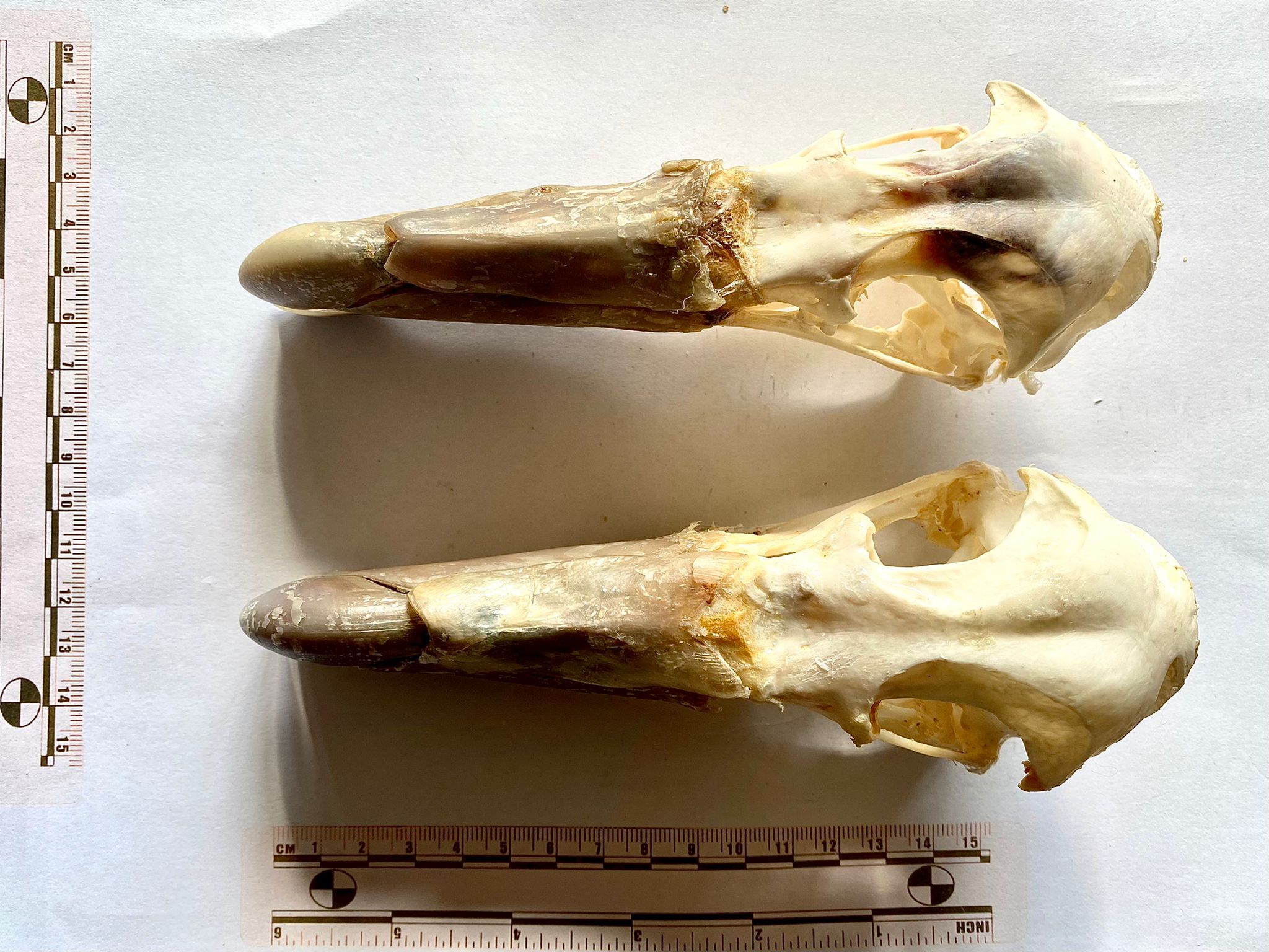
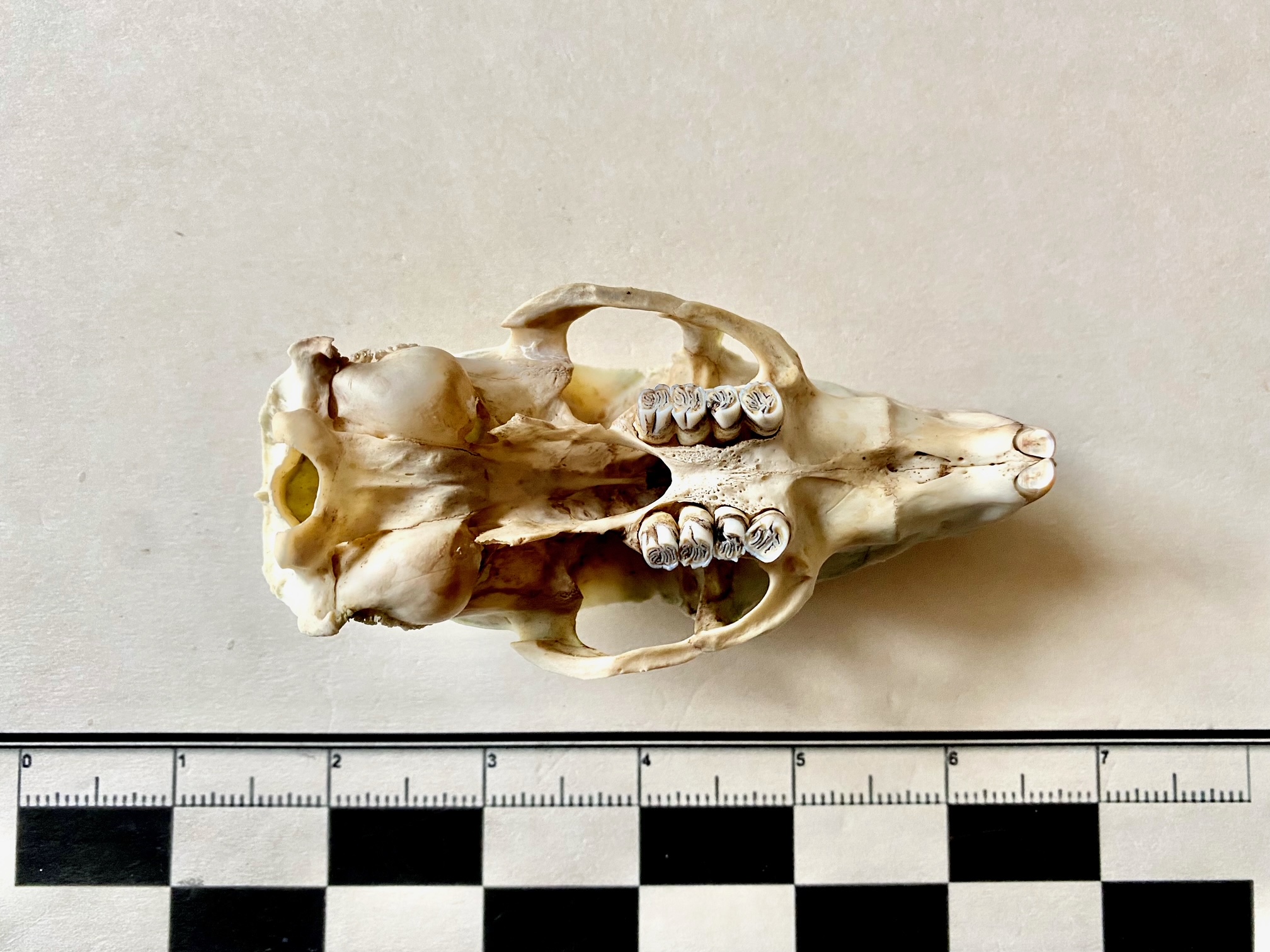
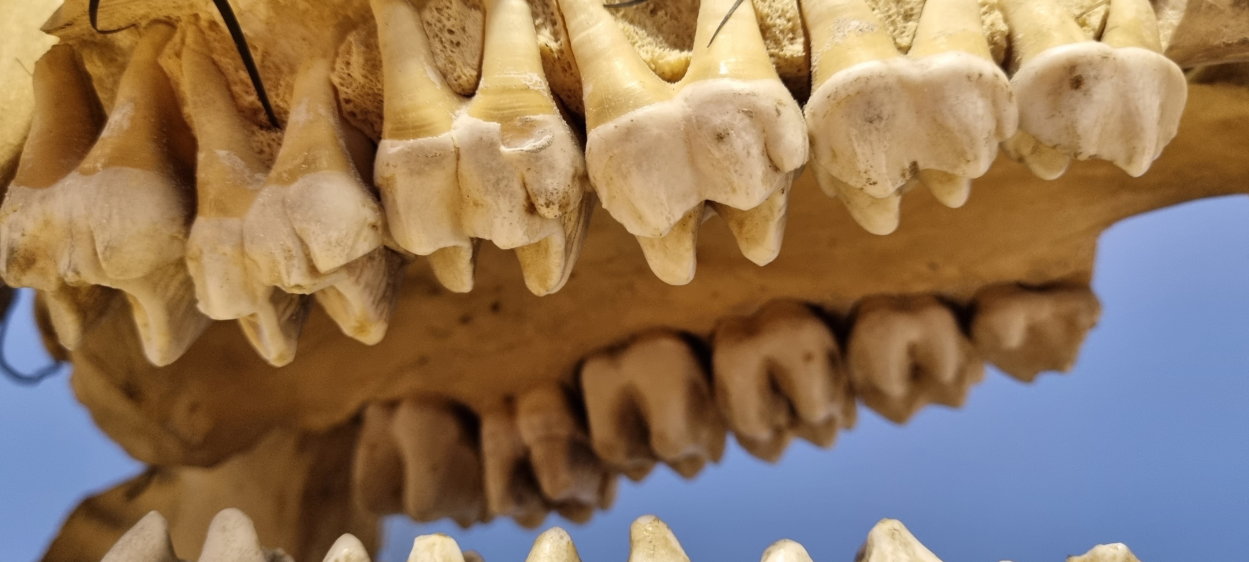
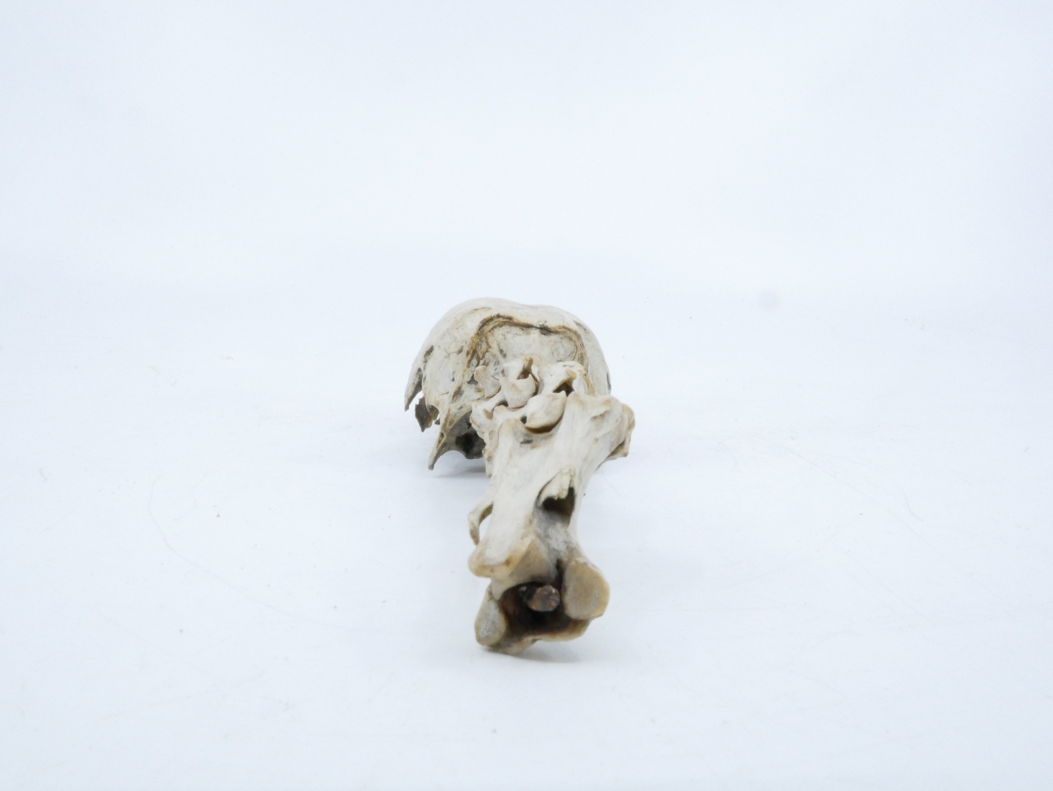
This is in fact the upper right incisor (or tusk) from the skull of an Indian Rhinoceros Rhinoceros unicornis Linnaeus, 1758:

African rhino species have lost their incisors, but the Indian Rhinoceros have incisors that form self-sharpening tusks with open roots, which are used in fighting.
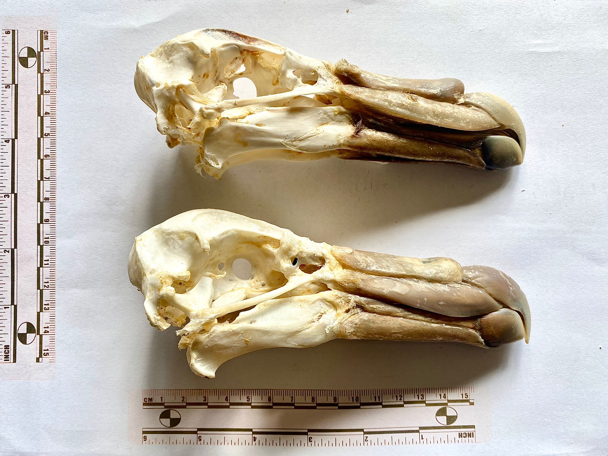
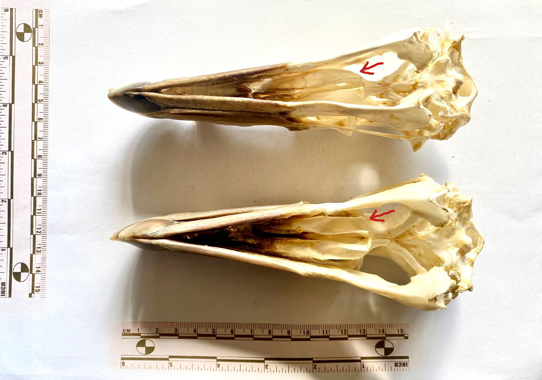
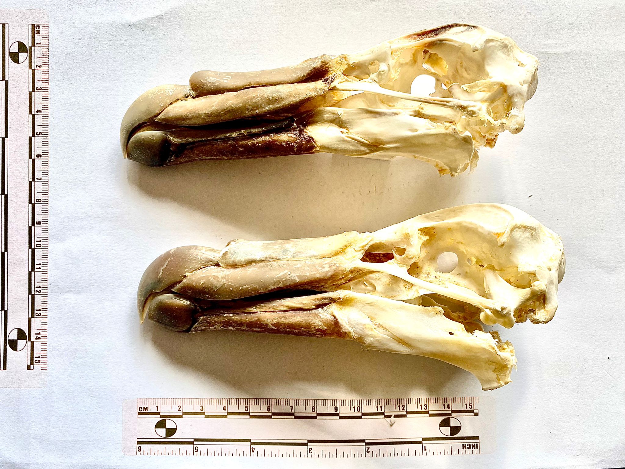
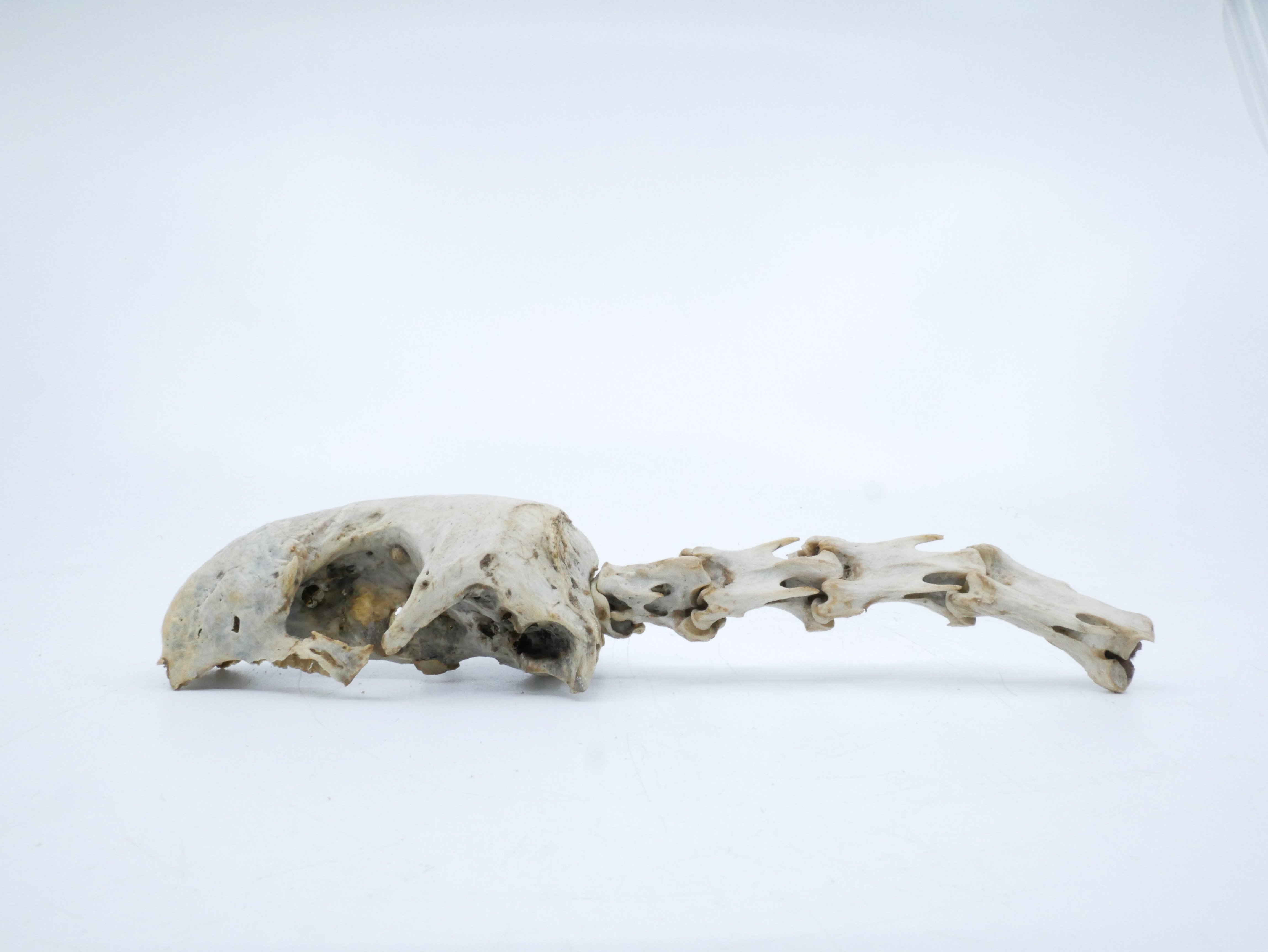
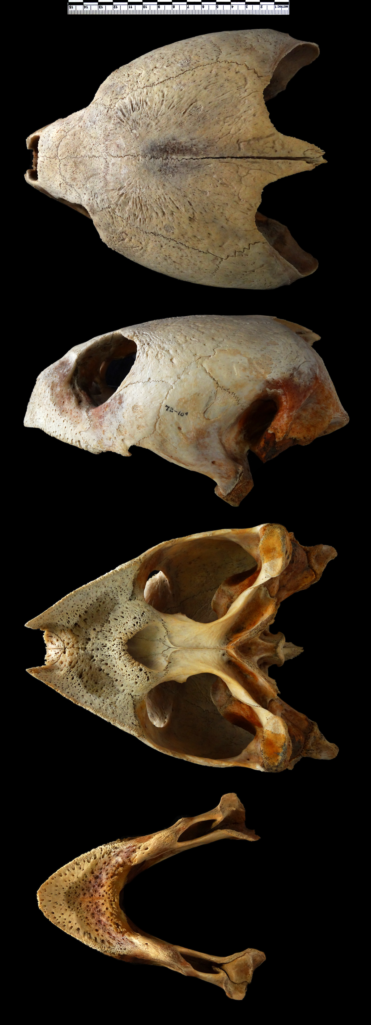
The full specimen that this is from is a mounted skeleton that was on display in the Dead Zoo for a century or so, and the animal clearly had a very tough life, as it shows a variety of pathologies that have healed.
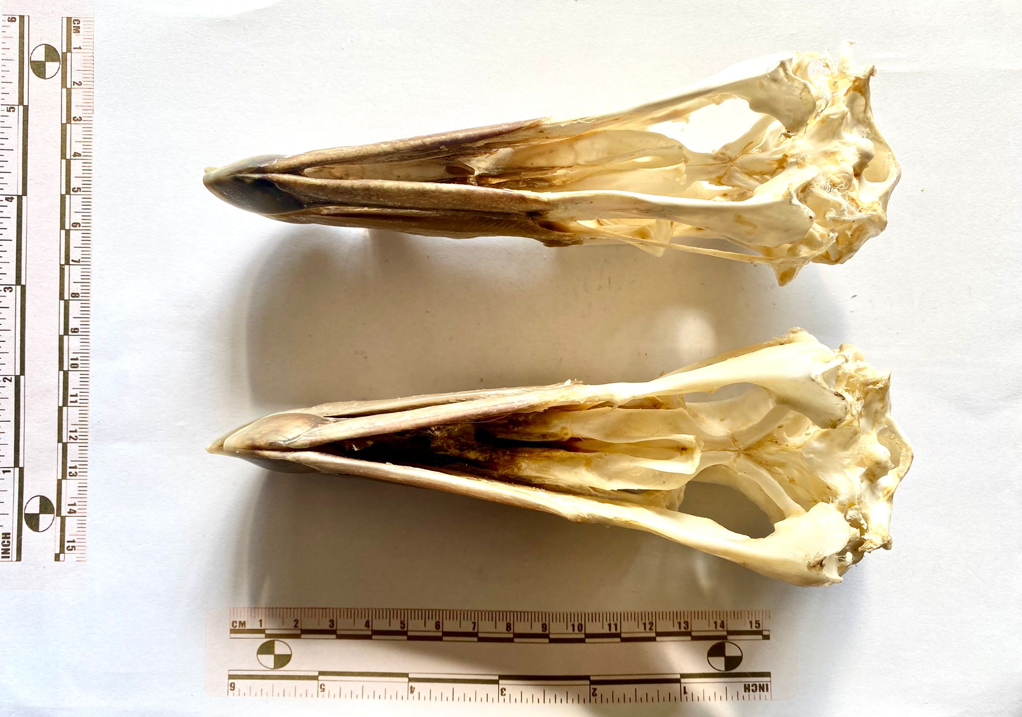
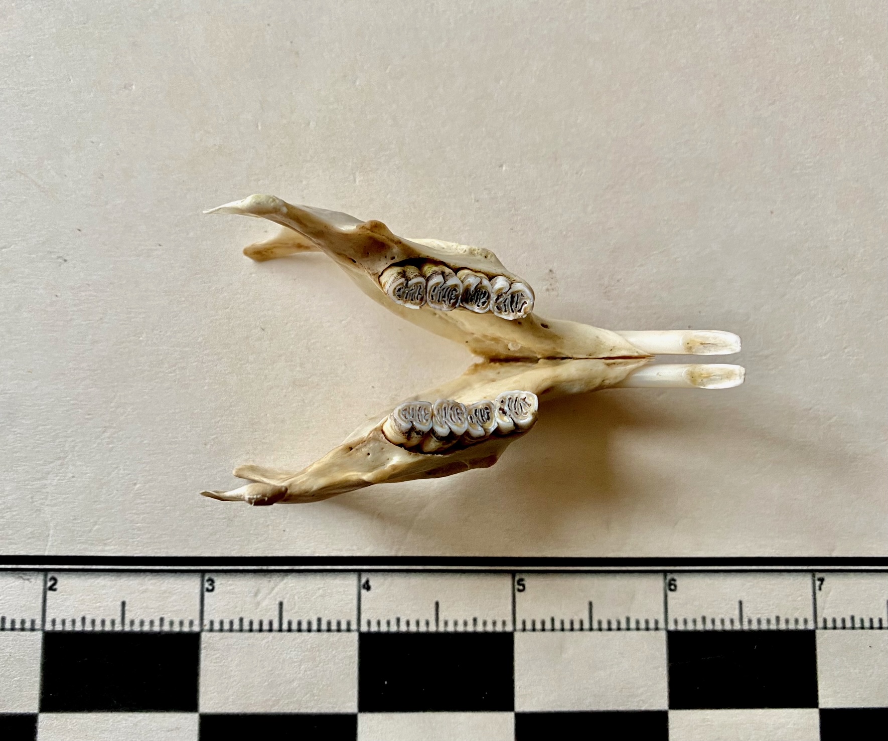
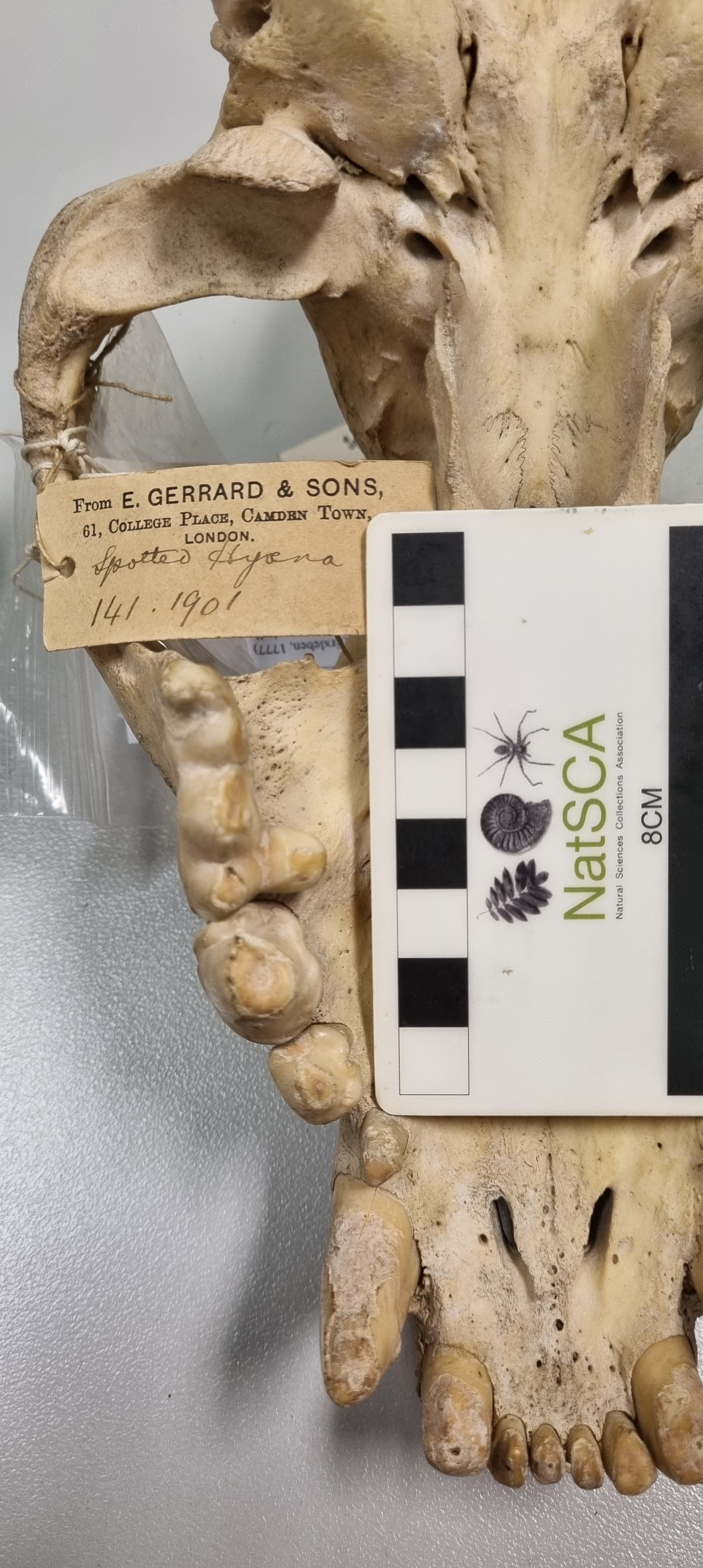
In the mandible there is a huge abscess at the base of the lower right tusk, possibly due to fighting or maybe a bullet wound:

The animal also has a variety of other injuries, including a bullet wound in the ribs:

and a major trauma to the lower front left leg, which has resulted in the humerus and ulna fusing together:

This would undoubtedly have been a very grumpy, very gnarly rhino. I’ve spoken a lot about rhinos in the past (including in a recent documentary series about the problem of rhino poaching and the wildlife trade), as they’re incredible animals that have been horribly exploited in the past, and still being horribly exploited today.

I have these photos of the specimen as over the last couple of weeks we have started the decant of specimens from the Dead Zoo in earnest, as we’re getting moving on a major capital developement project in the Museum, to solve a number of issues with access for the public, problems with the museum environment, and conservation of the 168 year old building.
Stay tuned for more updates on the project, as I suspect it will be keeping me busy for the next few years!