Friday mystery object #534 answer

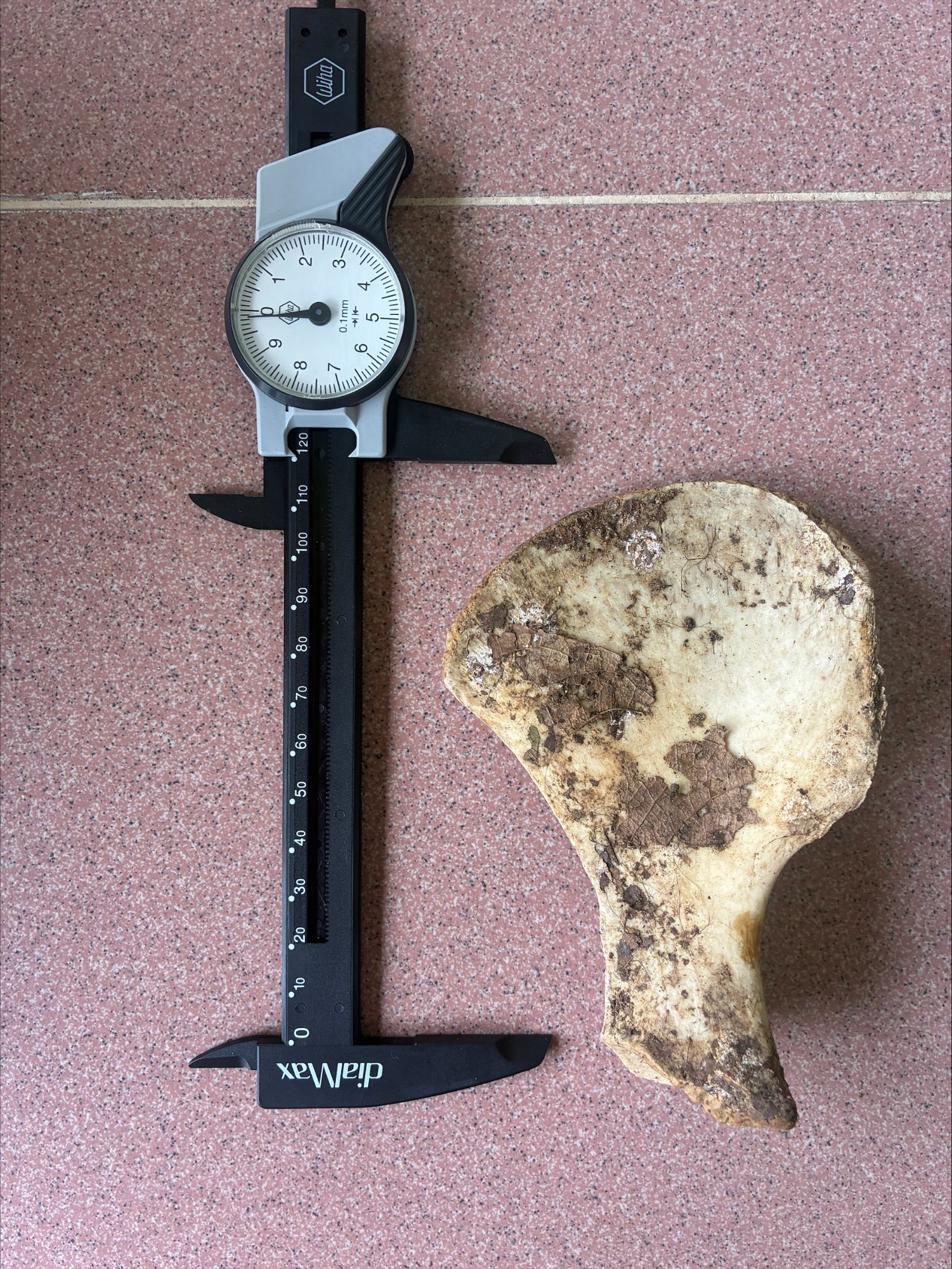
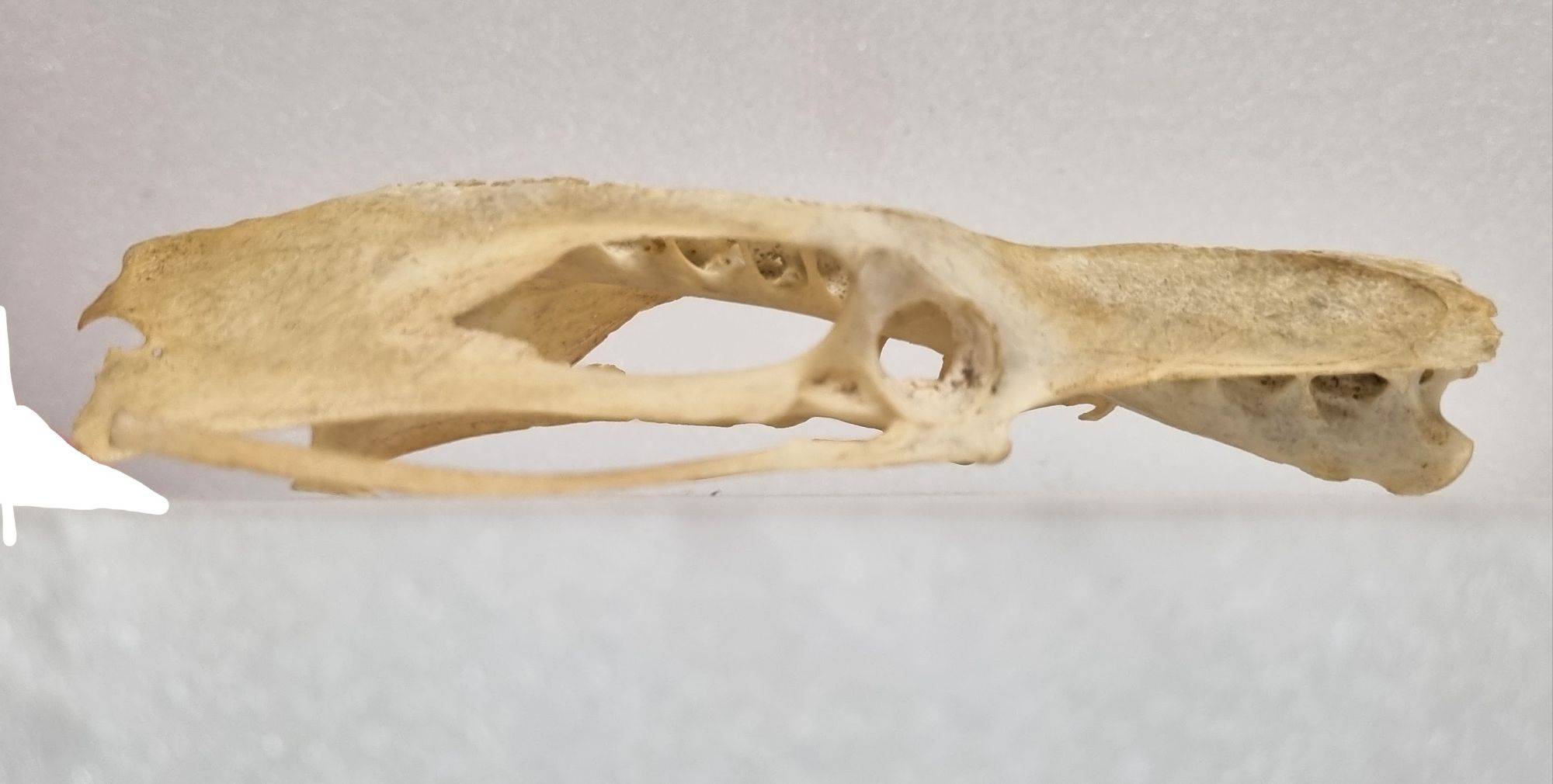
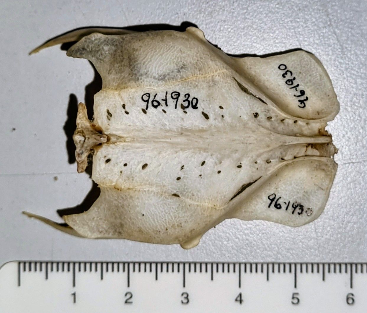
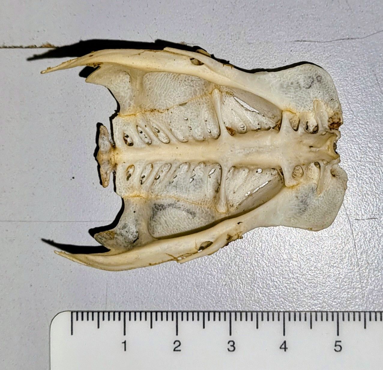
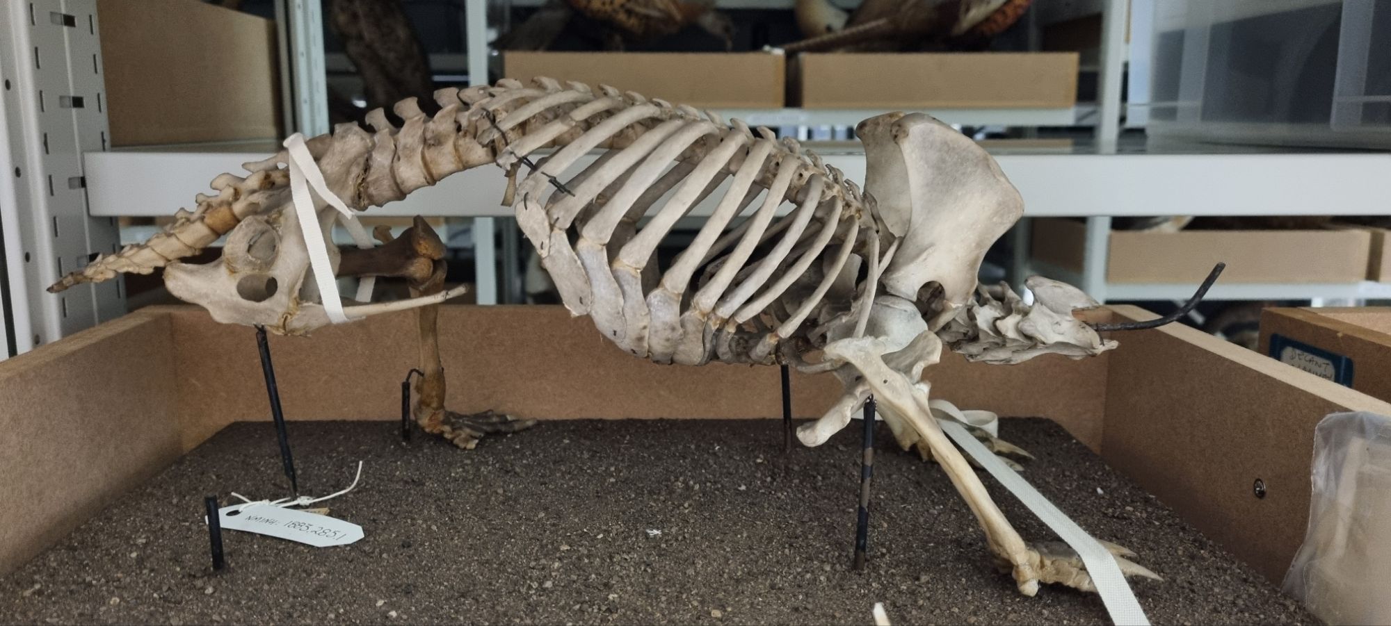
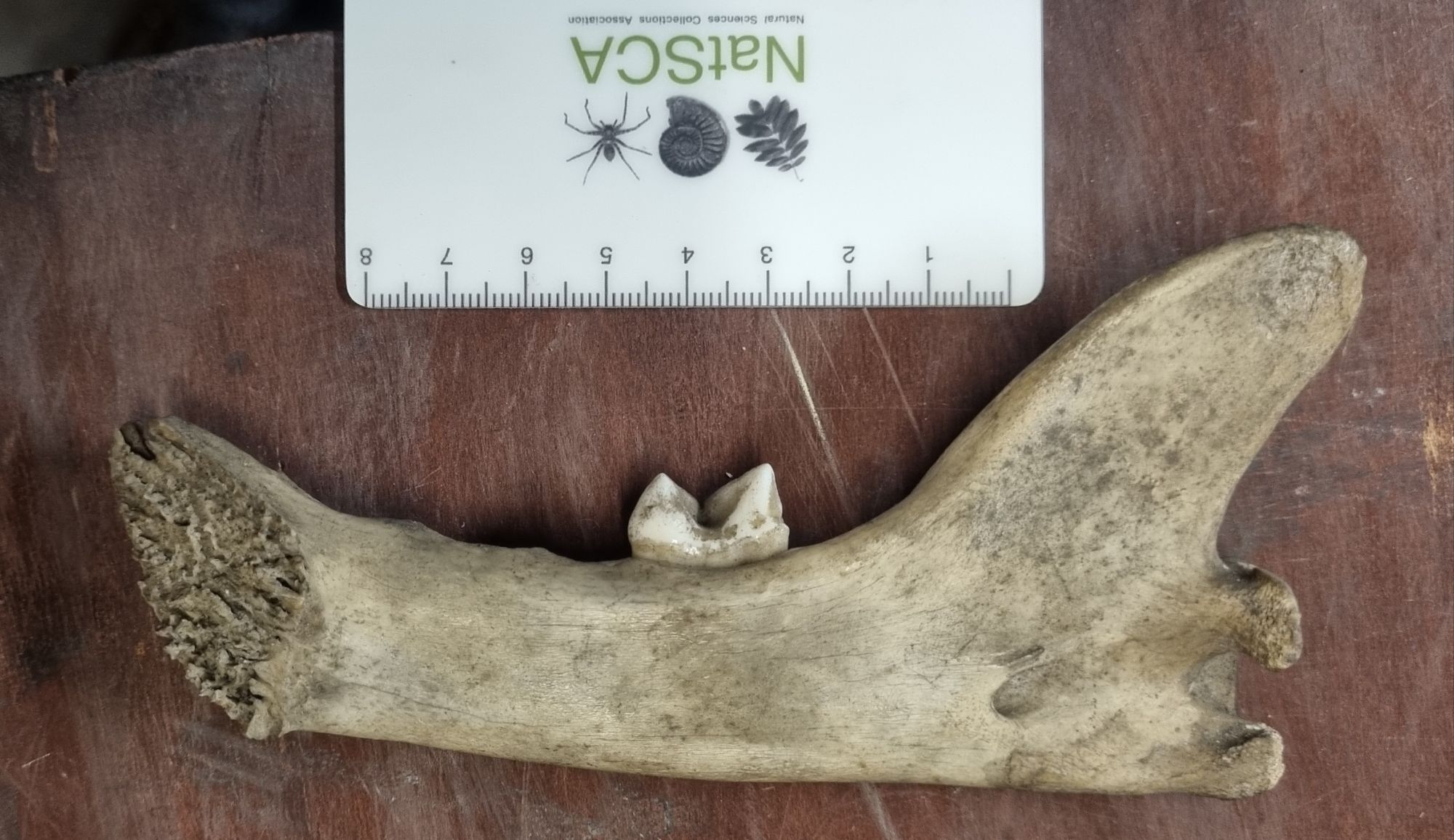
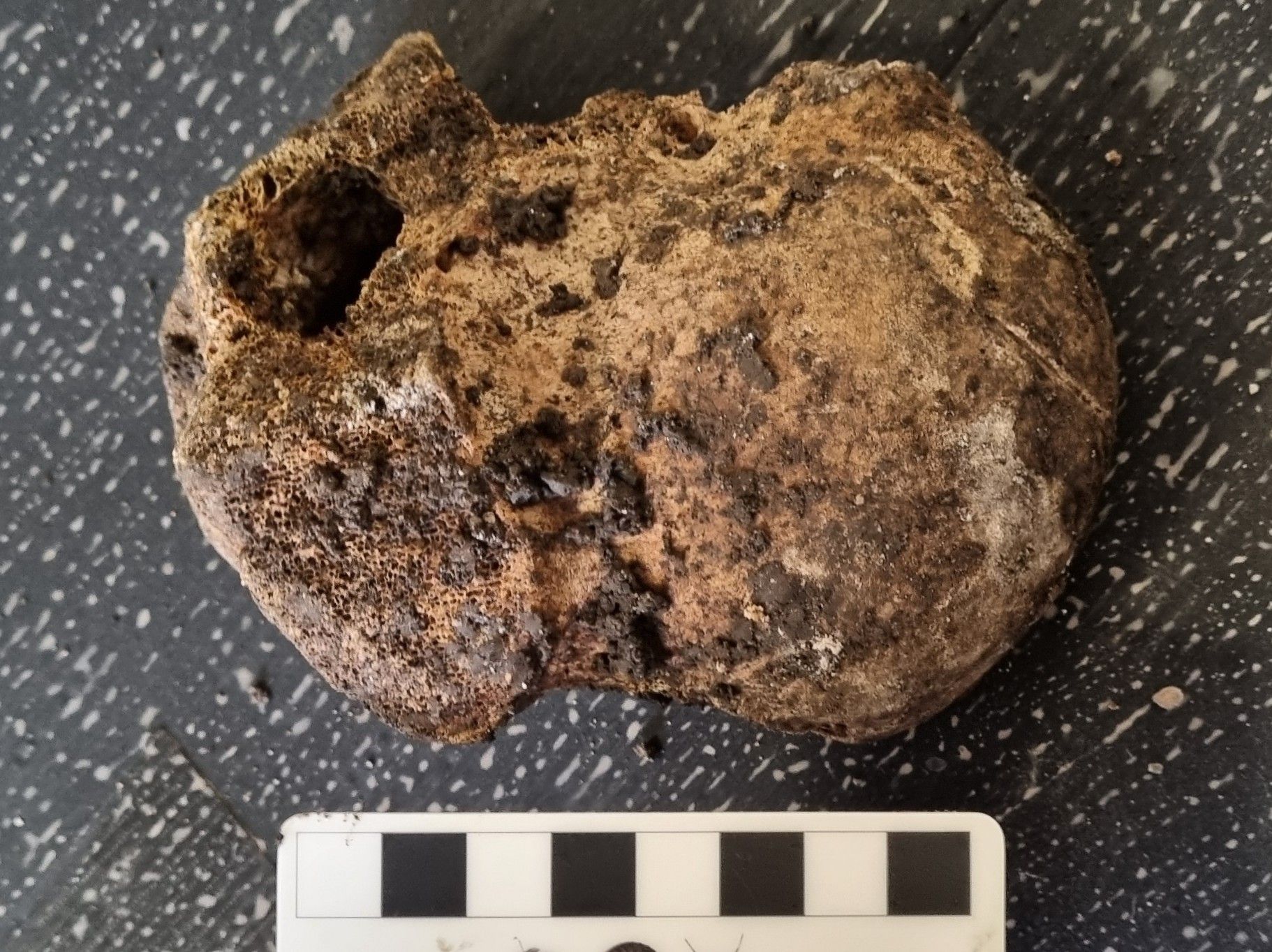
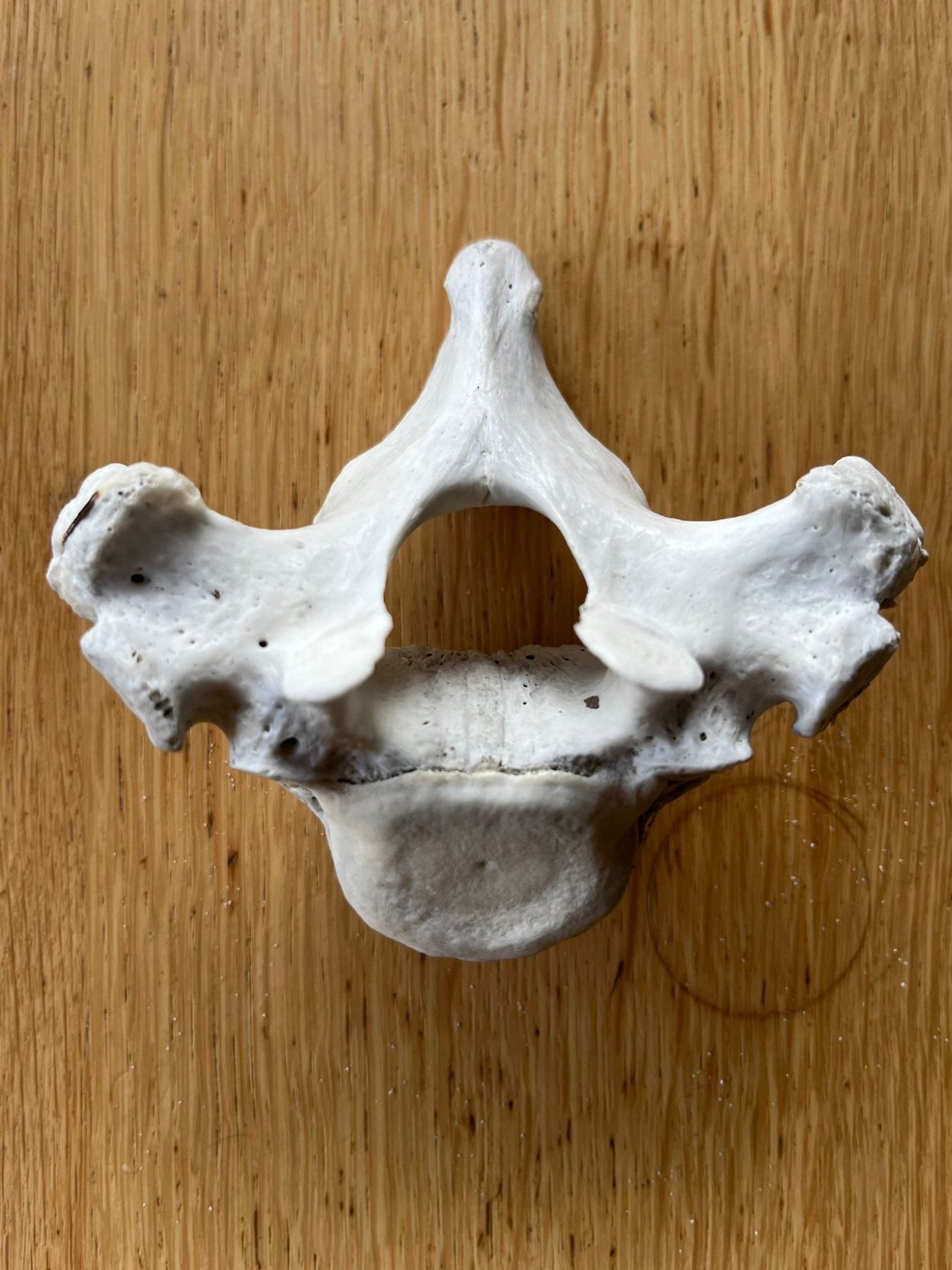
Last week I gave you this genuine mystery object from the forests of Borneo to get your thoughts on:

This was sent to me by Dave Hone (who runs Archosaur Musings), but it was found and photographed by his colleagues Lauryn and Tom of Queen Mary University of London, who are doing research in Borneo.

I had an idea of what it might be, and I’m pleased to say that everyone who responded – both here and on social media – came to the same conclusion as me.

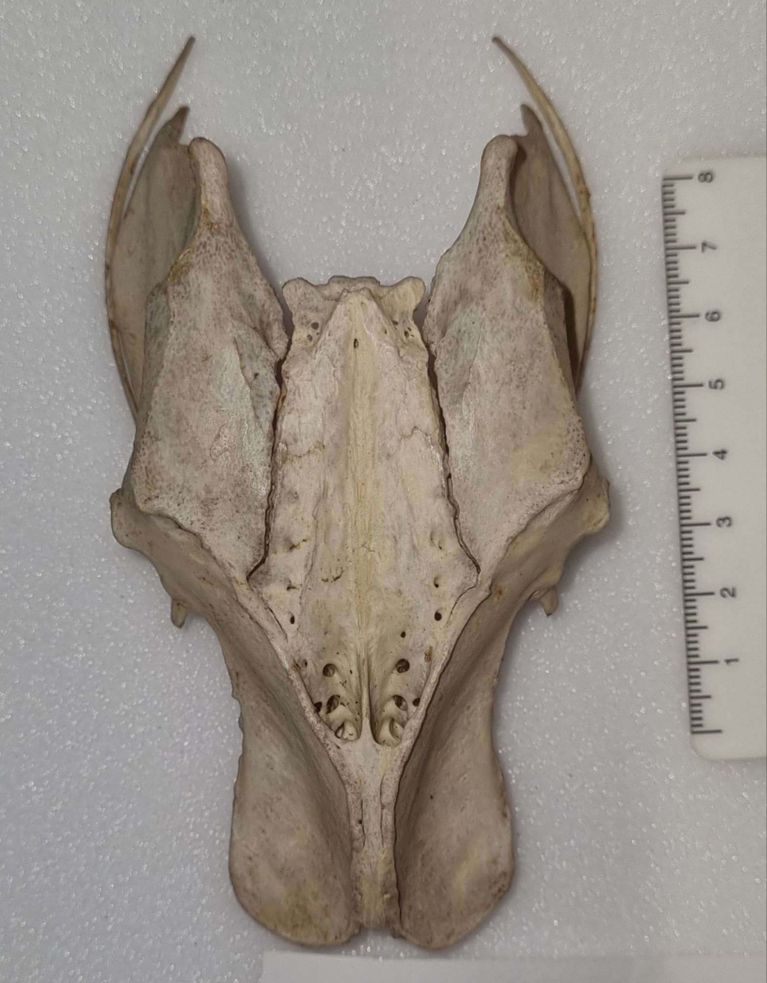
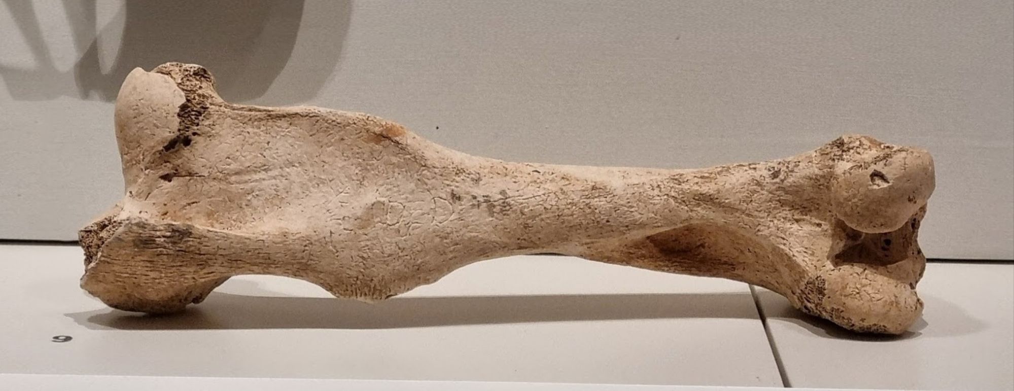
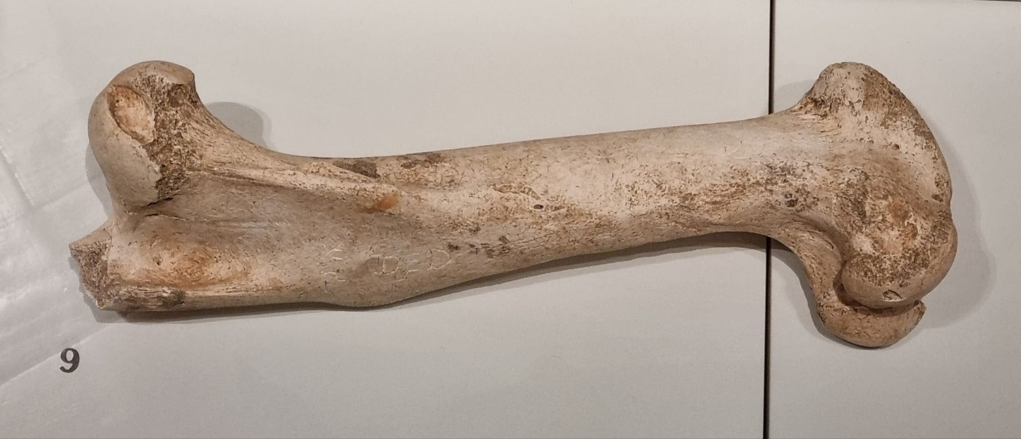
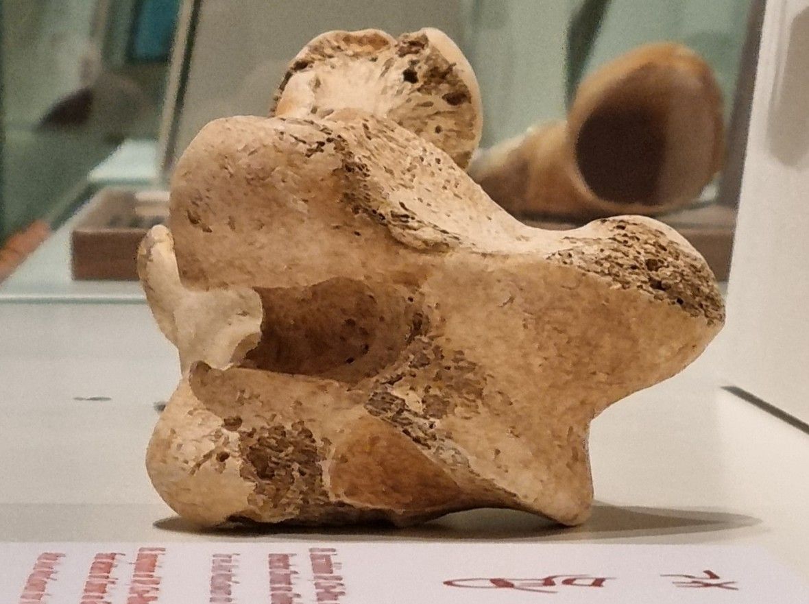
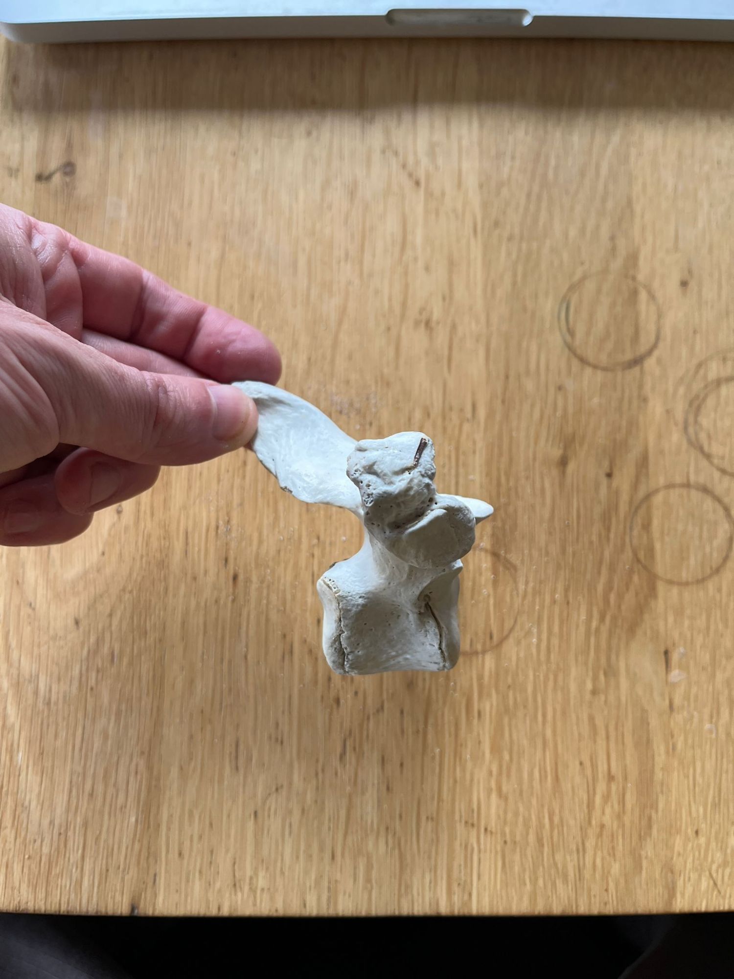
This is the left ilium crest from the pelvis of a juvenile animal. That much can be seen from the unfused sections where this would have connected to the sacral vertebrae and the pubic bone:

Photo of unfused surface of the ilium that would have connected to the pubic bone by Tom Fayle, 2026

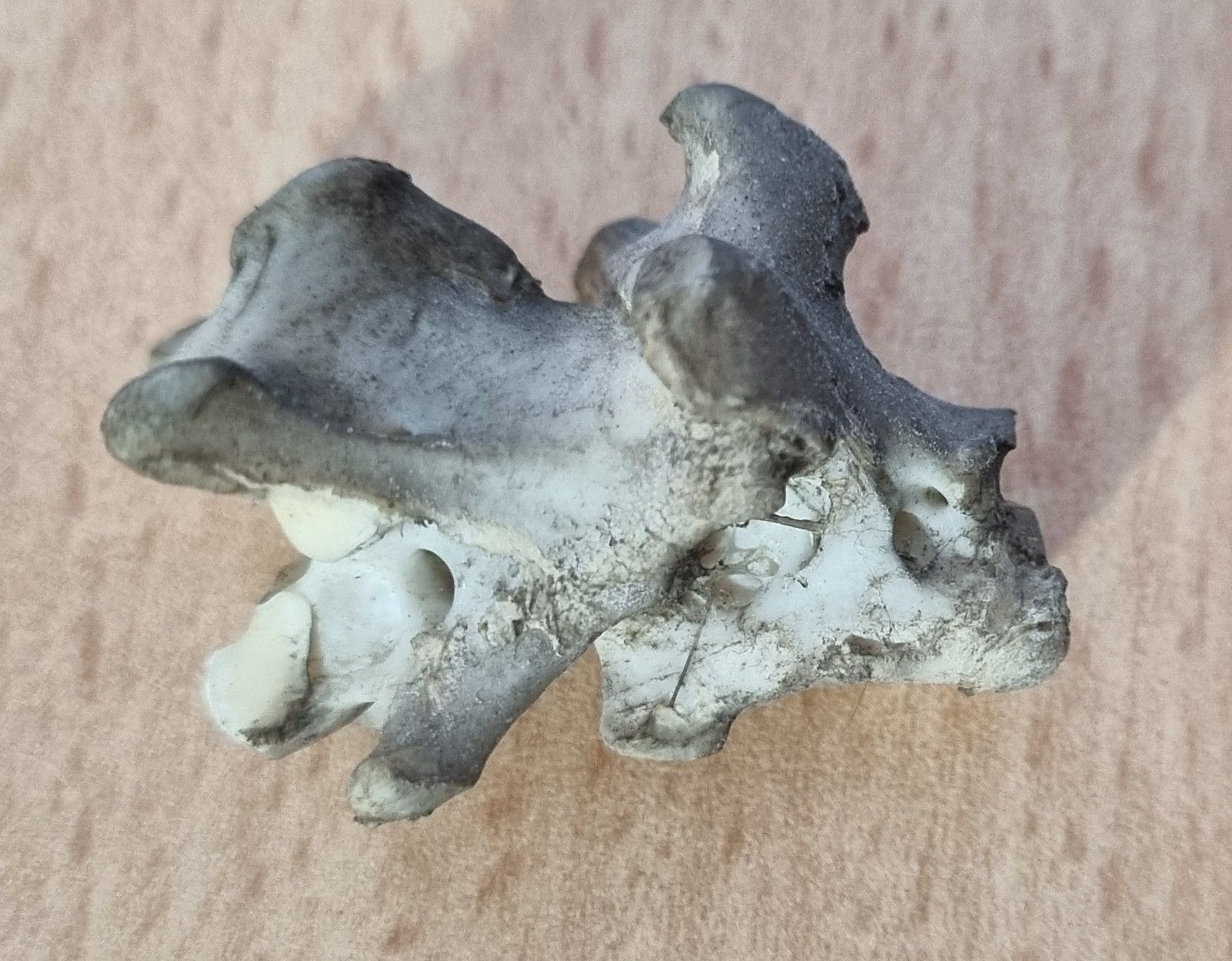
An unusual thing about this ilium is the position of the point of fusion with the sacrum:

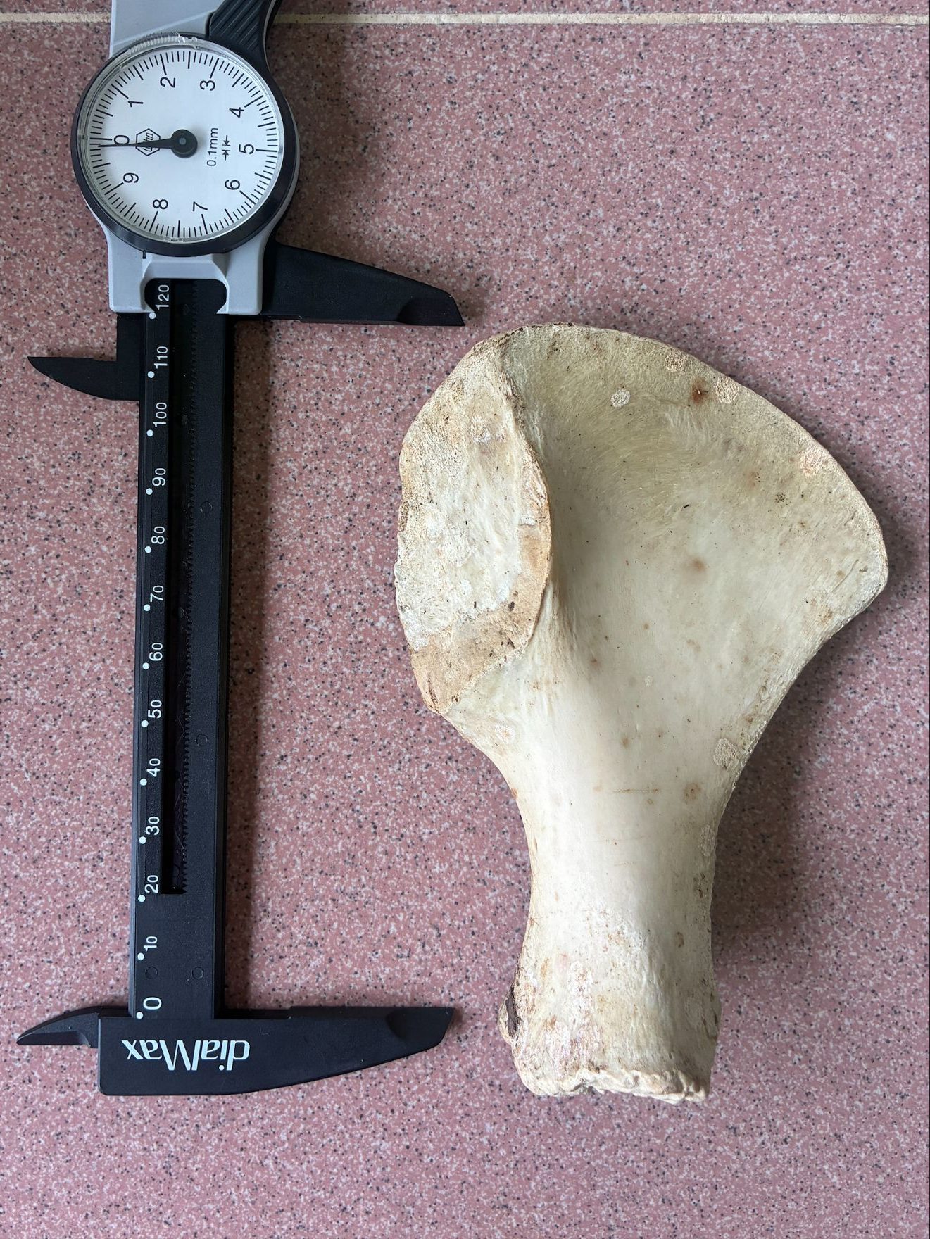
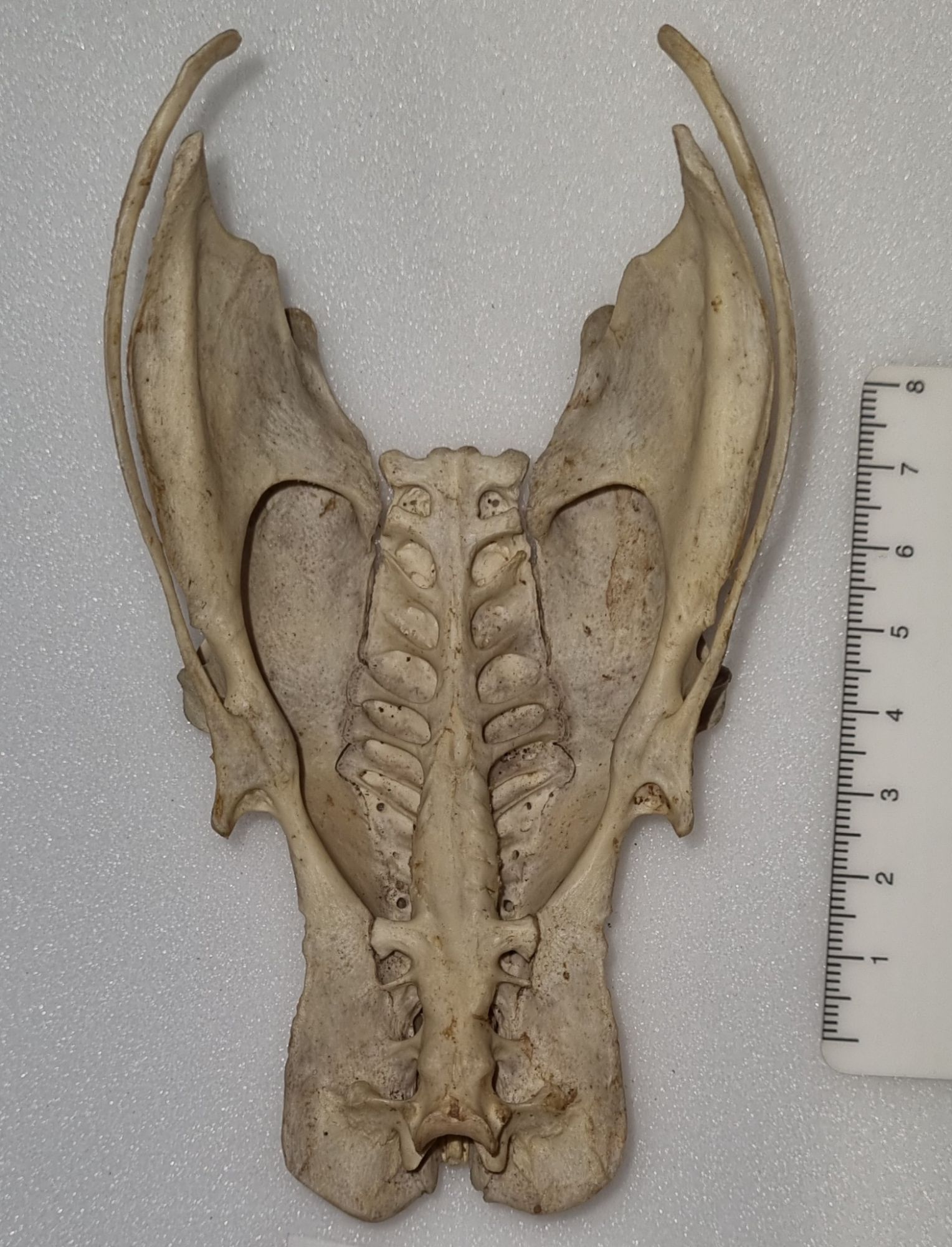
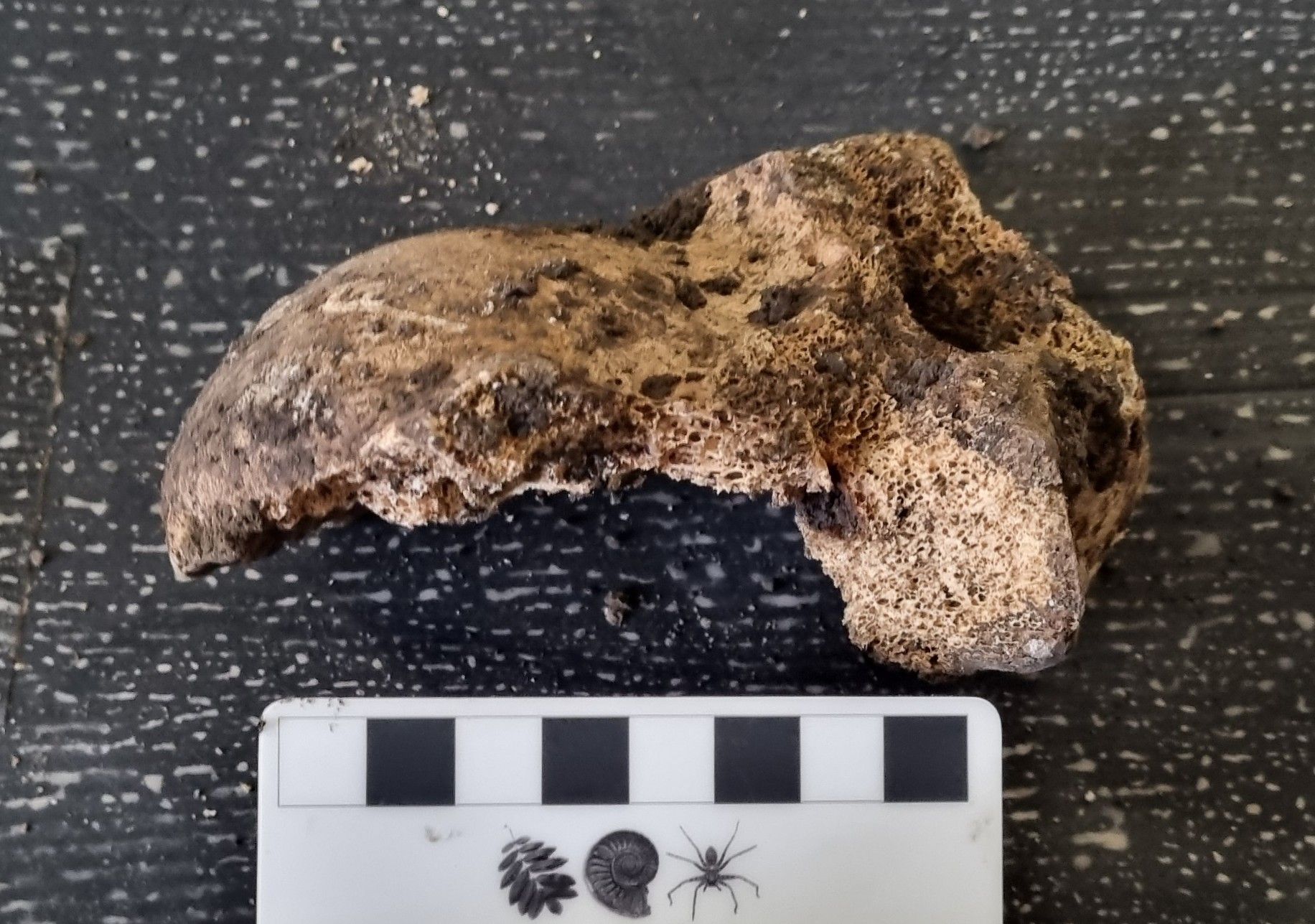
For most species the iliac crest (that’s the curved bit at the top) extends upward quite a bit before flaring out – and that flaring is often quite squared off and tends to be narrow and more blade-like. That configuration is what you would expect to see from most quadrupedal mammals – a notable exception is in the form of the human pelvis, due to our bipedalism.

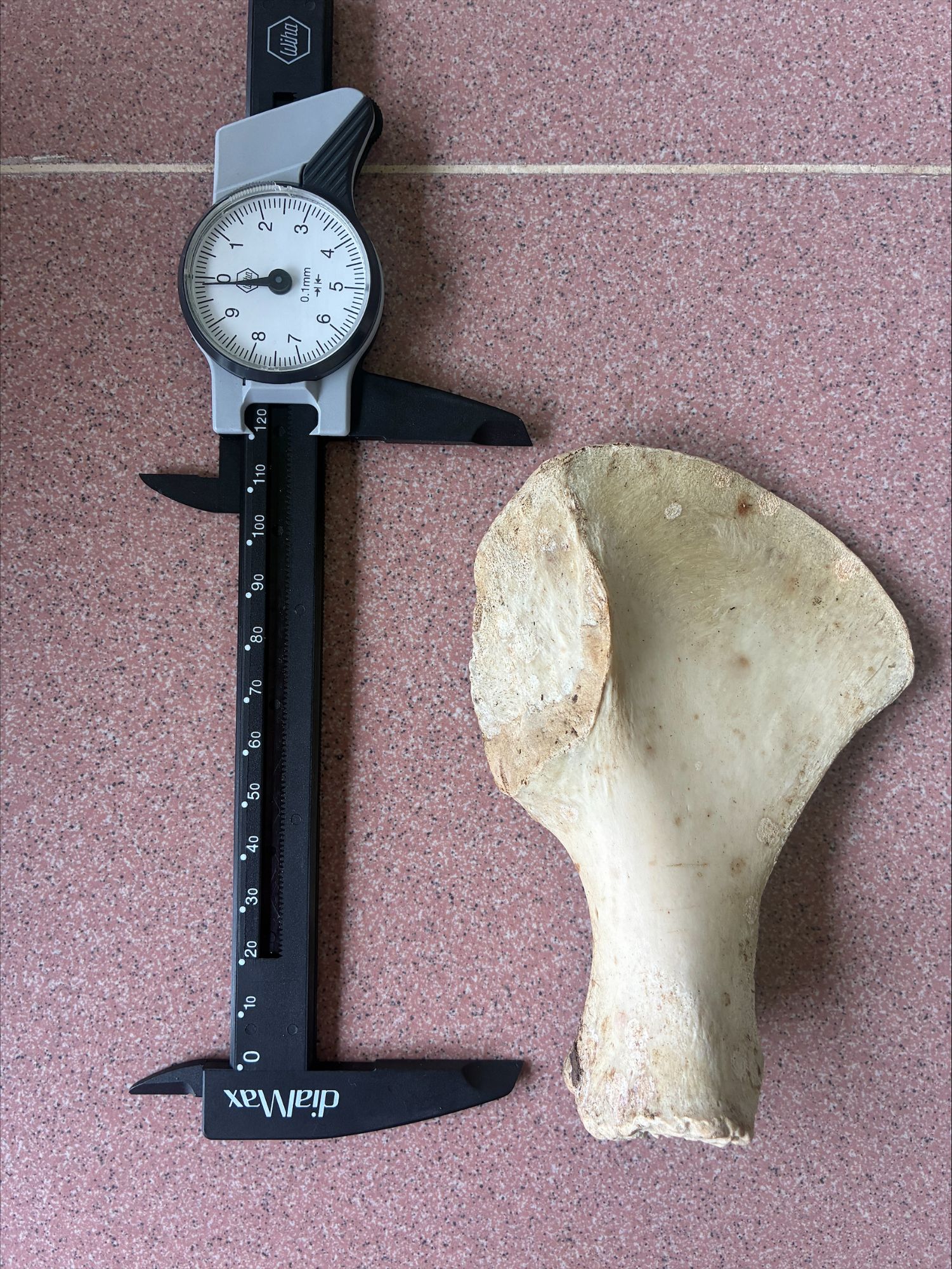
However, this pelvis doesn’t quite conform to the human shape, as it’s a little bit less curved and a little more blade-like. This is consistent with one of our close cousins – there are some useful comparisons of disarticulated primate pelvises on the Bone Clones website and while this is very similar to a female Chimpanzee that’s shown, the location of Borneo suggests a more likely option – a Bornean Orangutan Pongo pygmaeus (Linnaeus, 1760).

On BlueSky, osteoarchaeologist Terry O’Connor spotted this straight away, as did Adam Yates and Chris Jarvis here in the comments. 10 year old Viren, who is a new visitor to Zygoma, was also spot-on. So well done to everyone, and thanks for your thoughts – it’s great to have my conclusion supported!

Friday mystery object #534

This week I have a genuine mystery for you to help solve, courtesy of Lauryn and Tom – researchers working in the forest in Borneo, and passed on by Dave Hone (an old friend and fellow blogger who runs Archosaur Musings):

Photo by Tom Fayle, 2026

I think I know what this is and what it’s from, but I’d love to hear your thoughts on this Bornean bone in the comments below!

Friday mystery object #533 answer

Last week I gave you this bird pelvis to have a go at identifying, as part of a series of posts on that particular feature across birds utilising different environments:

It probably wasn’t the easiest challenge, since this pelvis is damaged. It’s missing part of its dorsal surface and with some small amount of damage to the posterior margin of the right ischium (that’s the broad bit that flares out on the bottom left of the image) and with both of the pubic bones snapped (those are the long skinny bits that stick out to either side).

Still, the overall shape still gives some useful clues. The long, narrow, triangular form offers some ideas about the distribution of mass and use of the legs, to give a sense of possible locomotion habits.

The pelvis is very flat and the points of articulation with the femur are quite far forward:

At least in comparison with something like a Chicken:

This suggests something that has a centre of mass that’s quite far forward compared the foot position. This immediately raises the question of why? The obvious answer for me is that this is a species that doesn’t brisky strut around, relying on more of a low-speed waddle to get around – or perhaps a paddle.

Waterbirds have legs located quite far back in the body to aid propulsion in the water. Perhaps a bit confusingly this requires the articulation of the femur with the pelvis to be pushed further forward due to the more horizontal orientation of the spine to allow the correct orientation of the legs to push water backwards and the body forwards – unlike terresrial species that are more interested in pushing the body upwards against gravity.

There are of course a lot of waterbirds, from the Pelecanimorphae mentioned in the comments by Adam Yates, to the Anseriformes. That’s what we’re looking at here.

There are a lot of ducks and geese, so narrowing it down isn’t easy. The size of this mystery specimen is in the right range for a large duck or small goose, but if you look at the left ischium (the undamaged one) you’ll notice a deep notch – this is something seen in geese.

You may also notice an additional extra notch – which I think offers a great clue to hint at the species. This is the pelvis of a Brent Goose Branta bernicla Linnaeus, 1758.

I’m not sure if the thoughts I’ve laid out here, and over the last couple of mystery objects are helpful for guiding future identifications of bird pelvises, but I hope they may be of use. Let me know your tips on working these things out – more ideas are always welcome!

Friday mystery object #531 answer

Last week I gave you this bony structure to identify:

I didn’t think this one would prove too difficult, since I went with something that most people have probably encountered on their dinner table or thrown in their domestic waste at some point. I wasn’t wrong and Chris Jarvis was first to drop a hint, with reference to Elvis the Pelvis and a famous brand of fried foodstuff from Kentucky.

This is of course the pelvis of a Chicken Gallus gallus domesticus (Linnaeus, 1758).

Bird pelvises are interesting structures, that are a bit more extensive than the usual mammalian equivalent1 due to the extended and fused vertebrae around the sacrum – the sacrum being the area of fused vertebrae where the hip bones attach to the spine. This extended region of fused vertebrae along the midline of the pelvis is referred to as the synsacrum.

In birds the fusion of the pelvis can be very extensive, and provide large areas for muscle attachment. If you look at the bottom of the photo above you can see where there are two scooped-looking sections, and this is where the “oysters” would be found in a roast Chicken. Those “oysters” are more technically referred to as the iliotrochantericus caudalis muscles and they attach to the femur and help stabilise the bird while walking.

The highly sculpted form of a bird’s pelvis creates quite a distinctive locomotor unit that reflects the way in which the bird uses its legs to walk, perch, paddle, swim or whatever else it may get up to. This means that the pelvis of a bird will usually reflect function very well and it will also carry a strong taxonomic signal since birds that are closely related will often share similar locomotion habits, lay similar sized eggs (that have to pass through the pelvis) and so on.

To my mind, the synsacrum provides an evolutionary mechanism to allow effective bipedalism while maintaining a horizontal spine – as opposed to the upright stance used in primates, which seems to come with some issues if my back is anything to go by. My background is in biomechanics and anatomy, so for me this is a topic that I find very interesting. So interesting that I may see if I can find another bird pelvis from a species with different habits to test your skills next week – let me know what do you think of that idea in the comments!

  1. Of course, nature being what it is, there is an exception to that generalisation with the Xenarthra, whose members have a similiarly fused synsacrum. ↩︎

Friday mystery object #527 answer

Last week I gave you this skeleton from the stores of the Dead Zoo to have a go at identifying:

The comments came flooding in, with some slightly off and some very much on target.

The robust skeleton and stocky build of this animal, combined with some interesting bony processes – especially in the pelvic region – offered up some pretty good indicators of the type of critter we’re looking at.

The forward-facing processes of the pelvis were initially mistaken for a baculum, but on a closer look their dual nature becomes more apparent:

These are epipubic bones, which aren’t found in Placental mammals – but this is definitely a mammal – so this is either a Marsupial or Monotreme.

The lack of a skull makes it a little harder to immediately figure out what this might be, but the feet are useful – very useful in fact:

These look like the feet of a digger with those big, robust, triangular claws – but not just a burrowing digger like a Wombat – more like an ant and termite specialist whocan break open their nests. That offers a key clue.

This is the skeleton of a species of spiny anteater – one of the four species of Echidna. The feet actually offer a further diagnostic clue to the species, since most species have five claws, while just one has three – the Three-toed or Western Long-beaked Echidna Zaglossus bruijnii (Peters & Doria, 1876).

This particular specimen was originally on display in the Dead Zoo under its old name Pro-echidna Bruijnii:

Photo of Three-toed Echidna skeleton NMINH:1883.285.1 at the National Museum of Ireland – Natural History taken by Illustratedjc, 2015.

The specimen has been taken off display along with everything else in the building over the last year or so, in preparation for a big refurbishment project.

When it was decanted, along with skull, the fragile right-hand-side rear limb was removed. In the photo above you can see where a claw is detaching – possibly as a result of incorrect foot positioning on the mount (Echidna feet point sideways and backwards, which seems to have confused some mounters). In other places, cotton tape was used to stabilise some of the more wobbly robust elements.

Being able to work through items like this while they’re in storage will be helpful, since it will allow us a chance to remove the worst of the dust from what may have been 140 years of display, and to make some small repairs to things like the detaching claw so it doesn’t get lost. Changing the foot position may be a bigger job, but it’s something to consider.

So while the Dead Zoo may be closed, we’re keeping busy checking the condition of the other 10,000 object we had on display, and working out what we need to do to put them back!

Friday mystery object #523 answer

Last week I gave you this mystery mandible to help me identify:

It was found in a box of mixed shells and bony fragments, with no label, and nothing to offer a clue to its origin. This is very unusual, since these sorts of boxes of miscellany were mainly addressed years ago, but this one was well tucked away and somehow escaped being dealt with.

It’s fairly easy to recognise this as being the right mandible of some kind of cat. The Felidae have a reduced tooth count, with those hyper-carnivorous bladed premolars forming a meat-shearing carnassial row that’s very distinctive. So that’s the easy bit done.

The hard bit comes next, since the cats all share this same dental configuration, making the specific kitty in question a bit harder to narrow down. However, the size is a useful clue. This is much too big to be something like a Domestic Cat, Serval or Ocelot, but it’s also too small to be one of the big Big Cats – like the Lion or Tiger:

Tiger skulls facing off – the scale of these skulls is much greater than the mystery mandible

This leaves some of the medium-large Big Cats and perhaps a very large Small Cat (i.e. the Cheetah). Let’s start by ruling out that last one – the size is still a bit on the big side, but also the Cheetah has a more gracile coronoid process that curves, and a relatively shorter toothrow. Pumas also have a more rounded coronoid process.

The Jaguar also has a more rounded coronoid process, and has a more robust mandible, that’s just built thicker to deal with the high bite forces these cats generate:

That leaves us with the Snow Leopard and the not-snow-Leopard, both of which have the more pointed coroid process that we see in the mystery object. However, the Snow Leopard has downwards inflected angular process, that’sI suspect may relate to the increased gape needed to use the excessively long canine teeth in this species:

The answer to last week's mystery mandible
Snow Leopard skull, showing the hypertrophied canines normal in this species.

So, by a process of elimination, we’re left with Leopard Pathera pardus (Linneaus, 1758). This species varies significantly in size and the morphology can be quite variable, since the species has a wide distribution, from Eastern Russia to as far West as Senegal.

This actually reminds me of a very similar mystery object from the Grant Museum of Zoology that I shared 10 years ago, so I’ hope that provided something useful for reference so I’m glad I trawled through some of my past posts to help solve this one. My thanks to everyone who offered their thoughts on this one – it’s great to see so many of you come to the same conclusion!

Friday mystery object #522 answer

Last week I presented this mystery bone from the Words on the Wave exhibition at the National Museum of Ireland on Kildare Street:

The bone is over a thousand years old, so it’s unsurprising that a few of the processes that could be useful for identification have been worn down a little:

That said, there was plenty of information remaining to allow several of you to identify the animal, bone type and side of the animal this is from. Adam Yates was first to the comments and he also left a very helpful Toot on Mastodon, detailing the diagnostic features:

@mike @PaoloViscardi 
The prominent third trochanter and really well-developed supracondylar fossa on the lateral caudal surface at the distal end of the shaft should get you there. You won't have to look beyond  familiar farmyard beasts to find it.

For me, the supracondylar fossa on the lateral caudal surface is the instant give-away. For those who don’t speak anatomese, that’s the bit that flares out from the side of the shaft of the bone (which is actually missing a bit in this example).

This is a feature I always associate with the Perissodactyla (the tapirs, rhinos and horses), and this one is nowhere near robust enough for anything other than one of the horses. It’s also fairly small (although the lack of a scale bar doesn’t make this obvious).

This a left femur that I suspect is from a Horse Equus ferus caballus Linnaeus, 1758, but it could also be from a Donkey Equus africanus asinus Linnaeus, 1758. It can be tricky to tell these species apart, especially when the bone is a little worn down and you only have a few photos to work from – so best to err on the side of caution and leave it at an identification of Equus sp.

If you’re in Ireland and wanted to take a look at this object – and a selection of remarkable mediaeval manuscripts from the Abbey of St Gall, Switzerland, some of which are returning to Ireland for the first time in 1000 years – you have until the 24th of this month, I’d definitely recommend it!

Friday mystery object #522

This week I have another bony mystery object for you:

This one is currently on display in the beautiful Words on the Wave exhibition at the National Museum of Ireland on Kildare Street, but the focus is on the inscription on the bone, rather than the species the bone came from:

So, I’d love to hear your thoughts on what this is? Have fun with it!

Friday mystery object #521 answer

Last week I had a genuine mystery object for you to identify that was freshly dug out of the ground:

While there were a few suggestions of baked goods on social media, plenty of you spotted what it actually is – with a nod to Adam Yates who was first with the correct identification.

The side view is probably the most useful for setting context, as it shows the smooth curve of the head of an articular surface, with an adjacent tuberosity (or sticky-up-bit, if you prefer):

This is what you expect to see at the proximal end of a humerus (that’s bit that works with the shoulder). It’s pretty big – the scale bar is 8cm and the head of this humerus is a good bit wider across, so that also tells us something.

Another key observation comes from the underside of the humerus head:

You can just make out a rugose pattern on the underside of this piece of bone, which tells us that this is an unfused epiphysis that has become detached from the rest of the humerus. That means the animal that this piece of bone came from was young enough to still be growing, as the bones hadn’t fused.

All of that information leads us to the conclusion that the animal this piece of humerus came from was large, but still young. For me, that says that this humerus is from a Cow Bos taurus Linnaeus, 1758.

I suspect this mystery object represents the remains of a Victorian meal, based on where it was found – just under the floor of the Dead Zoo. It came to light during the investigation works on the building, which are currently underway. The building work started in 1856, so that gives us a good idea of when this beef shoulder was probably eaten.

Friday mystery object #514 answer

Last week I gave you another guest mystery object from regular contributor Andy Taylor FLS FBNA:

Image by Andy Taylor FLS FBNA, 2025

This proved to be a tricky challenge. The large size and the lack of a halux (the rear-facing small toe found in many birds) makes this seem like the feet of some sort of Ratite, which is the group of birds that includes a variety of flightless examples, including Ostriches, Emus, Cassowarys, Rheas, and Kiwis – many of which lack the hallux.

Image by Andy Taylor FLS FBNA, 2025

However, these feet do not belong to a Ratite – and in the comments, many of you seemed to notice that there was something funky with the arrangement of the toes and bones that may have offered a clue to indicate that fact. Even though they’re not from a Ratite, there are some useful pointers that can be taken from the similarities, since form follows function and if we know Ratite feet can look similar to these, then perhaps it’s worth asking “why is that?”

Many (although not all) Ratites have three forward-facing robust toes, although the Ostriches are a bit of an exception, as they only have two toes per foot. In the evolutionary push to be fast on your feet, too many toes can make you slow – just look at how horses have lost all but one of theirs.

Ratites also lack a hallux, and their claws don’t curve. These features all reflect the fact that Ratites don’t perch on branches, they’re heavy, and they spend most, if not all, of their time walking around on dry grasslands (actually, this isn’t true for Kiwis or the extint Moas that have more scrubby or habitats, but these Ratites do have a hallux). These are all useful things to consider when thinking about potential species that these feet could be from.

If you think of large and heavy birds that don’t perch and have a grassland habitat the list of possible options gets narrowed down quite quickly. Sereimas and Secretary Birds are out, as they have a hallux and more curved claws. Storks, Megapodes and Galliformes all also have a hallux. But one type of bird matches the description very well – the Bustards (or Otididae if you prefer).

The size means it must be one of the larger species of Bustard, but I suspect that it would be difficult to confirm the identification to species level without good quality reference material, since there are quite a few species. But in this case we’re luck to know the species, since Andy has the information – these feet belonged to a Great Bustard Otis tarda Linneaus, 1758.

It’s a species I’ve talked about before in a previous post and I really appreciate Andy sharing the images and information from his collection, and all the thoughts and comments that came in about the mystery object from the community here on Zygoma. Thanks for taking part!

Friday mystery object #514

This week I have another guest mystery object for you from regular contributor Andy Taylor FLS FBNA:

Image by Andy Taylor FLS FBNA, 2025

I’m keen to hear your thoughts on what the owner of this magnificent pair of feet might have been in the comments below. If it proves very difficult then myself of Andy might be tempted to offer up some clues, but I have a suspicion that some of you might work it out yourselves. Either way, I hope you enjoy the challenge!

Friday mystery object #513 answer

Last week I gave you this mystery object found by the family of one of my colleagues to have a go at identifying:

Image by Andrew Brady, May 2025

Adam Yates got the ball rolling with some key observations about this skeletal element that provides a handy set of clues to look for when assessing vertebra:

The element is easier than than the taxon. It is a thoracic vertebra from a mammal because it has zygapophyses (so therefore not a fish), flat centrum faces (so therefore not bird or reptile), and rib articulation facets (so therefore not from the neck, lumbar or tail regions). Which mammal is less easy, I’m pretty sure it is isn’t a relative of flipper, but that’s all I’ve got now.

Adam was also right in flagging that it’s not from a cetacean – their transverse processes are longer and less robust than we see in this mystery specimen. However, this bone was found on the beach on Achill Island, so there’s a good chance it’s from a species that’s frequently in the water.

Then Joe Vans, Kat Edmonson and Adam got stuck in with consideration of the possible species and specific thoracic vertebra, and as part of that there was reference to images from John Rochester’s Flickr pages. As far as I’m concerned, John’s pages are some of the best resources available for osteological comparison and I use them regularly.

The discussion ended up converging on the 5th thoracic vertebra (or T5 as it would be commonly referred to) of a Harbour Seal, based on specimens imaged by John here: https://www.flickr.com/photos/jrochester/14181517647 – best compared to this image of the mystery specimen:

Image by Andrew Brady, May 2025

This could well be correct, but I personally think it may be from a different species of seal. The size is fairly large (based on the hand that provides some scale), yet the epiphyses of the articular surfaces are not fully fused, suggesting that this is from a subadult animal:

This suggests to me that the animal had some more growing to do before reaching full size, which may impact on some of the features, but it also makes me think that the vertebra is probably on the smaller end of the size range for the species.

For me that would suggest a Grey Seal Halichoerus grypus (O. Fabricius, 1791) and looking at another of John’s pages: https://www.flickr.com/photos/jrochester/24441096790/in/photostream/ I would expect it to be in the T6 or T7. This also tracks for me in terms of the shape of the neural canal, which seems narrower and taller (generally more square) in the Grey Seal compared to a broader and perhaps more triangular neural canal in the Harbour Seal.

I suspect a measurement of the size would help confirm one way of the other, so I’ll see if I can get that to be sure. I hope you enjoyed the challenge of working this out, and I’d love to hear your thoughts if you think it could be something else!

Friday mystery object #513

This week I have another guest mystery object for you, found on a beach by the family of one of my museum colleagues:

Image by Andrew Brady, May 2025

Let me know your thoughts about the species, where in the body this particular bone came from, and any other deductions you might have about the animal? I look forward to seeing if you agree with my thoughts!