
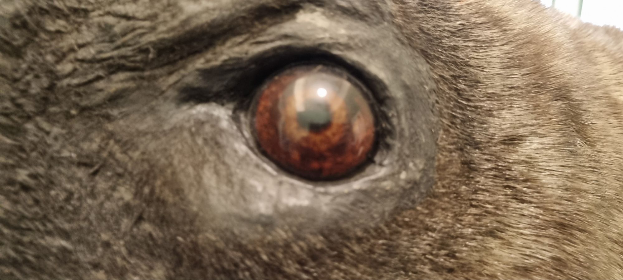
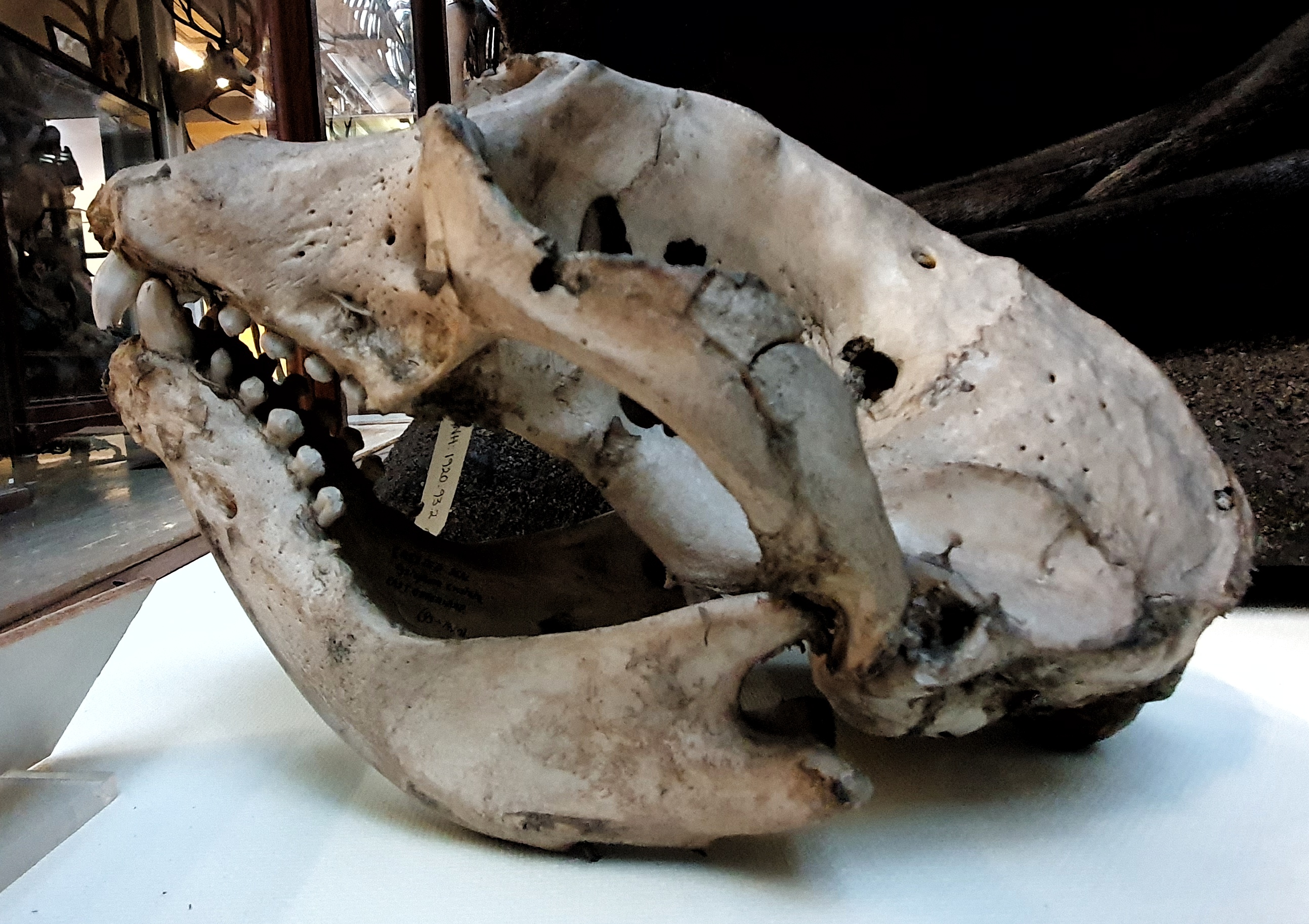
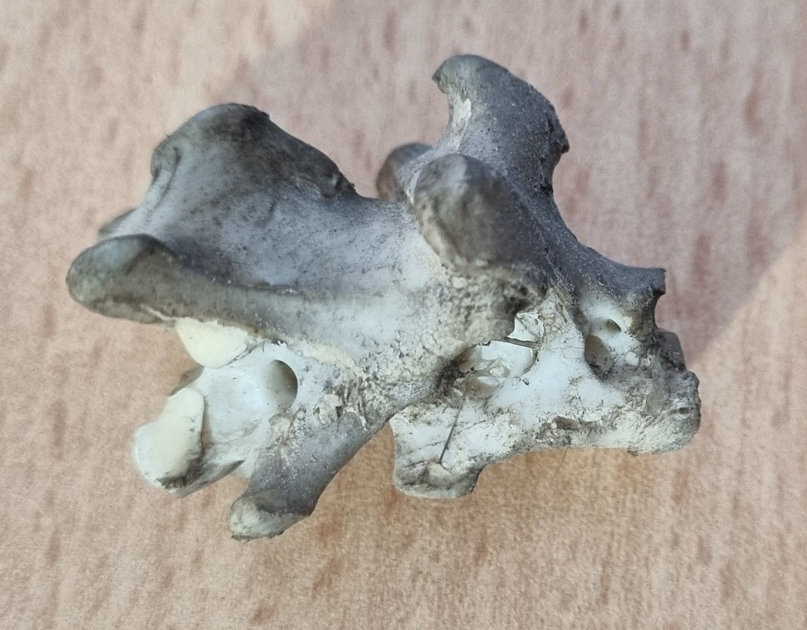
Last week I gave you this guest mystery object to try identifying, from the collection of Andy Taylor FLS FBNA:

Andy had brought this particular specimen along to the NatSCA Conference, which was held in the Manchester Museum this year. It’s not the only one he brought – there were several fantastic specimens from his incredible collection, my favourite being the Bathynomus Giant Marine Isopod:

It was great to see these specimens, and it’s surprisingly rare to be able to see specimens like this at a conference these days, since UK museums have very strict controls on how accessioned collections are transported and used, which can make it hard to support use in outreach. While this is good for the care of the collections, it does reduce their ability to be used to educate and inspire.
Andy doesn’t have this limitation, since the specimens are from his own collection, which has been built up over 40 years of interest in natural history and through a network of contacts researching life in the deep seas and a variety of other environments. Andy does this because he’s passionate about nature, and his collection is available for use for research, loans to support exhibitions, and use in training and outreach activities.
Many natural history collections in museums came from the activities of passionate naturalists like Andy – in fact quite a lot of local and regional museums in the UK were originally founded to house the collections of local naturalist organisations, that sprung up to support the learning of working people who didn’t have the opportunity to follow an academic route into the natural sciences, but who made huge contributions to our understanding of local biodiversity in the areas in which they were active. These people were true amateurs – doing this work for the love of it, rather than for profit.
Various organisations still support the activities of passionate naturalists (both amateur and professional), such as the British Naturalists’ Association and the Linnean Society of London (of which I am a Fellow) and I think they have huge value for furthering our understanding of natural science and supporting the activities of taxonomists and conservationists who do the vital work needed to protect our threatened natural world.
However, none of this offers an answer to last week’s mystery object, so let’s get to that.
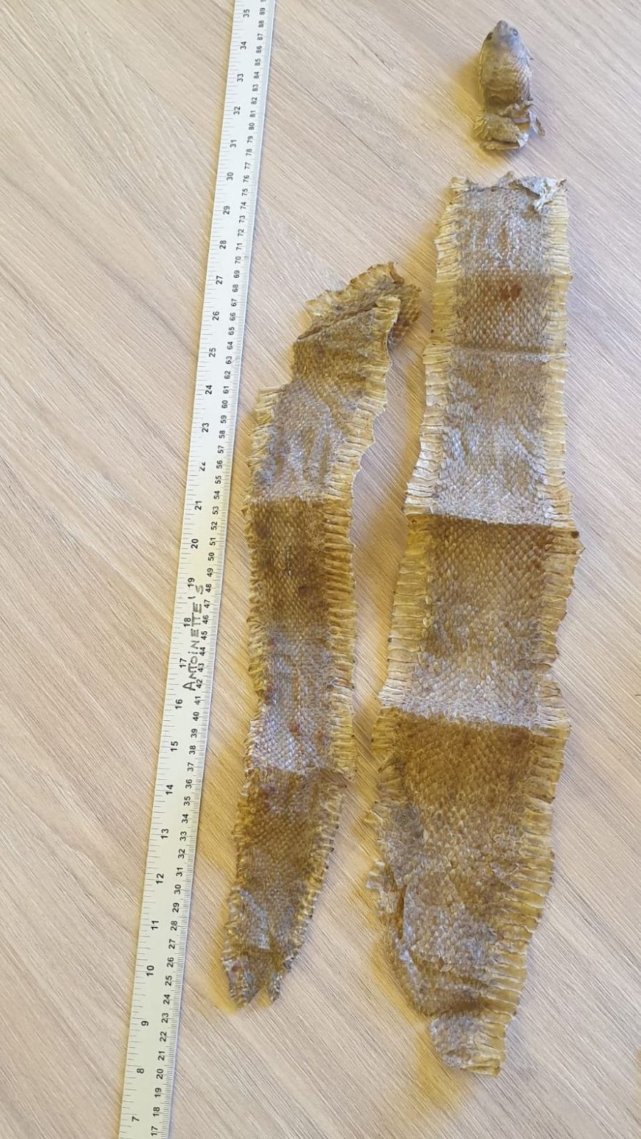
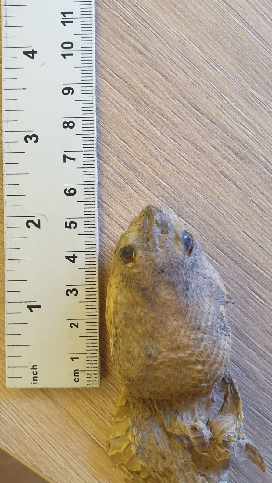
The photos I provided weren’t great, but that didn’t stop the Zygoma regulars from figuring it out. Chris Jarvis indicated that it was a type of mussel, Sallie Reynolds got the clue I offered in last week’s post (“you may need to dive deep to work it out”) and figured it was from very deep in the ocean, and Adam Yates suggested the wrong species in the right genus, and then dialled in on the correct species after a helpful comment from Andy.
This is a specimen of Bathymodiolus thermophilus Kenk & Wilson, 1985 a type of mussel that lives around hydrothermal vents. This particular specimen was collected at a depth of 2.2km from a hydrothermal vent on the East Pacific Rise by Alvin Deep-Submergence Vehicle (DSV) as part of research by the Woods Hole Oceanographic Institution.

The distribution of specimens like this to a variety of people and organisations is an important activity, since it helps ensure examples of species can be made more available for interested parties around the globe – decentralising collections so local events (from earthquakes to funding cuts by anti-science governments) stand less chance of taking specimens out of circulation for research into our global biodiversity.
I would like to offer up my thanks to Andy – his support for this blog has always been enthusiastic and I appreciate it greatly!


