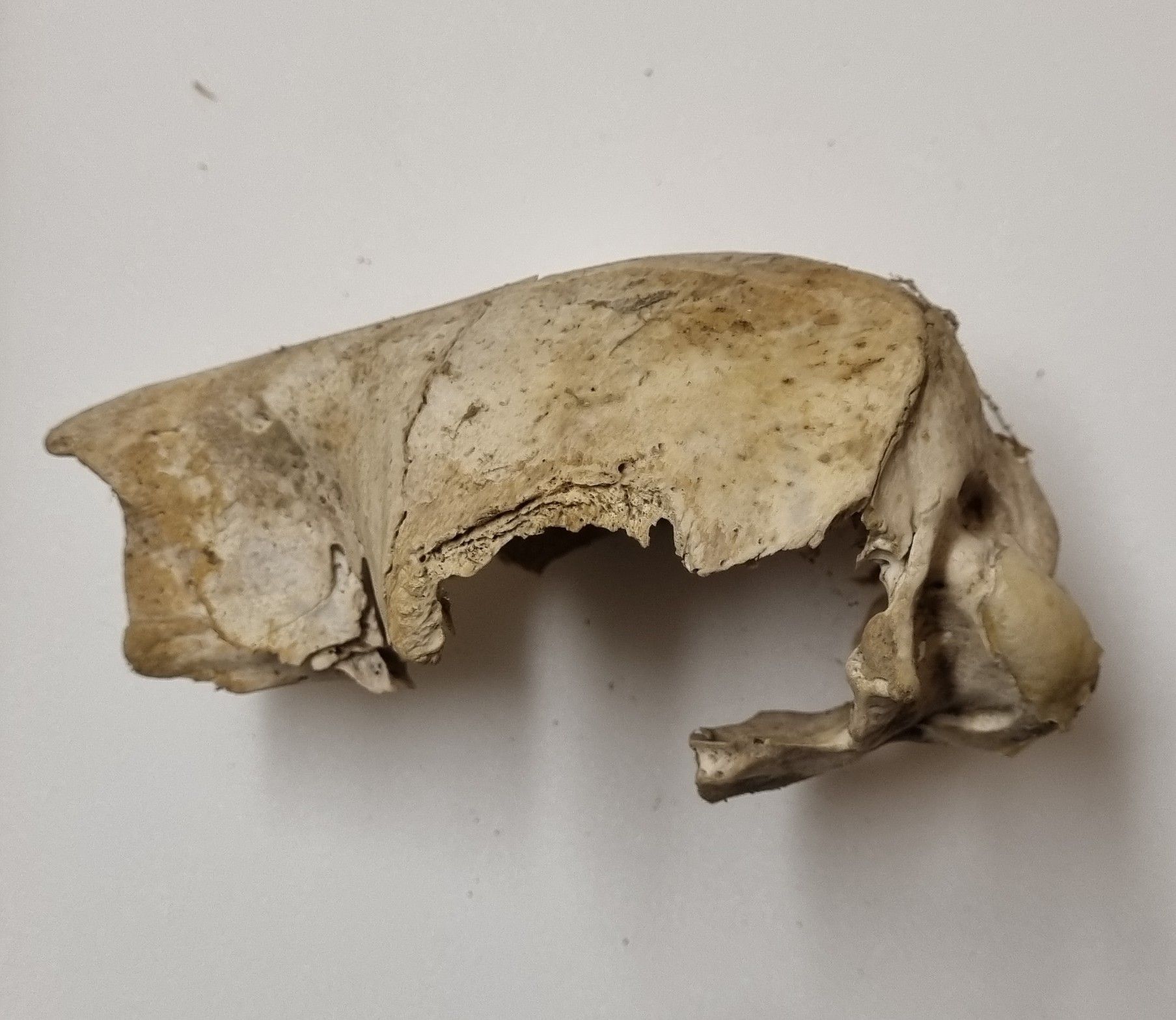
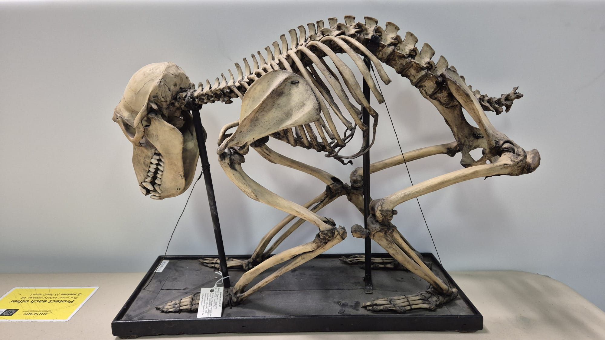
Last week I gave you this mystery object from the Dead Zoo to try your hand at identifying:
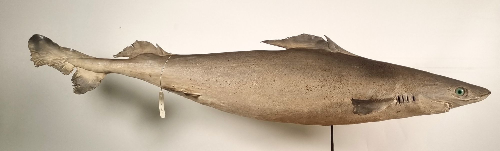
There were some muttering about this being a squalid in the comments and on social media (I left Twitter some time ago, but I’m on Bluesky, Mastodon, and LinkedIn), and those mutterings were of course correct, since this is a shark in the Order Squaliformes. A useful point for identification of this Genus is the lack of an anal fin.
There are a lot of Squaliformes, but this one has a profile that best fits the form of either the Squalidae (the Dogfish) or the Centrophoridae (the Gulper sharks). The tail offers a clue to distinguish though, as it has a full tail notch and a secondary partial notch that gives a squared off section of the tail tip that isn’t seen in the Squalidae (although some do have a squared off tail tip, but with just a single partial notch).
Within the family Centrophoridae you could spend a bit of time going through about 20 species to rule some out, or you can take a shortcut and guess that this specimen is from the waters around Ireland and start searching for likely candidates there. This approach helped Adam Yates get the identification.
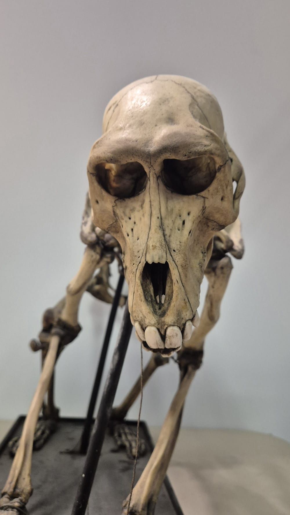
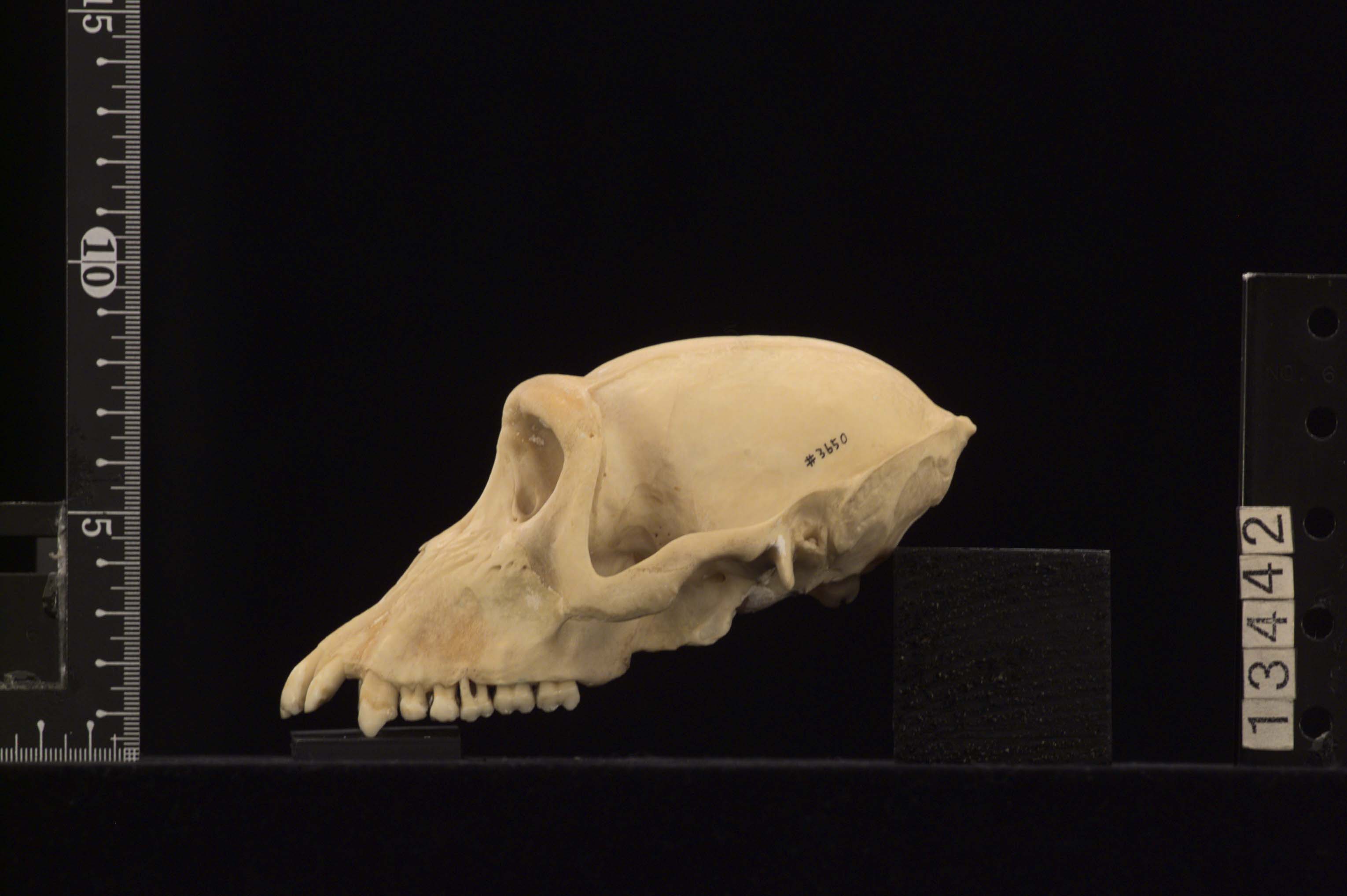
I think the taxidermy of our specimen may have made this harder, since I don’t think the taxidermist had a big enough eye for the species (if you look at the specimen’s head you can see how hollow the orbit appears around the eye):
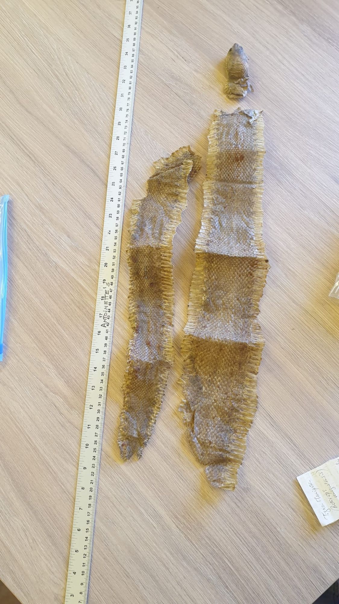
The large eyes are one of the features of this deep sea species that does indeed occur in Irish waters, but the scales are probably the most useful feature, although unfortunately they aren’t easy to see clearly on the specimen (although if you click the photo you do get a bigger version).
As Adam recognised, this is a Leafscale Gulper Centrophorus squamosus Bonnaterre, 1788.
This particular specimen was collected in May 1906, 70 miles off Bull Rock in County Kerry, in a trawl at 110 fathoms. It came to the Dead Zoo via Irish Fisheries, along with many other specimens that have helped lay the foundations of our understanding of the marine life found in Irish waters.
We still receive new specimens from trawlers around the island, with new records of species and interesting variations within species being donated quite frequently. With the dizzying rate of technological developments, it’s easy to assume that we already know everything about the life on our planet, but in reality we’re still discovering new things all the time, and our knowledge is very incomplete.
So thanks to everyone who joins in the mystery object – it gives me a chance to learn new things from you, and I hope it helps you to test your skills, and maybe learn some new things about the animals we share the planet with!


