This week I have an invertebrate for you to have a go at identifying for a change:
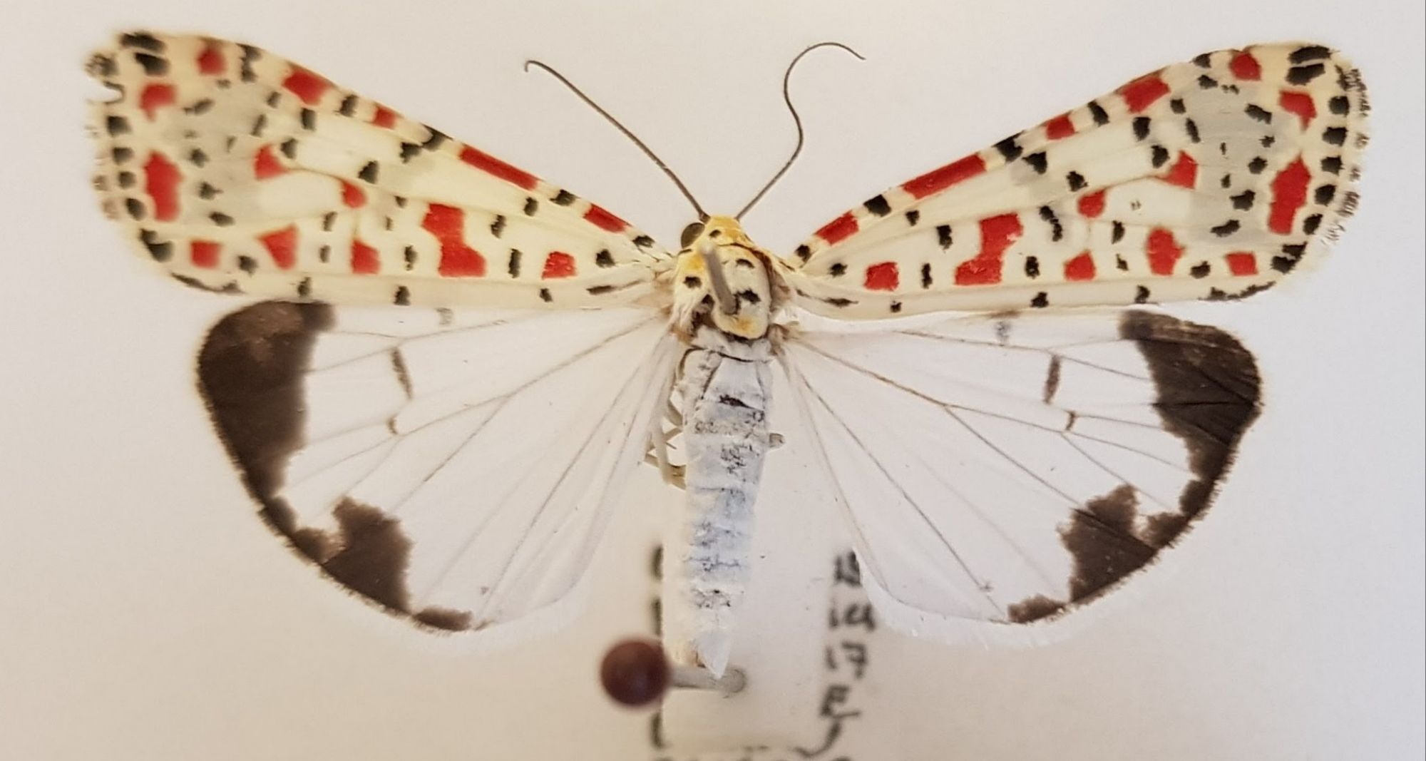
Any idea what this rather beautiful beastie might be?
Let me know what you think it might be in the comments below. I hope you have fun working it out!

This week I have an invertebrate for you to have a go at identifying for a change:
Any idea what this rather beautiful beastie might be?
Let me know what you think it might be in the comments below. I hope you have fun working it out!
Last week I gave you this genuine mystery object from the forests of Borneo to get your thoughts on:

This was sent to me by Dave Hone (who runs Archosaur Musings), but it was found and photographed by his colleagues Lauryn and Tom of Queen Mary University of London, who are doing research in Borneo.
I had an idea of what it might be, and I’m pleased to say that everyone who responded – both here and on social media – came to the same conclusion as me.
This is the left ilium crest from the pelvis of a juvenile animal. That much can be seen from the unfused sections where this would have connected to the sacral vertebrae and the pubic bone:
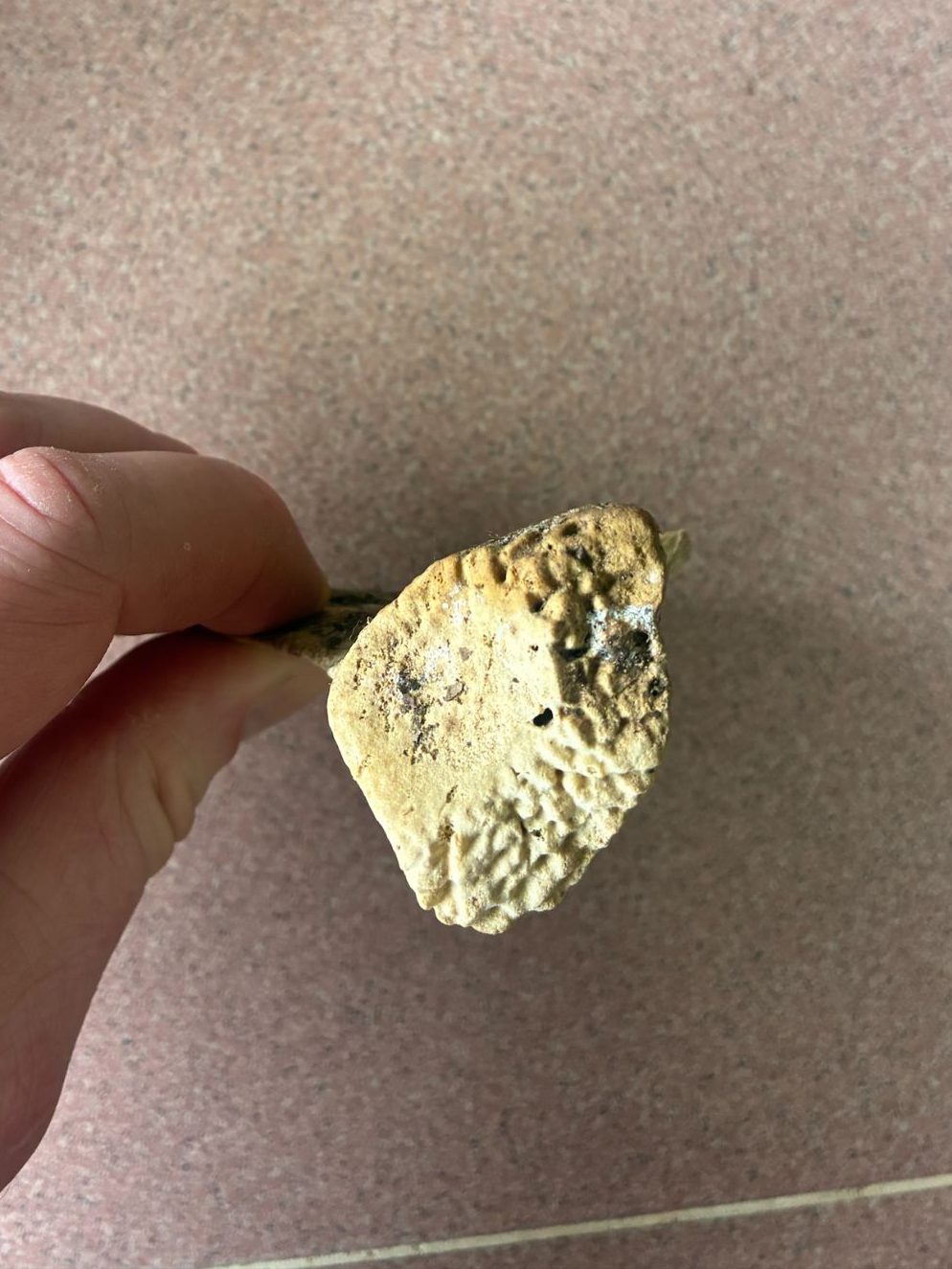
An unusual thing about this ilium is the position of the point of fusion with the sacrum:
For most species the iliac crest (that’s the curved bit at the top) extends upward quite a bit before flaring out – and that flaring is often quite squared off and tends to be narrow and more blade-like. That configuration is what you would expect to see from most quadrupedal mammals – a notable exception is in the form of the human pelvis, due to our bipedalism.
However, this pelvis doesn’t quite conform to the human shape, as it’s a little bit less curved and a little more blade-like. This is consistent with one of our close cousins – there are some useful comparisons of disarticulated primate pelvises on the Bone Clones website and while this is very similar to a female Chimpanzee that’s shown, the location of Borneo suggests a more likely option – a Bornean Orangutan Pongo pygmaeus (Linnaeus, 1760).
On BlueSky, osteoarchaeologist Terry O’Connor spotted this straight away, as did Adam Yates and Chris Jarvis here in the comments. 10 year old Viren, who is a new visitor to Zygoma, was also spot-on. So well done to everyone, and thanks for your thoughts – it’s great to have my conclusion supported!
This week I have a genuine mystery for you to help solve, courtesy of Lauryn and Tom – researchers working in the forest in Borneo, and passed on by Dave Hone (an old friend and fellow blogger who runs Archosaur Musings):
I think I know what this is and what it’s from, but I’d love to hear your thoughts on this Bornean bone in the comments below!
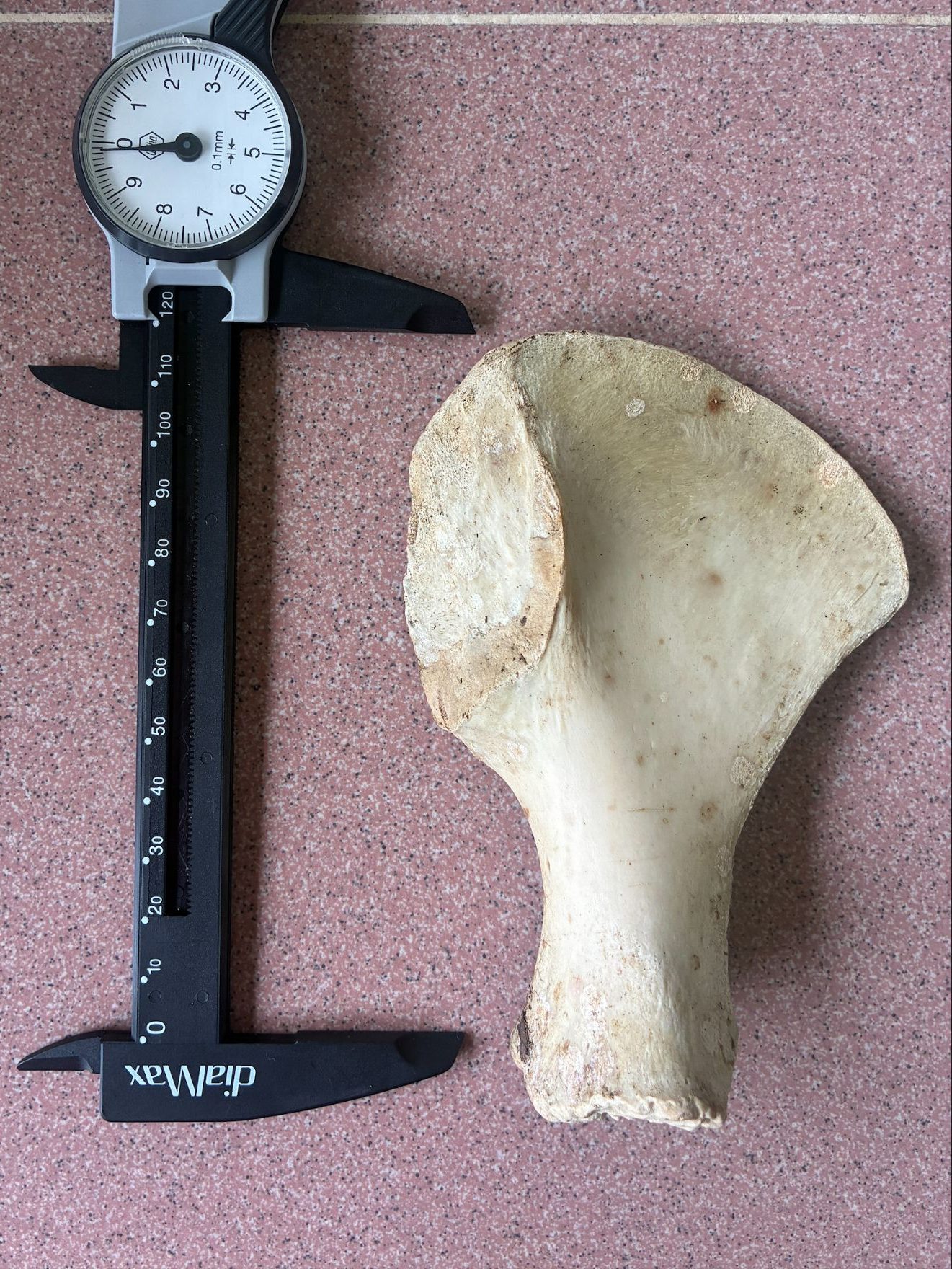
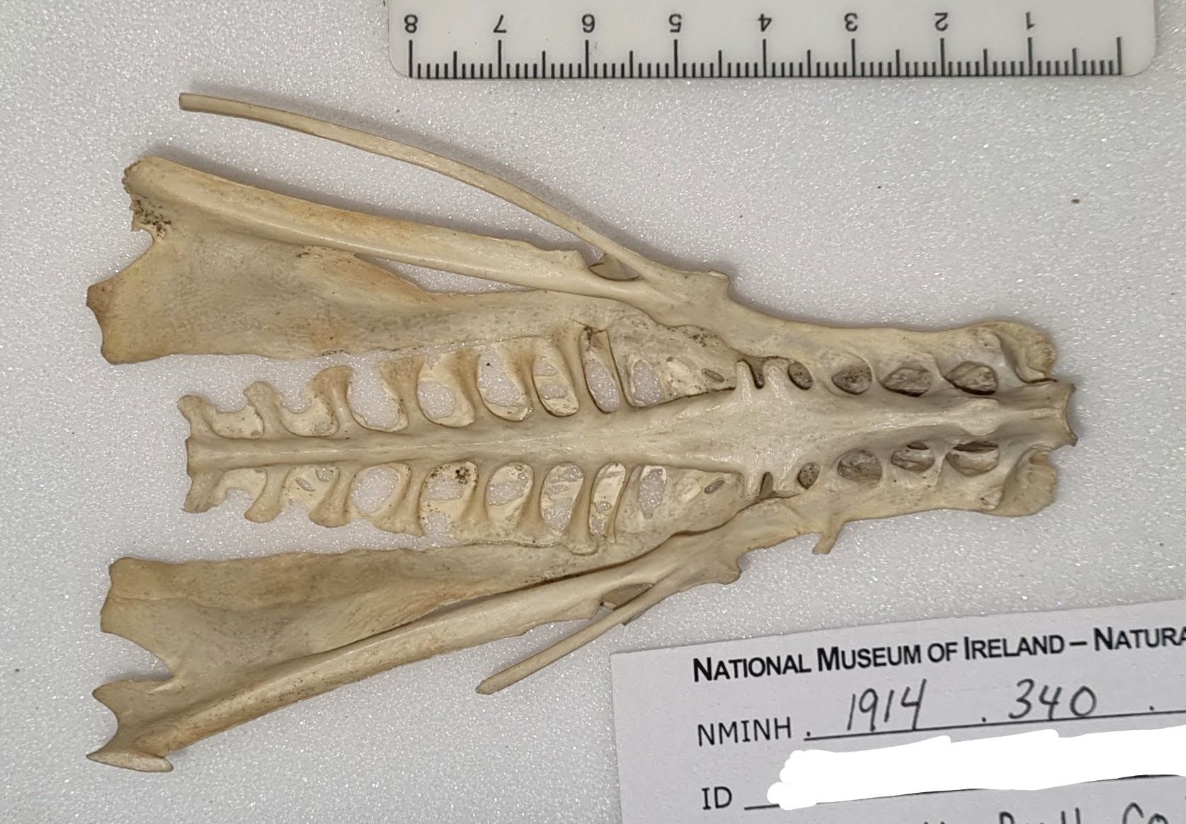
Last week I gave you this bird pelvis to have a go at identifying, as part of a series of posts on that particular feature across birds utilising different environments:
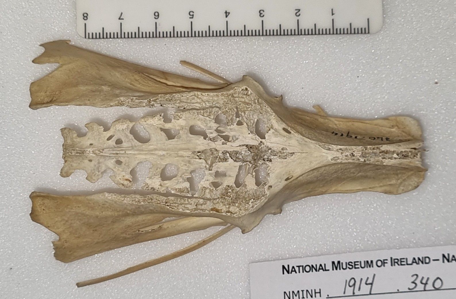
It probably wasn’t the easiest challenge, since this pelvis is damaged. It’s missing part of its dorsal surface and with some small amount of damage to the posterior margin of the right ischium (that’s the broad bit that flares out on the bottom left of the image) and with both of the pubic bones snapped (those are the long skinny bits that stick out to either side).
Still, the overall shape still gives some useful clues. The long, narrow, triangular form offers some ideas about the distribution of mass and use of the legs, to give a sense of possible locomotion habits.
The pelvis is very flat and the points of articulation with the femur are quite far forward:
At least in comparison with something like a Chicken:
This suggests something that has a centre of mass that’s quite far forward compared the foot position. This immediately raises the question of why? The obvious answer for me is that this is a species that doesn’t brisky strut around, relying on more of a low-speed waddle to get around – or perhaps a paddle.
Waterbirds have legs located quite far back in the body to aid propulsion in the water. Perhaps a bit confusingly this requires the articulation of the femur with the pelvis to be pushed further forward due to the more horizontal orientation of the spine to allow the correct orientation of the legs to push water backwards and the body forwards – unlike terresrial species that are more interested in pushing the body upwards against gravity.
There are of course a lot of waterbirds, from the Pelecanimorphae mentioned in the comments by Adam Yates, to the Anseriformes. That’s what we’re looking at here.
There are a lot of ducks and geese, so narrowing it down isn’t easy. The size of this mystery specimen is in the right range for a large duck or small goose, but if you look at the left ischium (the undamaged one) you’ll notice a deep notch – this is something seen in geese.
You may also notice an additional extra notch – which I think offers a great clue to hint at the species. This is the pelvis of a Brent Goose Branta bernicla Linnaeus, 1758.
I’m not sure if the thoughts I’ve laid out here, and over the last couple of mystery objects are helpful for guiding future identifications of bird pelvises, but I hope they may be of use. Let me know your tips on working these things out – more ideas are always welcome!
This week I have a third mystery bird pelvis for you to try your hand at identifying:
Let me know what you think this might be from – I’d be keen to hear your reasoning!
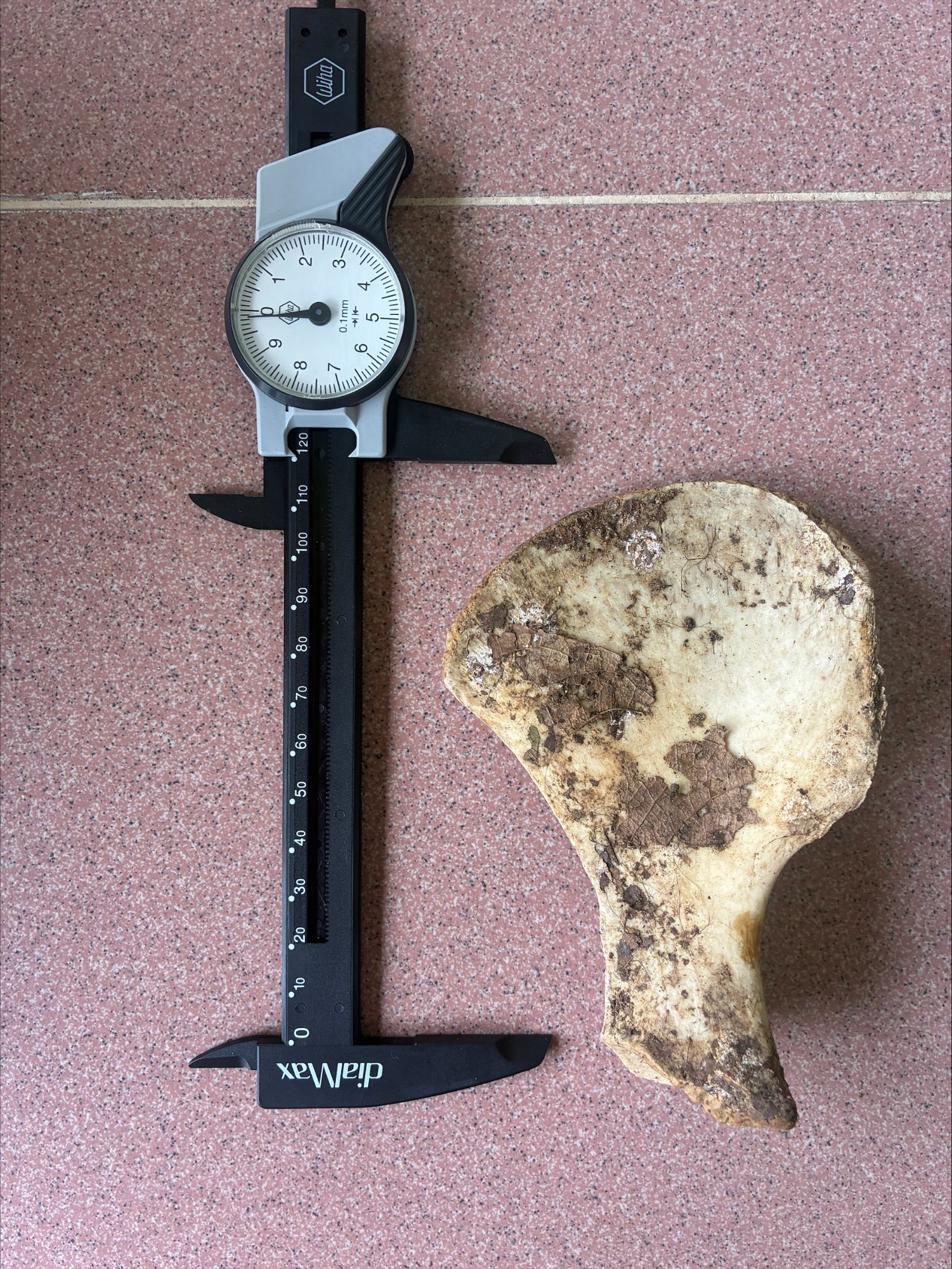
Last week I gave you the second of a series of bird pelvises to try your hand at identifying:
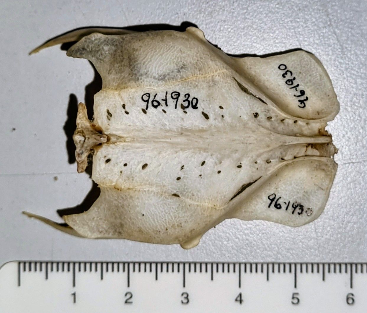
This one proved a bit more difficult than the previous example I offered up, which was from a Chicken – here’s that one for comparison:
As you can see, our mystery pelvis is a good bit smaller than that of the Chicken, and in terms of the shape, while it has some similarities, but it’s more compact and almost square. I’m not 100% sure why, but that makes me think of something that has a more upright body orientation than a Chicken.
Part of the reason it’s more square is that the areas of muscle attachment for the iliotrochantericus caudalis muscles (which I talked about a couple of weeks ago) don’t extend far forward, which suggests its femur isn’t being stabilised to enable a long stride. The attachment is quite wide though, so I suspect it may be optimised to cope with large forces in a burst instead.
There are a few species that would fit the bill (if you’ll excuse the pun) including Grouse – which was suggested by Chris Jarvis and it is remarkably similar – but this is the pelvis of a Rock Dove (in this case the domestic version) Columba livia Gmelin, JF, 1789 which was correctly spotted by Adam Yates.
I thought this one might be nice to use, as it lets me share a blast from the past that looks at the explosive take-off of a Pigeon, from Ben Garrod’s TV series Secrets of Bones, which I had the privilege to be scientific advisor for back in 2014. I hope you find the clip interesting!
The last mystery object was a bit of bird bone that I personally found really interesting, and it’s inspired me to offer up the same bit of bone, but from a different bird:
Any idea of the species that this came from? I’d love to hear your thoughts in the comments box below!
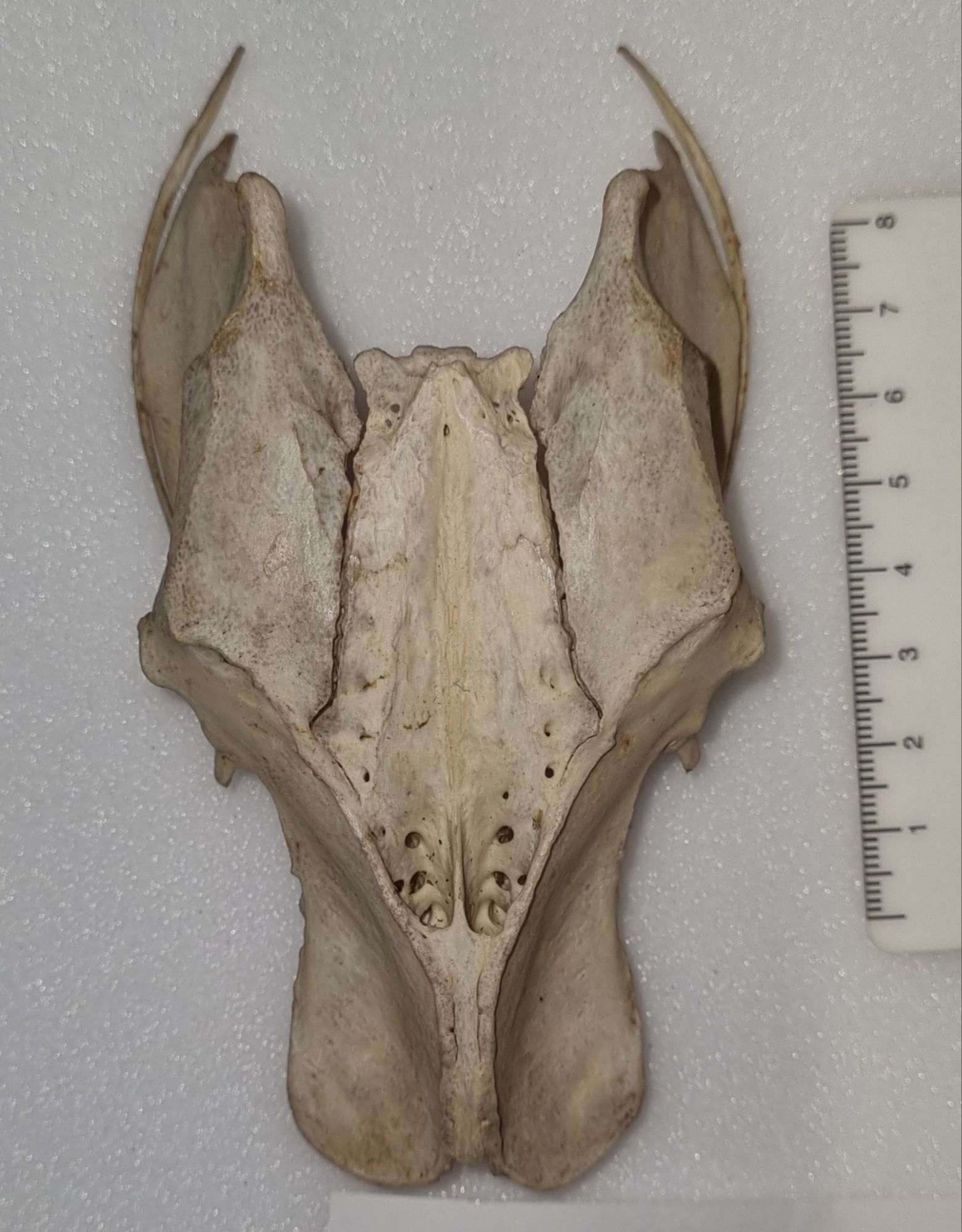
Last week I gave you this bony structure to identify:
I didn’t think this one would prove too difficult, since I went with something that most people have probably encountered on their dinner table or thrown in their domestic waste at some point. I wasn’t wrong and Chris Jarvis was first to drop a hint, with reference to Elvis the Pelvis and a famous brand of fried foodstuff from Kentucky.
This is of course the pelvis of a Chicken Gallus gallus domesticus (Linnaeus, 1758).
Bird pelvises are interesting structures, that are a bit more extensive than the usual mammalian equivalent1 due to the extended and fused vertebrae around the sacrum – the sacrum being the area of fused vertebrae where the hip bones attach to the spine. This extended region of fused vertebrae along the midline of the pelvis is referred to as the synsacrum.
In birds the fusion of the pelvis can be very extensive, and provide large areas for muscle attachment. If you look at the bottom of the photo above you can see where there are two scooped-looking sections, and this is where the “oysters” would be found in a roast Chicken. Those “oysters” are more technically referred to as the iliotrochantericus caudalis muscles and they attach to the femur and help stabilise the bird while walking.
The highly sculpted form of a bird’s pelvis creates quite a distinctive locomotor unit that reflects the way in which the bird uses its legs to walk, perch, paddle, swim or whatever else it may get up to. This means that the pelvis of a bird will usually reflect function very well and it will also carry a strong taxonomic signal since birds that are closely related will often share similar locomotion habits, lay similar sized eggs (that have to pass through the pelvis) and so on.
To my mind, the synsacrum provides an evolutionary mechanism to allow effective bipedalism while maintaining a horizontal spine – as opposed to the upright stance used in primates, which seems to come with some issues if my back is anything to go by. My background is in biomechanics and anatomy, so for me this is a topic that I find very interesting. So interesting that I may see if I can find another bird pelvis from a species with different habits to test your skills next week – let me know what do you think of that idea in the comments!
This week I have bony mystery object for you.:
Do you have any idea of the species that this came from?
As ever, you can leave your questions, observations and suggestions in the comments section below. Have fun with it!
One type of enquiry we get in the museum relates to the identification of natural materials. Often these come from law enforcement or customs officials, who may need an expert eye cast over a material to check whether it’s been imported or sold illegally.
Here’s a worked piece of natural material – I’d be keen to hear your thoughts on what it might be:
As ever, you can leave your suggestions in the comments section below. I look forward to hearing your thoughts!
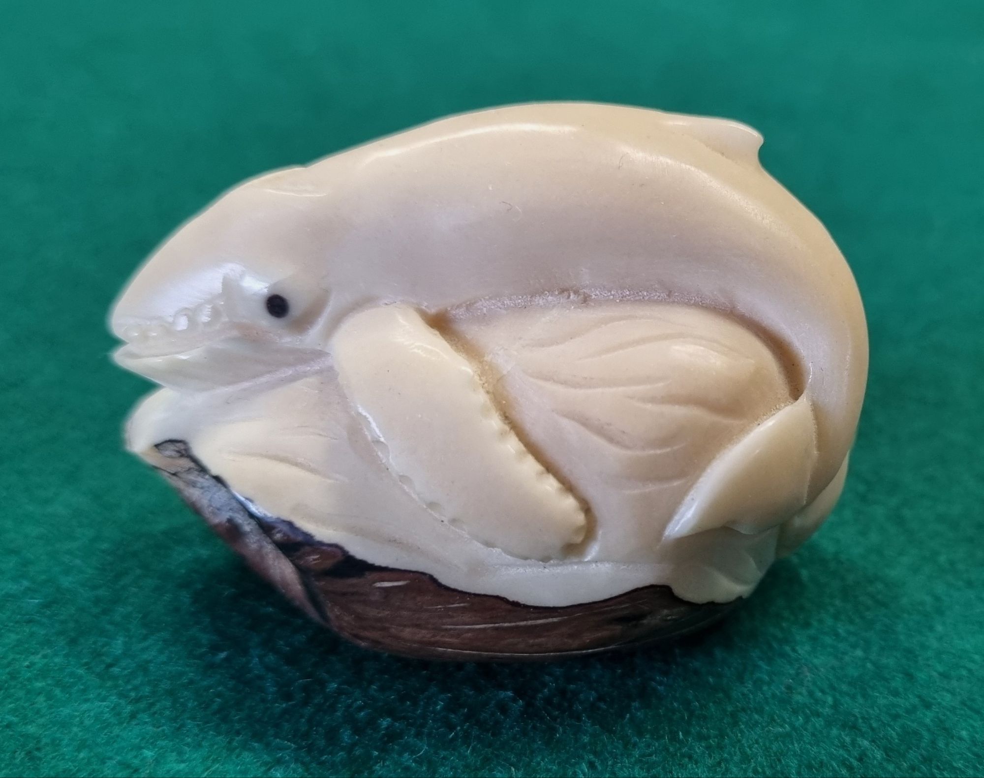
Last week I gave you this somewhat faded and oddly shaped fish to have a go at identifying as a New Year challenge:
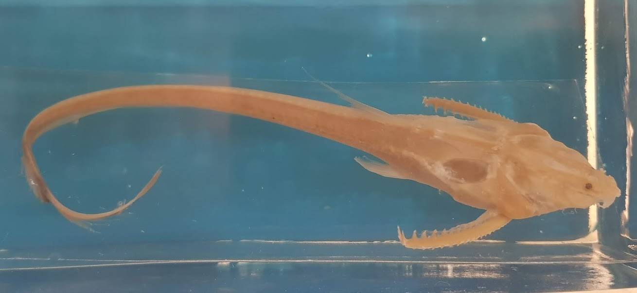
It was a very big ask, as the image isn’t really detailed enough to allow a species identification, but it was great to hear your various thoughts.
Adam Yates got very close with the suggestion it could be one of the Loricariidae – a family containing the armoured suckermouth catfish, but while this is from the same Order (the Siluriformes) this particular catfish is from a different Family.
While it has an armoured appearance, and shares those ornamented pectoral fin spines that are found in many catfish, this one has a filamentous tail and dorsal fin (that you can only just make out). What you can’t really see are the eight pectoral fin rays and the seven barbels present on the head.
This long thin tail is a hint that this is one of the Banjo Catfish, and the details of the barbels and pectoral fin rays I mentioned above let us know that it’s the Sevenbarbed Banjo Aspredinichthys filamentosus (Valenciennes, 1840).
For some reason the name Banjo Catfish always makes me think of this scene from the film Deliverance:
Musical shenanigans aside, these South American fish are bottom feeders in brackish waters, and have the unusual reproductive trait of the female attaching her eggs to her underside, so they can be moved around in the muddy waters in order to keep them oxygenated during their development.
That was certainly a challenging mystery object to start 2026, so I may see if I can find a slightly easier, but hopefully no less interesting specimen for the next mystery object!
Happy New Year! I hope you enjoyed seeing in 2026 and I wish you all a very healthy and enjoyable year ahead.
This Friday’s mystery object is a specimen from the spirit collections in the Dead Zoo:
Do you know what this mystery aquatic beastie might be?
Let me know your thoughts in the comments below – I hope you enjoy the challenge!
I hope you had an enjoyable Christmas! Last week I gave you this chonky skull as a mystery object to have a go at identifying:
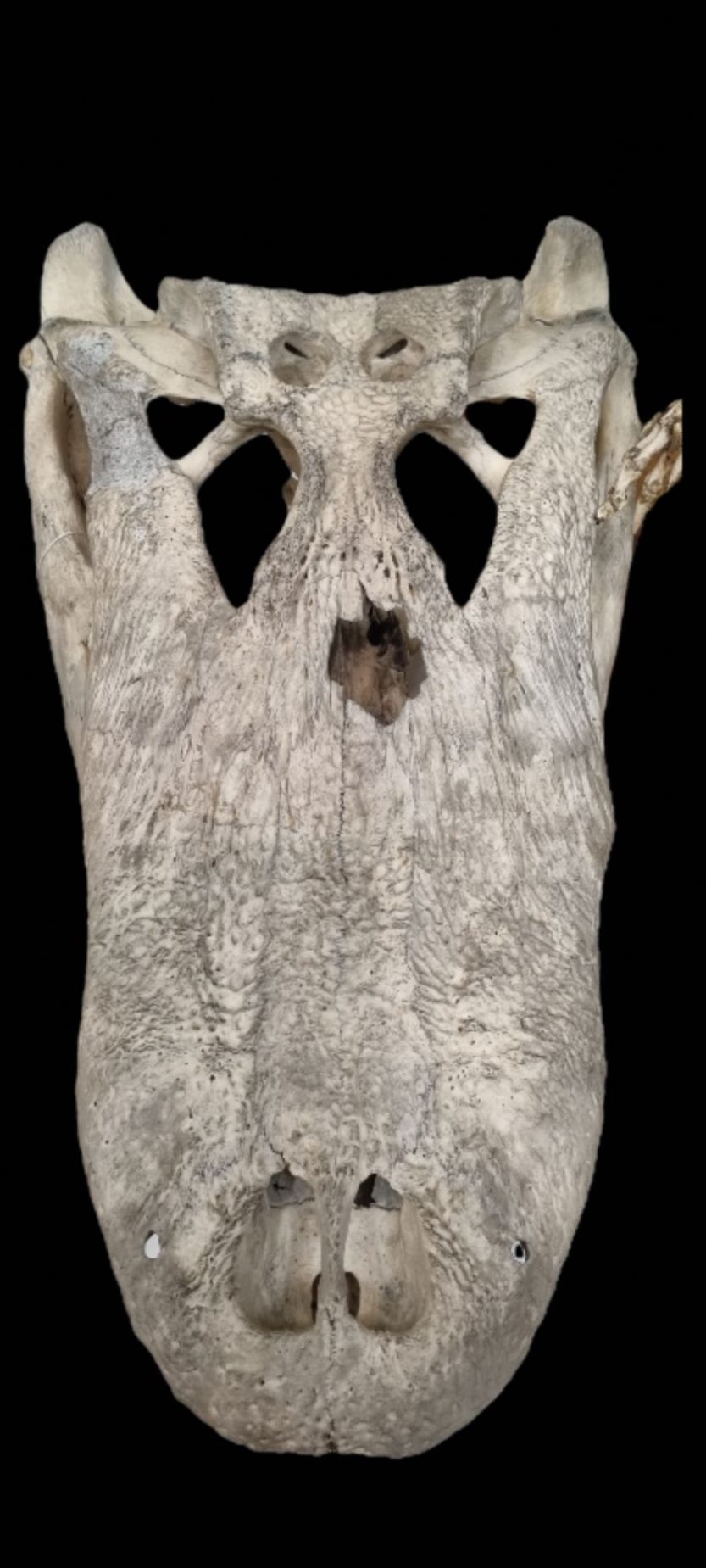
It wasn’t anything particularly challenging or unusual, but it’s a very cool skull that we recently put on display in the Dead Zoo Lab in Dublin, and it’s nice to have a chance to share it here.
While Caimans did get a mention, pretty much everyone worked out that this is the skull of an Alligator – although which of the two living species it might be sparked some conversation.
In the words of Adam Yates:
Clues include snout shape and length, divided nostrils and lack of a lateral notch for receiving the big fangs from the lower jaw.
Excellent advice, although the simplest way I use to tell the difference is to check how much of a U-shape the maxilla forms. In this case it’s very U-shaped, which says American Alligator Alligator mississippiensis (Daudin, 1802).

I’m keeping this answer brief for this mystery object, since I’m sitting at a dinner table with family after a fantastic – and very large – meal, so I’m struggling to be creative with all my blood rushing to my stomach rather than my brain.
I hope you had a great Christmas, and I look forward to sharing more mystery objects for you in 2026!
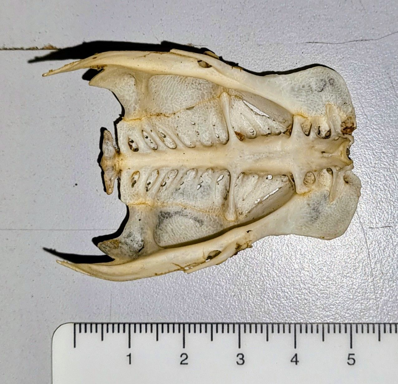
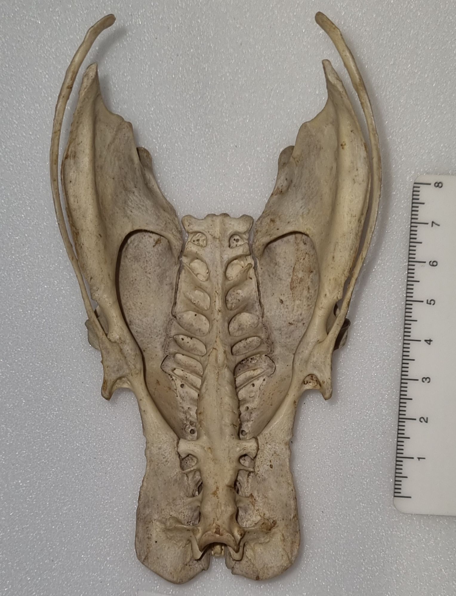
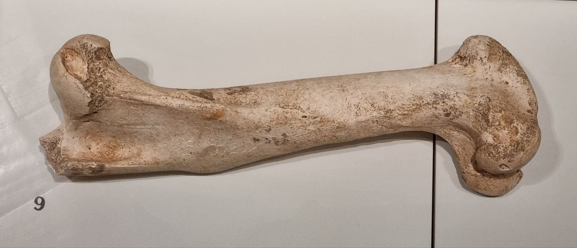
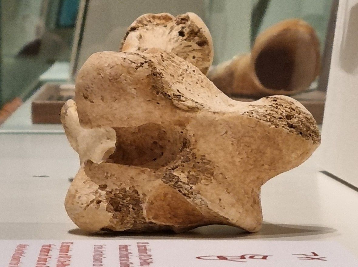
Last week I gave you this skeleton from the stores of the Dead Zoo to have a go at identifying:
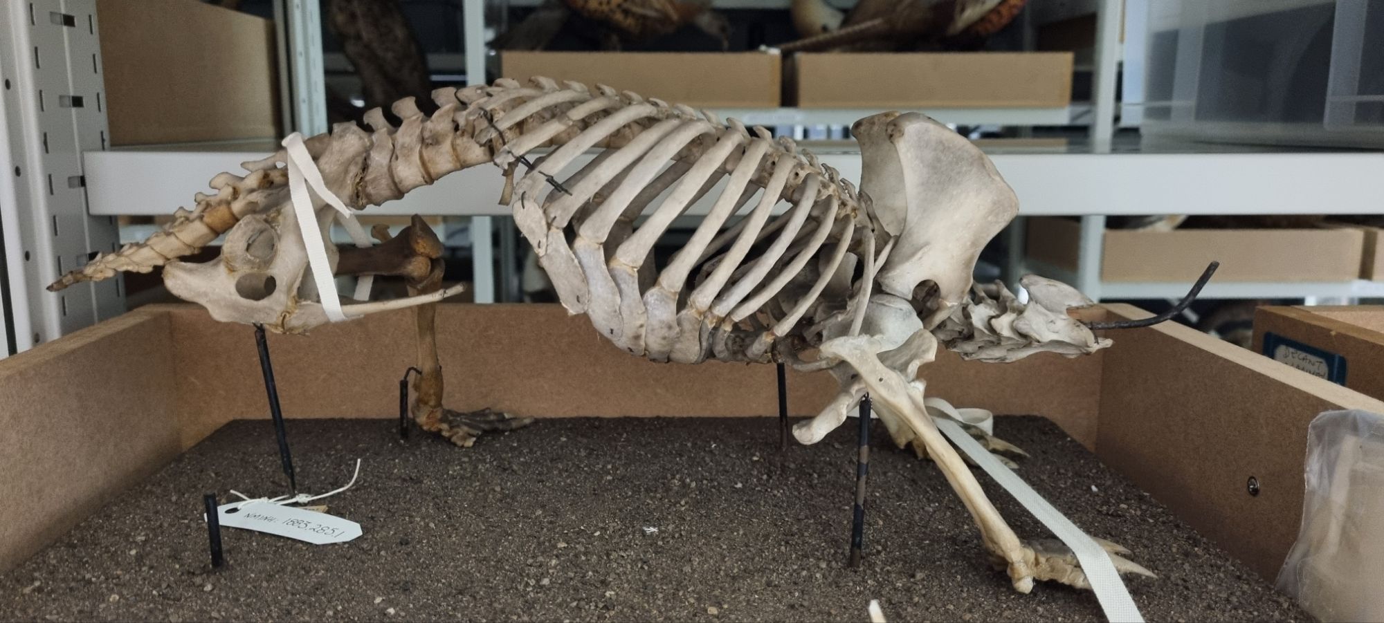
The comments came flooding in, with some slightly off and some very much on target.
The robust skeleton and stocky build of this animal, combined with some interesting bony processes – especially in the pelvic region – offered up some pretty good indicators of the type of critter we’re looking at.
The forward-facing processes of the pelvis were initially mistaken for a baculum, but on a closer look their dual nature becomes more apparent:
These are epipubic bones, which aren’t found in Placental mammals – but this is definitely a mammal – so this is either a Marsupial or Monotreme.
The lack of a skull makes it a little harder to immediately figure out what this might be, but the feet are useful – very useful in fact:
These look like the feet of a digger with those big, robust, triangular claws – but not just a burrowing digger like a Wombat – more like an ant and termite specialist whocan break open their nests. That offers a key clue.
This is the skeleton of a species of spiny anteater – one of the four species of Echidna. The feet actually offer a further diagnostic clue to the species, since most species have five claws, while just one has three – the Three-toed or Western Long-beaked Echidna Zaglossus bruijnii (Peters & Doria, 1876).
This particular specimen was originally on display in the Dead Zoo under its old name Pro-echidna Bruijnii:

The specimen has been taken off display along with everything else in the building over the last year or so, in preparation for a big refurbishment project.
When it was decanted, along with skull, the fragile right-hand-side rear limb was removed. In the photo above you can see where a claw is detaching – possibly as a result of incorrect foot positioning on the mount (Echidna feet point sideways and backwards, which seems to have confused some mounters). In other places, cotton tape was used to stabilise some of the more wobbly robust elements.
Being able to work through items like this while they’re in storage will be helpful, since it will allow us a chance to remove the worst of the dust from what may have been 140 years of display, and to make some small repairs to things like the detaching claw so it doesn’t get lost. Changing the foot position may be a bigger job, but it’s something to consider.
So while the Dead Zoo may be closed, we’re keeping busy checking the condition of the other 10,000 object we had on display, and working out what we need to do to put them back!
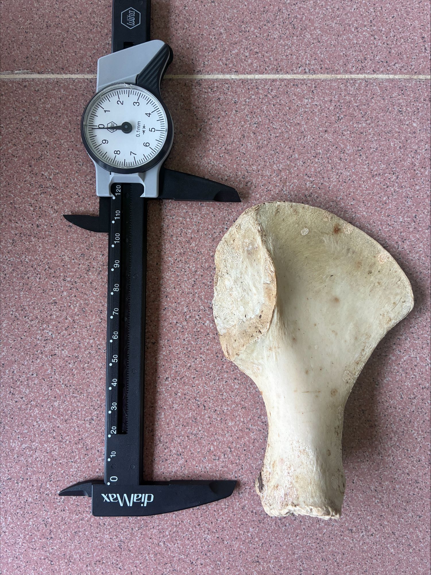
This week I have a bony mystery object for you to have a go at identifying:
This is one that may be very easy, or it may prove quite tricky, and I’m looking forward to seeing how this goes!
I hope you have fun with this one.
This week I have a mystery object I spotted when I was visiting Copenhagen earlier this year:


I’d love to hear your thoughts on which species this big specimen could be. I hop you have fun with it!
N.B. if you click the images, they should open in a larger size.
Last week I gave you these specimens that are new to the Dead Zoo to have a go at identifying:
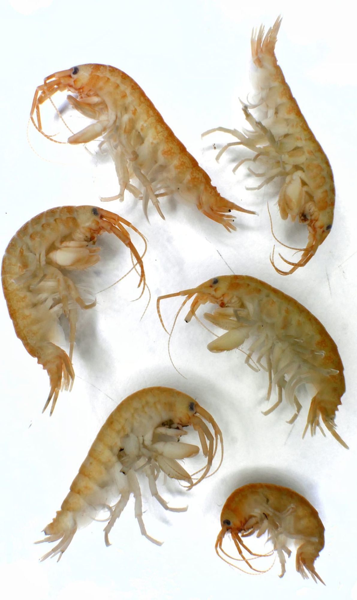
These are specimens that were lodged in the collection as part of research identifying them as a new invasive alien species to Ireland.
Adam Yates and Chris Jervis both worked out which invasive this happens to be, and it’s a bad one. These are examples of the Demon Shrimp Dikerogammarus haemobaphes (Sowinsky, 1894).
This species is a real problem, as it’s predatory and voracious – able to predate species much larger than themselves. They are very difficult to differentiate from other amphipods, although they do have some distinguishing features that can provide an identification.
There’s some very useful information about the species on the National Biodiversity Data Centre’s Invasives.ie page, which highlights some of the features for identification and details the risk posed by the species in Ireland.
Invasive species like this can have a huge environmental impact – altering food chains, introducing diseases that related taxa may be less able to cope with, and ultimately disrupting ecosystems that are already under pressure from multiple other impacts.
Managing the introduction and spread of species like this requires vigilence – the Check Clean Dry campaign offers some useful advice on how to help stop the spread:
Well done to everyone who worked out this demonic little mystery – I hope you can avoid finding these in your area!
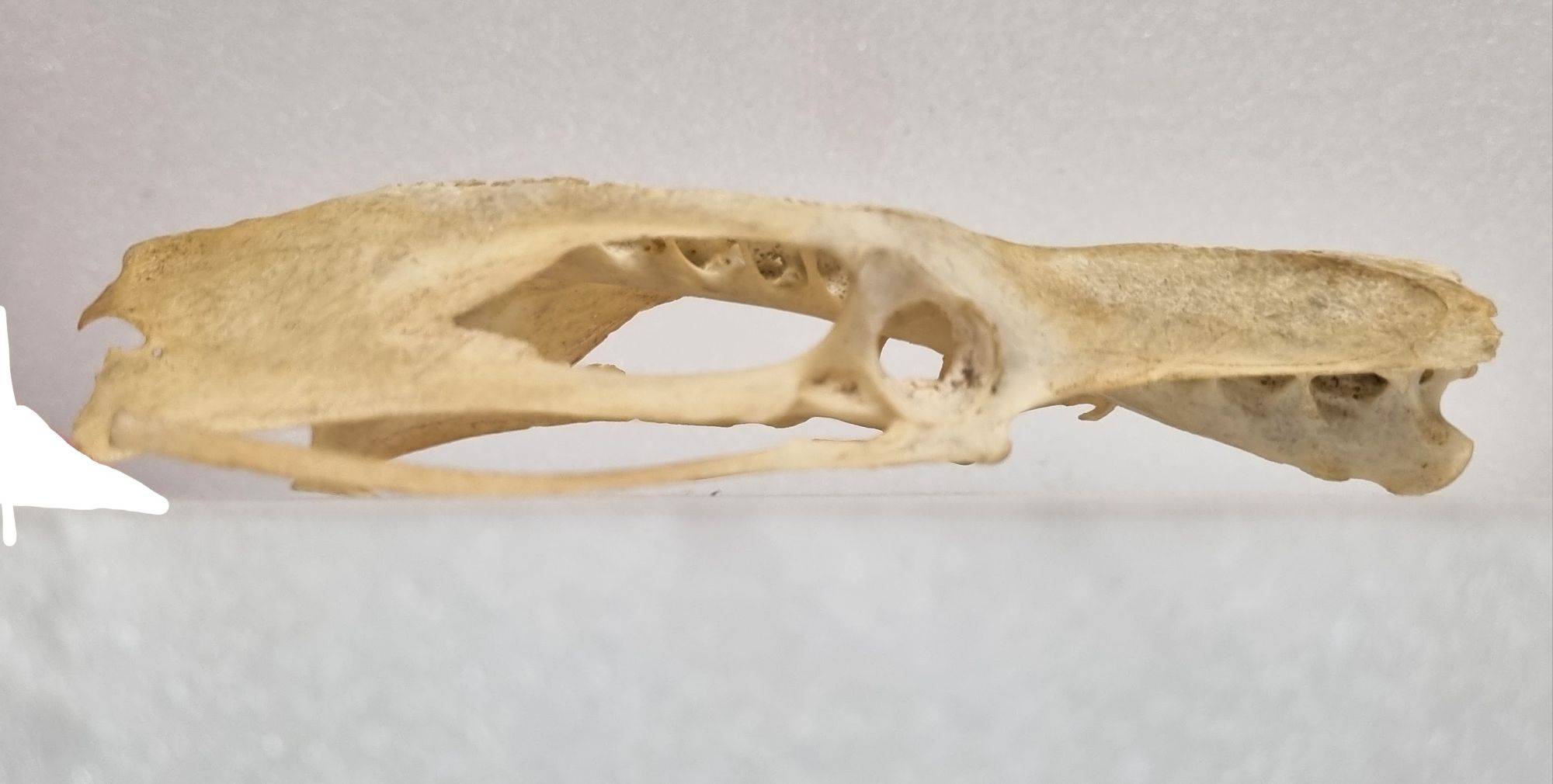
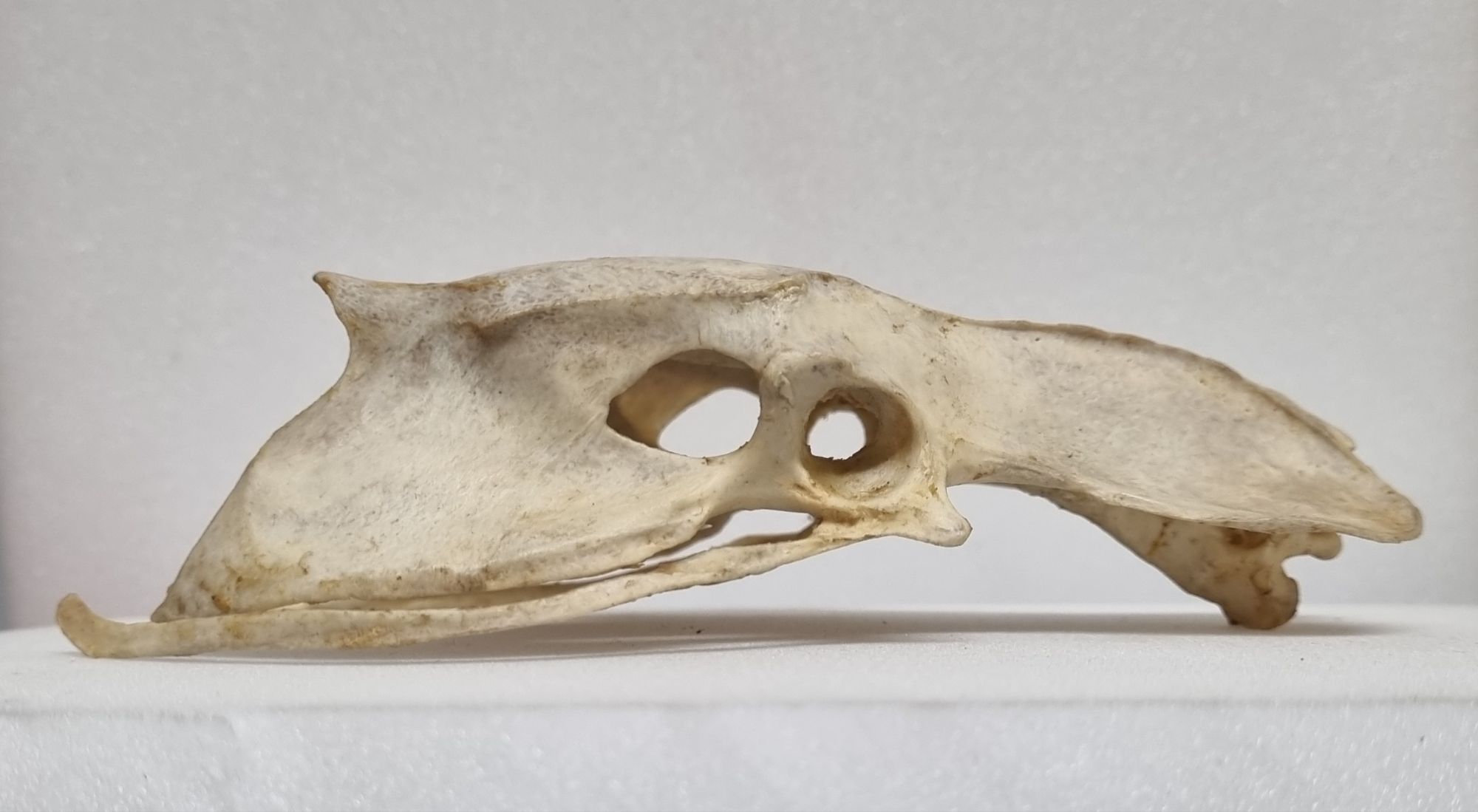
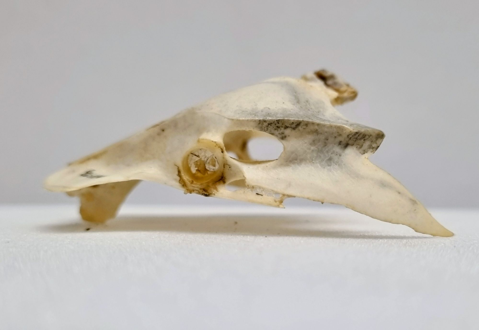
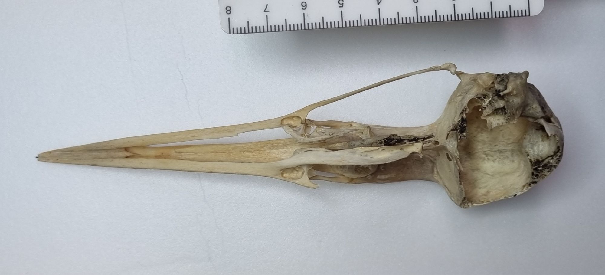
Last week I gave you this skull from the collections of the Dead Zoo to have a go at identifying:
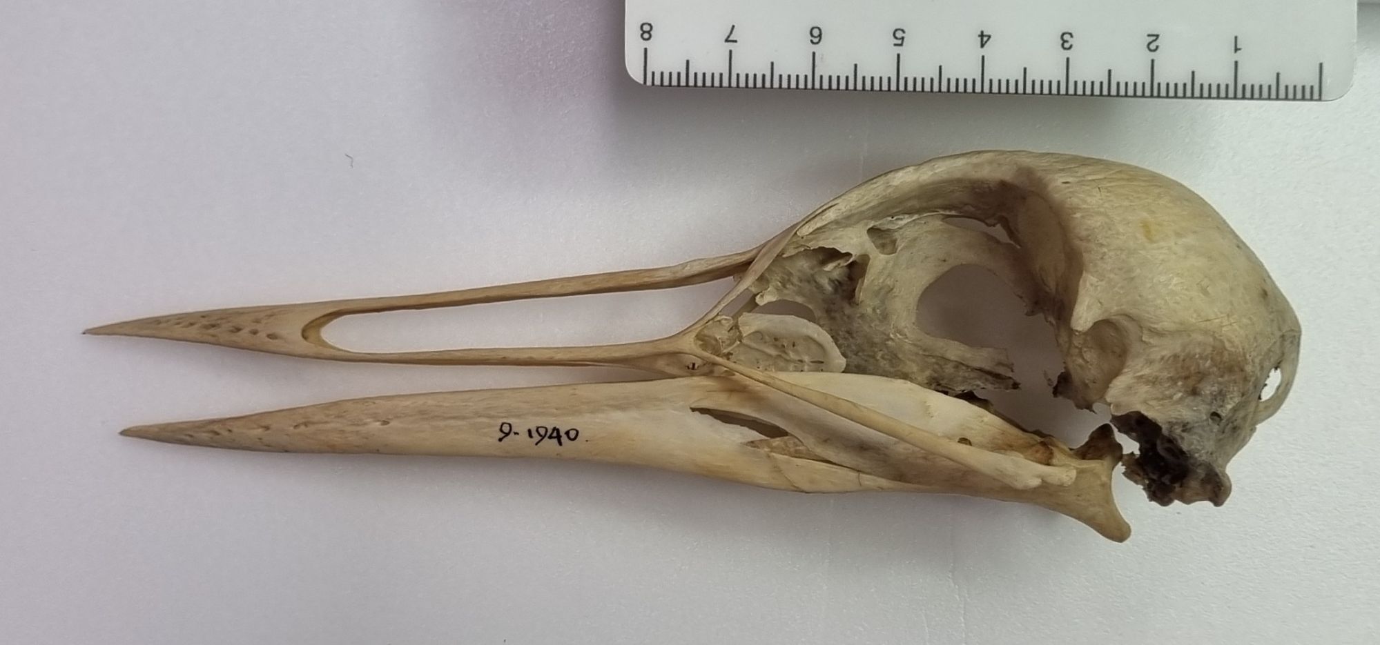
Bird skull identification can take a bit of work, until you get your eye in on things like the bill morphology (especially without the clues provided by the keratin sheath). Resources like the excellent Skullsite.com certainly help a lot, by providing a huge range of images of different species for comparison, and with tools to help narrow down options based on skull size and bill morphology.
As it turns out, Adam Yates certainly had his eye in, and he was first to comment with a correct identification for this specimen. It’s a Common or European Crane Grus grus (Linnaeus, 1758).
I chose this specimen as it’s one that we recently put on display in the Dead Zoo Lab as part of a community curated project called Our Irish Natural History. Eight community groups involved with iCAN (the Irish Community Archive Network) contributed to the work, which was coordinated by Adriana Ballinger – a fantastic postgraduate humanities researcher who has been working with us for the past year on a project with a focus on the wider cultural context surrounding natural history collections. The community groups involved each explored a different areas of interest, illustrating and exploring some of the connections between objects and local communities.
The Common Crane offered a fascinating topic explored by the Woodlawn Heritage Group and Galway Community Archaeology, who delved into the past history of the Crane in Ireland, and its importance to Bronze Age people, also touching on their recent return, with a pair of these fantastic birds recently recorded nesting in rewetted boglands. There’s too much information for me to cover it all here, so I recommend taking a look at the work for yourself on the Galway Community Archaeology web resource about the project.
These sorts of projects, that connect our ostensibly scientific objects back into local communities through a cultural link are a fantastic way to broaden the relevance and interest in our collections. As a scientist it can be easy to focus on one aspect of an object – but every item we look after can be viewed from multiple perspectives – all of which add value and relevance.
I look forward to working on similar projects in the future, while hopefully taking the opportunity to share more of the collection here, for those of you with an interest in identification!
For this week’s mystery object, I have a skull from the collections of the Dead Zoo to test your identification skills:

I look forward to hearing what you think it might be. Have fun with it!
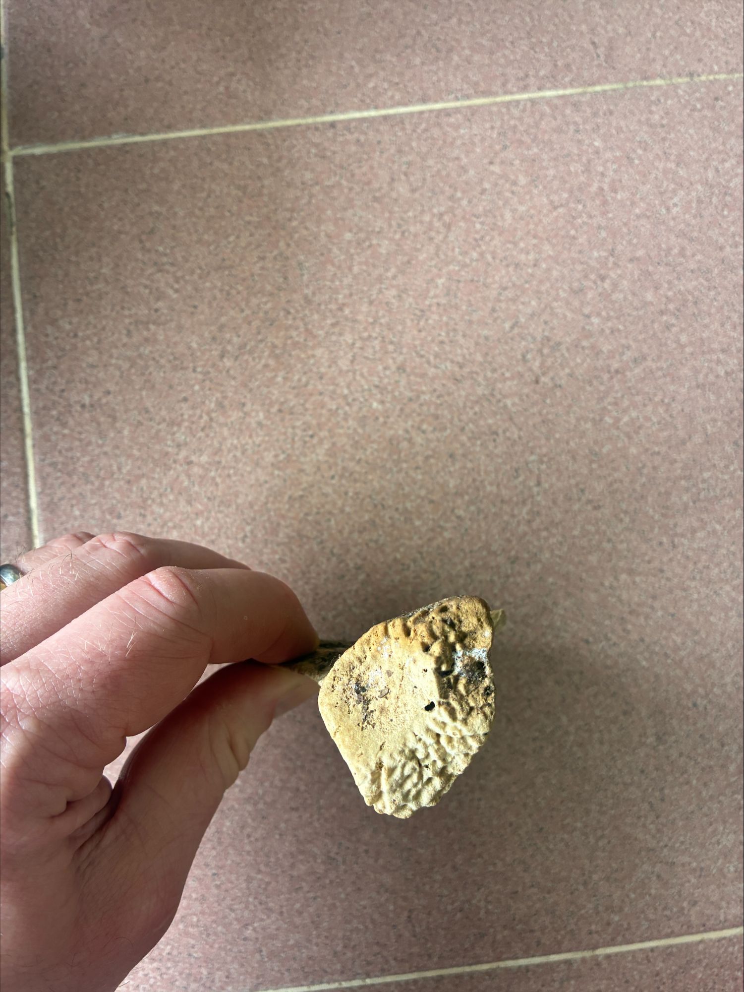
This week I have another bony mystery object for you:

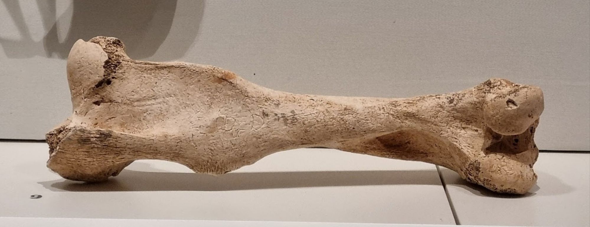
This one is currently on display in the beautiful Words on the Wave exhibition at the National Museum of Ireland on Kildare Street, but the focus is on the inscription on the bone, rather than the species the bone came from:

So, I’d love to hear your thoughts on what this is? Have fun with it!
