The last mystery object was a bit of bird bone that I personally found really interesting, and it’s inspired me to offer up the same bit of bone, but from a different bird:
Any idea of the species that this came from? I’d love to hear your thoughts in the comments box below!
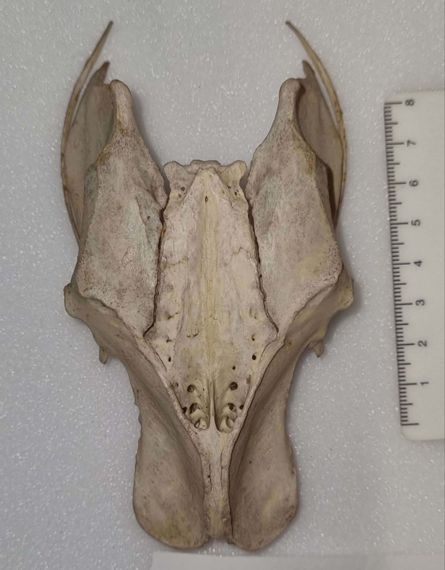
Last week I gave you this bony structure to identify:
I didn’t think this one would prove too difficult, since I went with something that most people have probably encountered on their dinner table or thrown in their domestic waste at some point. I wasn’t wrong and Chris Jarvis was first to drop a hint, with reference to Elvis the Pelvis and a famous brand of fried foodstuff from Kentucky.
This is of course the pelvis of a Chicken Gallus gallus domesticus (Linnaeus, 1758).
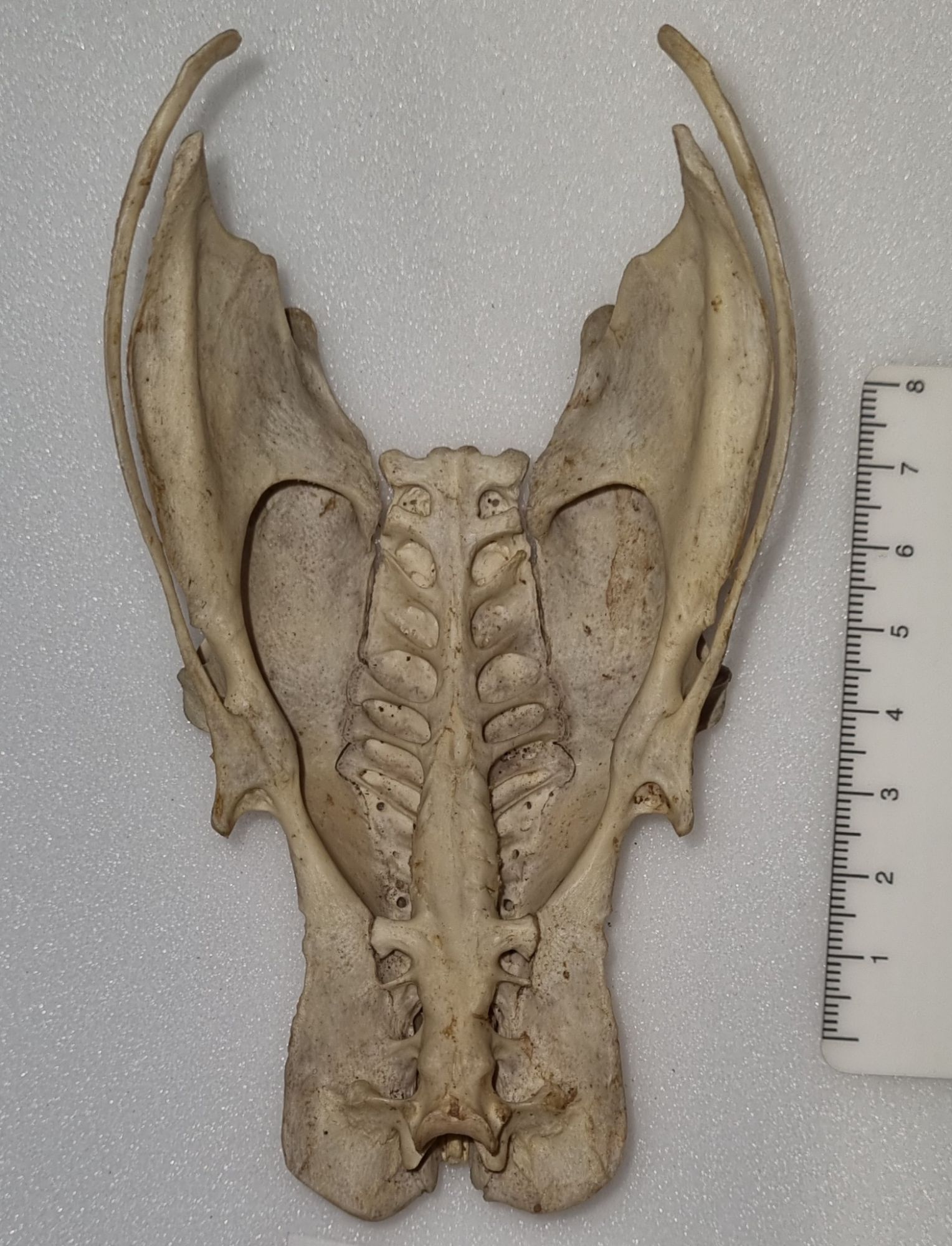
Bird pelvises are interesting structures, that are a bit more extensive than the usual mammalian equivalent1 due to the extended and fused vertebrae around the sacrum – the sacrum being the area of fused vertebrae where the hip bones attach to the spine. This extended region of fused vertebrae along the midline of the pelvis is referred to as the synsacrum.
In birds the fusion of the pelvis can be very extensive, and provide large areas for muscle attachment. If you look at the bottom of the photo above you can see where there are two scooped-looking sections, and this is where the “oysters” would be found in a roast Chicken. Those “oysters” are more technically referred to as the iliotrochantericus caudalis muscles and they attach to the femur and help stabilise the bird while walking.
The highly sculpted form of a bird’s pelvis creates quite a distinctive locomotor unit that reflects the way in which the bird uses its legs to walk, perch, paddle, swim or whatever else it may get up to. This means that the pelvis of a bird will usually reflect function very well and it will also carry a strong taxonomic signal since birds that are closely related will often share similar locomotion habits, lay similar sized eggs (that have to pass through the pelvis) and so on.
To my mind, the synsacrum provides an evolutionary mechanism to allow effective bipedalism while maintaining a horizontal spine – as opposed to the upright stance used in primates, which seems to come with some issues if my back is anything to go by. My background is in biomechanics and anatomy, so for me this is a topic that I find very interesting. So interesting that I may see if I can find another bird pelvis from a species with different habits to test your skills next week – let me know what do you think of that idea in the comments!
Of course, nature being what it is, there is an exception to that generalisation with the Xenarthra, whose members have a similiarly fused synsacrum. ↩︎
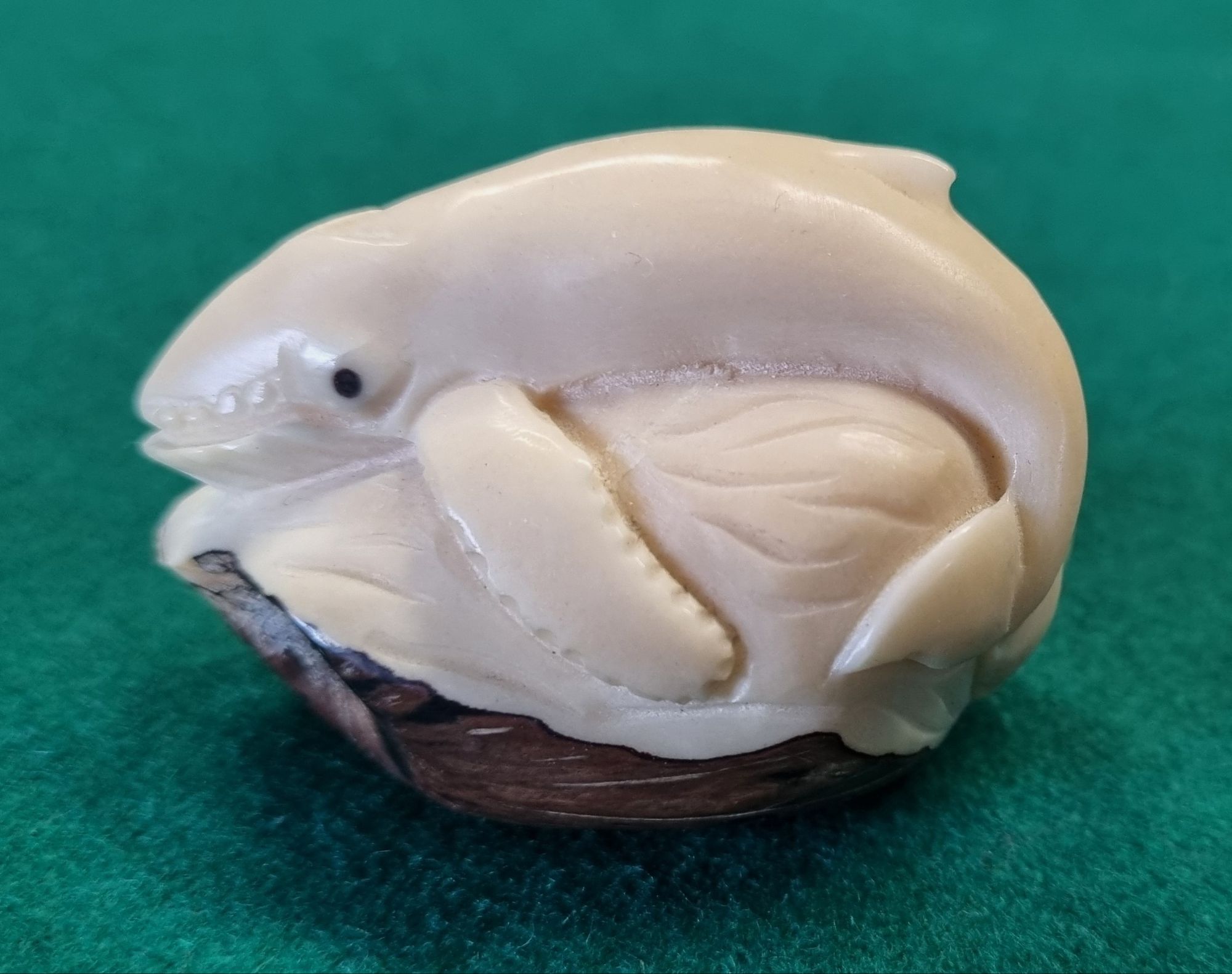
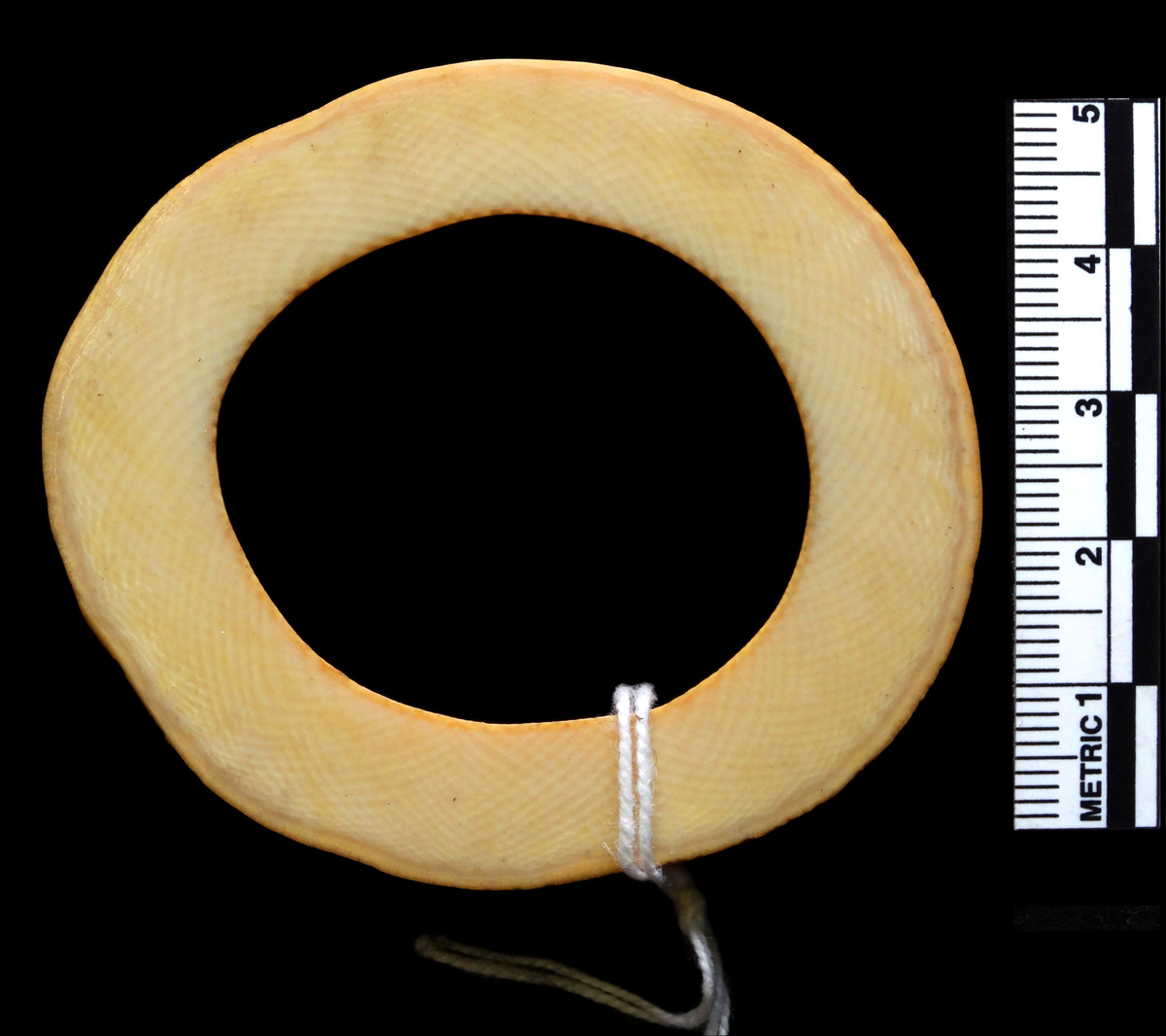
Last week I gave you this cute little netsuke (pronounced nets-keh) to see if you could identify what it was carved from:
I was hoping it might offer a bit of a challenge, with a few options being put forward for a variety of ivories, and perhaps some pondering on the topic of the carving, but I was disappointed by how quickly and comprehensively everyone told me exactly what this is.
I suppose I shouldn’t be surprised, since the community here on Zygoma are really good at identifying things (I suspect there’s a bit of self selection going on there!) The shape is quite distinctive, but more there’s also a small section on the base that’s just visible, which is very characteristic if you know what you’re looking for.
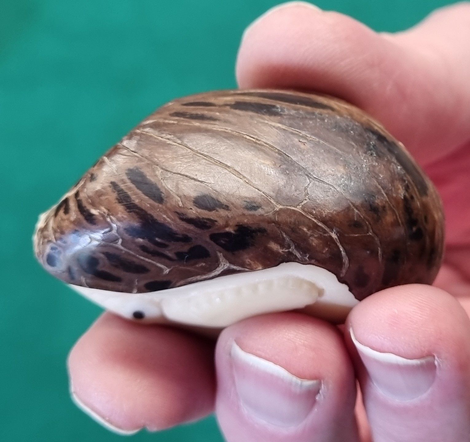
Here it is pictured from underneath:
This is an example of vegetable ivory – a material derived from the nut of a Tagua Palm in the genus Phytelephas Ruiz & Pav. These large seeds have a creamy, ivory coloured flesh that is easily carved when wet, but which become hard when dried.
The vegetable ivory lacks the Schreger lines found in proboscidean ivories, such as mammoth or elephant:
Section through elephant ivory, clearly showing the Schreger lines – an optical effect caused by light scattering caused by dentine fibrils laid down during the growth of the tusk.
There are plenty of other ivories of course, some of which have similarly distinctive aspects to look for, like Walrus ivory that has a central section that looks almost like clouds due to secondary dentine secretion in the root space of the tusk, and Hippopotamus canine ivory that has a subtle discoloured arch in the middle that results from the flattened triangular shape of the tooth.
In the case of the Tagua nut there is a hole in the centre, but this will often be avoided by not carving too deeply, or by incorporating the natural void into the design of the carving.
So well done to everyone – your skills are still top notch! You can test them again soon, as there will be more mysteries to come next week.
One type of enquiry we get in the museum relates to the identification of natural materials. Often these come from law enforcement or customs officials, who may need an expert eye cast over a material to check whether it’s been imported or sold illegally.
Here’s a worked piece of natural material – I’d be keen to hear your thoughts on what it might be:
As ever, you can leave your suggestions in the comments section below. I look forward to hearing your thoughts!
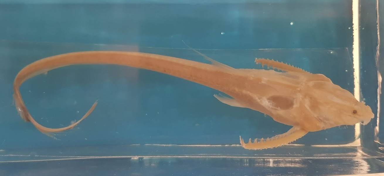
Last week I gave you this somewhat faded and oddly shaped fish to have a go at identifying as a New Year challenge:
It was a very big ask, as the image isn’t really detailed enough to allow a species identification, but it was great to hear your various thoughts.
Adam Yates got very close with the suggestion it could be one of the Loricariidae – a family containing the armoured suckermouth catfish, but while this is from the same Order (the Siluriformes) this particular catfish is from a different Family.
While it has an armoured appearance, and shares those ornamented pectoral fin spines that are found in many catfish, this one has a filamentous tail and dorsal fin (that you can only just make out). What you can’t really see are the eight pectoral fin rays and the seven barbels present on the head.
This long thin tail is a hint that this is one of the Banjo Catfish, and the details of the barbels and pectoral fin rays I mentioned above let us know that it’s the Sevenbarbed Banjo Aspredinichthys filamentosus (Valenciennes, 1840).
For some reason the name Banjo Catfish always makes me think of this scene from the film Deliverance:
Musical shenanigans aside, these South American fish are bottom feeders in brackish waters, and have the unusual reproductive trait of the female attaching her eggs to her underside, so they can be moved around in the muddy waters in order to keep them oxygenated during their development.
That was certainly a challenging mystery object to start 2026, so I may see if I can find a slightly easier, but hopefully no less interesting specimen for the next mystery object!
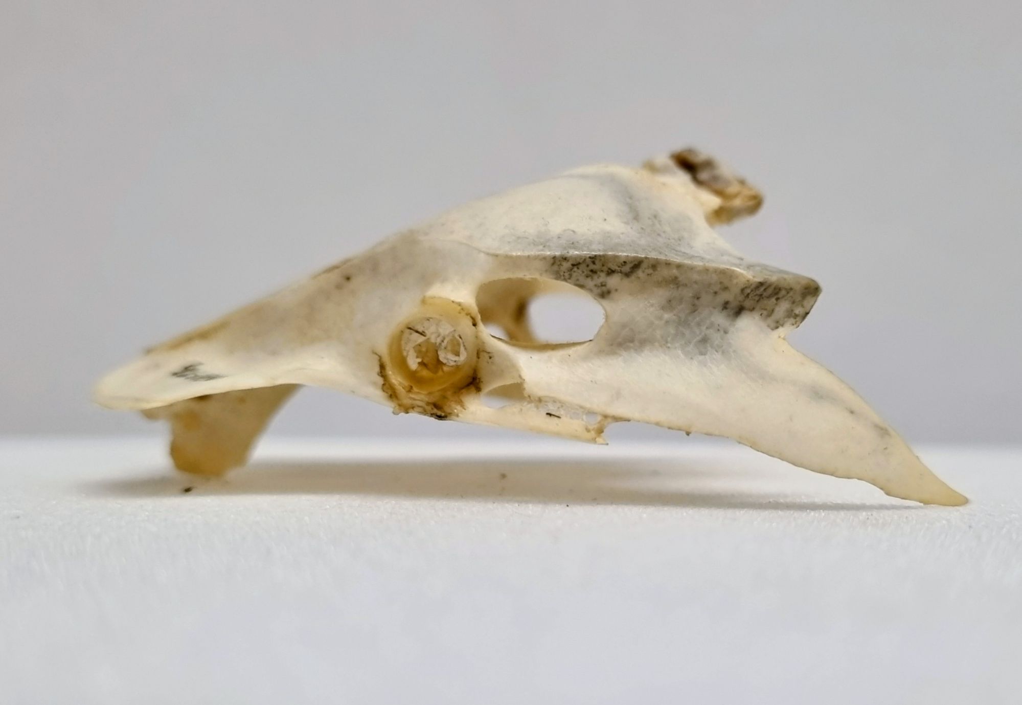
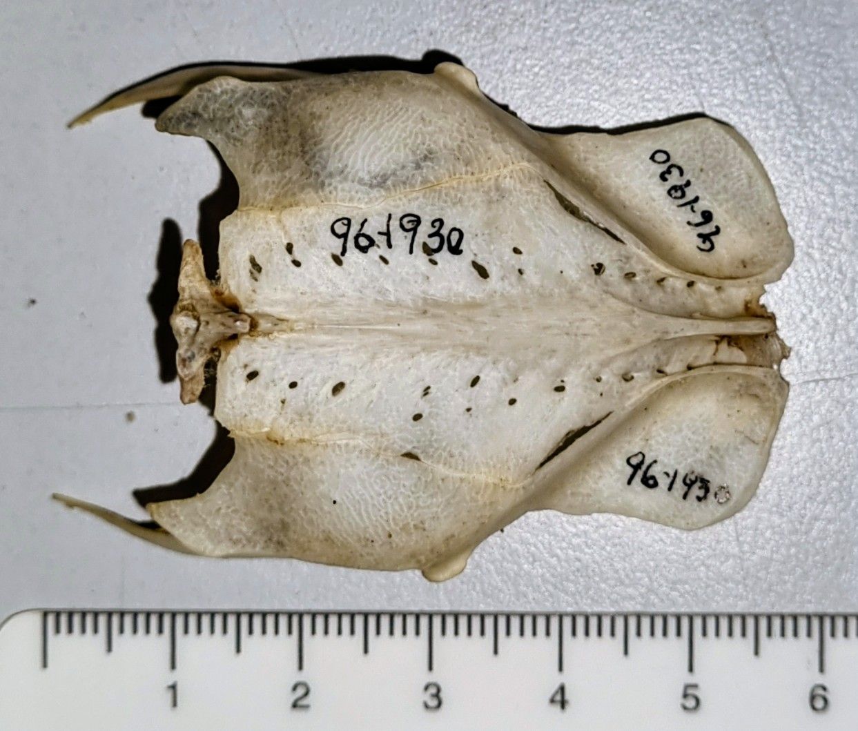
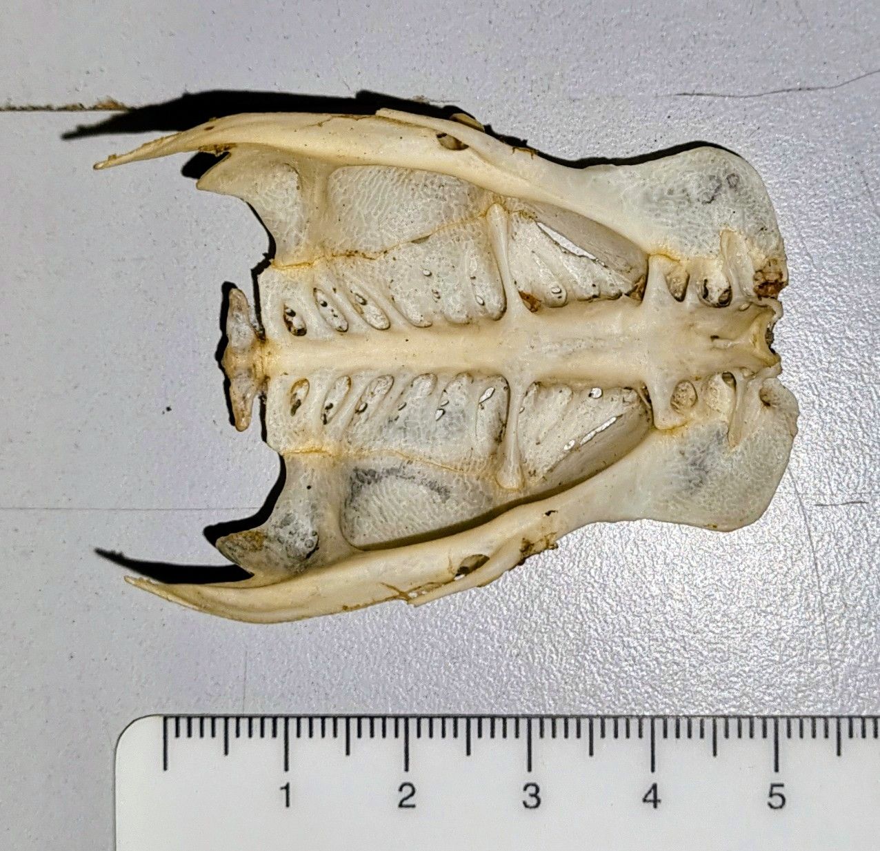
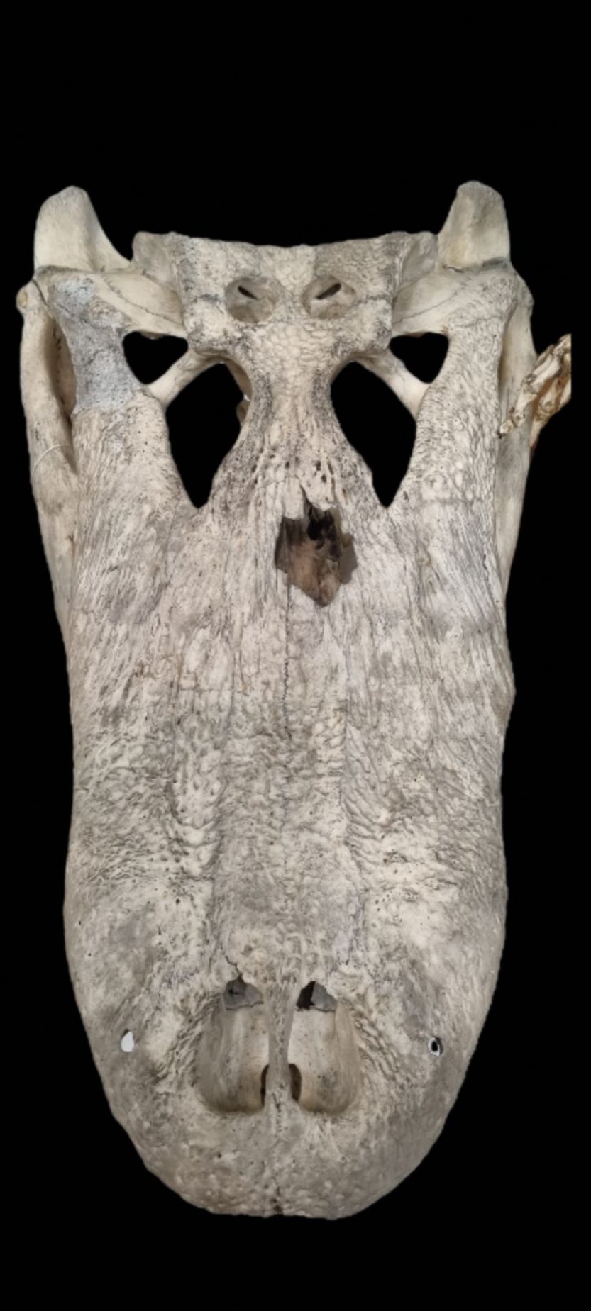
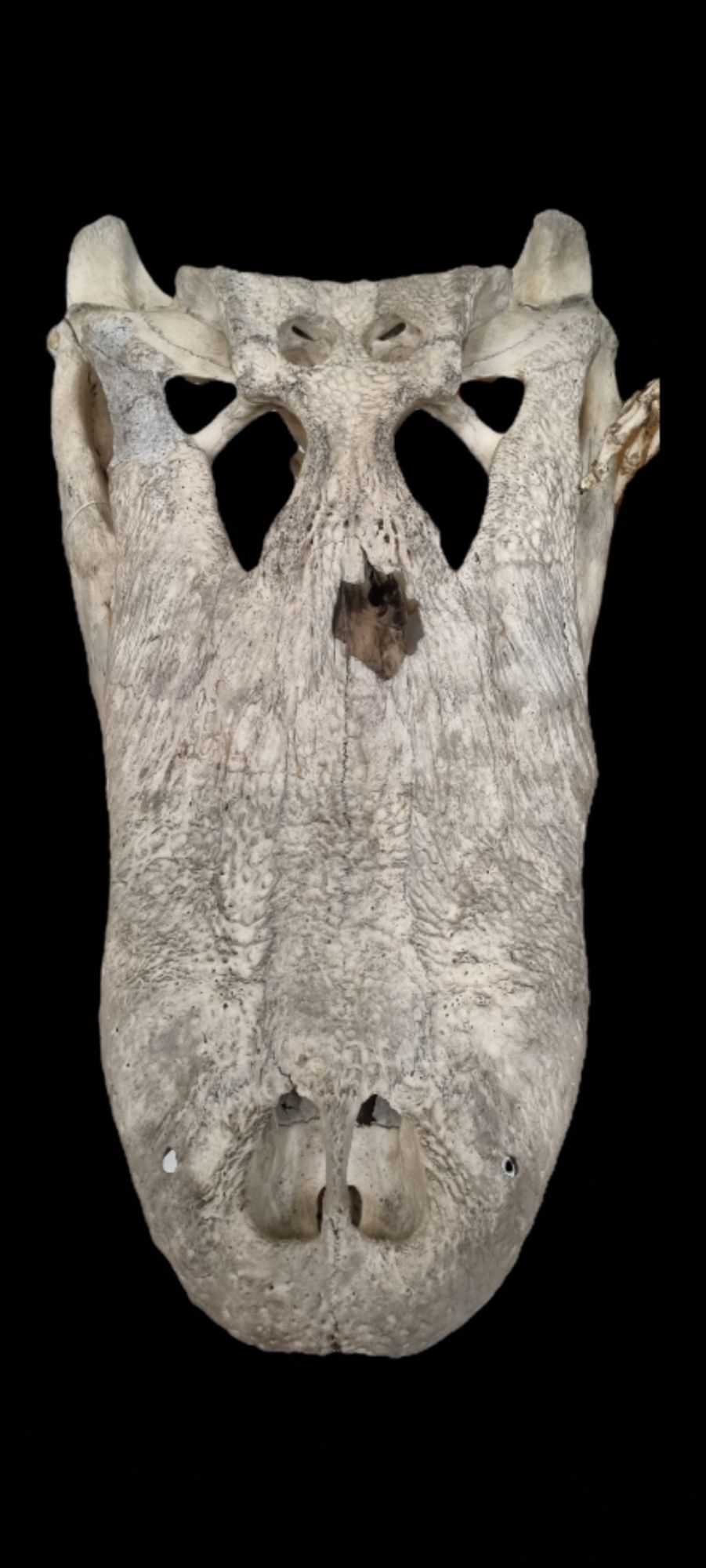
I hope you had an enjoyable Christmas! Last week I gave you this chonky skull as a mystery object to have a go at identifying:
It wasn’t anything particularly challenging or unusual, but it’s a very cool skull that we recently put on display in the Dead Zoo Lab in Dublin, and it’s nice to have a chance to share it here.
While Caimans did get a mention, pretty much everyone worked out that this is the skull of an Alligator – although which of the two living species it might be sparked some conversation.
In the words of Adam Yates:
Clues include snout shape and length, divided nostrils and lack of a lateral notch for receiving the big fangs from the lower jaw.
Excellent advice, although the simplest way I use to tell the difference is to check how much of a U-shape the maxilla forms. In this case it’s very U-shaped, which says American Alligator Alligator mississippiensis (Daudin, 1802).
I’m keeping this answer brief for this mystery object, since I’m sitting at a dinner table with family after a fantastic – and very large – meal, so I’m struggling to be creative with all my blood rushing to my stomach rather than my brain.
I hope you had a great Christmas, and I look forward to sharing more mystery objects for you in 2026!
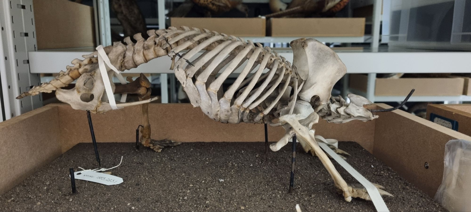
Last week I gave you this skeleton from the stores of the Dead Zoo to have a go at identifying:
The comments came flooding in, with some slightly off and some very much on target.
The robust skeleton and stocky build of this animal, combined with some interesting bony processes – especially in the pelvic region – offered up some pretty good indicators of the type of critter we’re looking at.
The forward-facing processes of the pelvis were initially mistaken for a baculum, but on a closer look their dual nature becomes more apparent:
These are epipubic bones, which aren’t found in Placental mammals – but this is definitely a mammal – so this is either a Marsupial or Monotreme.
The lack of a skull makes it a little harder to immediately figure out what this might be, but the feet are useful – very useful in fact:
These look like the feet of a digger with those big, robust, triangular claws – but not just a burrowing digger like a Wombat – more like an ant and termite specialist whocan break open their nests. That offers a key clue.
This is the skeleton of a species of spiny anteater – one of the four species of Echidna. The feet actually offer a further diagnostic clue to the species, since most species have five claws, while just one has three – the Three-toed or Western Long-beaked Echidna Zaglossus bruijnii (Peters & Doria, 1876).
This particular specimen was originally on display in the Dead Zoo under its old name Pro-echidna Bruijnii:
Photo of Three-toed Echidna skeleton NMINH:1883.285.1 at the National Museum of Ireland – Natural History taken by Illustratedjc, 2015.
The specimen has been taken off display along with everything else in the building over the last year or so, in preparation for a big refurbishment project.
When it was decanted, along with skull, the fragile right-hand-side rear limb was removed. In the photo above you can see where a claw is detaching – possibly as a result of incorrect foot positioning on the mount (Echidna feet point sideways and backwards, which seems to have confused some mounters). In other places, cotton tape was used to stabilise some of the more wobbly robust elements.
Being able to work through items like this while they’re in storage will be helpful, since it will allow us a chance to remove the worst of the dust from what may have been 140 years of display, and to make some small repairs to things like the detaching claw so it doesn’t get lost. Changing the foot position may be a bigger job, but it’s something to consider.
So while the Dead Zoo may be closed, we’re keeping busy checking the condition of the other 10,000 object we had on display, and working out what we need to do to put them back!
Last week I gave you this mystery object that I found dead in the street when visiting Copenhagen earlier this year:
Clearly this is a species of insect in the Order Odonata, which are the dragonflies and damselflies. Damselflies are much slimmer than this, and their wings fold back along their body at rest, so this one is a dragonfly.
Everyone who commented had worked out the species, since the metallic green-bronze body and fuzzy body are quite distinctive.
This is an example of a Downy Emerald Cordulia aenea (Linnaeus, 1758) which is a species that is fairly widespread across northern Europe. While this species does occurs in Ireland, it has only been recorded from a few localities.
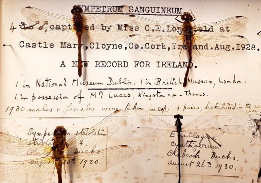
We only have one example of this species in the collections of the Dead Zoo, collected in 1978 from County Cork. I was a little surprised that it wasn’t collected by Madame Dragonfly herself, Cynthia Longfield (1896-1991), since she lived in Cork in the 1970s and was a hugely experienced and prolific expert on dragonflies.
Longfield was an adventurous and trailblazing female scientist – the first woman to be a member of the Royal Entomological Society, with expeditions in the 1920s to the Andes, Amazon, Galapagos, Egypt and a host of other localities that she undertook as part of a team from London Zoo, collecting specimens for the Natural History Museum in London, where she later became a voluntary cataloguer and was credited with saving the Museum from fire following bombing during WWII thanks to her actions as part of the Auxillary Fire Service.
Most of Longfield’s collections are held in the NHM, London, but we do have specimens collected by her in the National Museum of Ireland, donated after her retirement and move from South Kensington to Cloyne in County Cork. The Downy Emerald is not one of the specimens we received, but her specimens include new records for Ireland and a number of Paratypes of African species that she collected on her adventures both near and far.
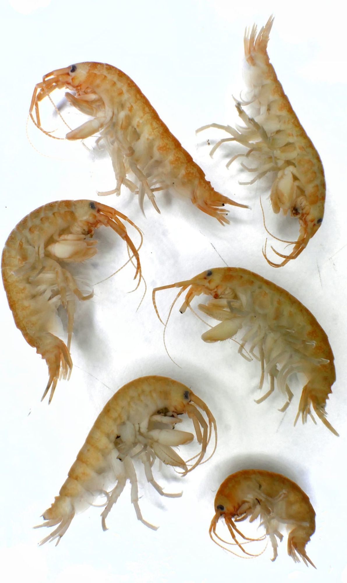
Last week I gave you these specimens that are new to the Dead Zoo to have a go at identifying:
Image taken by Jamie Maxwell, 2025
These are specimens that were lodged in the collection as part of research identifying them as a new invasive alien species to Ireland.
Adam Yates and Chris Jervis both worked out which invasive this happens to be, and it’s a bad one. These are examples of the Demon Shrimp Dikerogammarus haemobaphes (Sowinsky, 1894).
This species is a real problem, as it’s predatory and voracious – able to predate species much larger than themselves. They are very difficult to differentiate from other amphipods, although they do have some distinguishing features that can provide an identification.
Invasive species like this can have a huge environmental impact – altering food chains, introducing diseases that related taxa may be less able to cope with, and ultimately disrupting ecosystems that are already under pressure from multiple other impacts.
Managing the introduction and spread of species like this requires vigilence – the Check Clean Dry campaign offers some useful advice on how to help stop the spread:
Well done to everyone who worked out this demonic little mystery – I hope you can avoid finding these in your area!
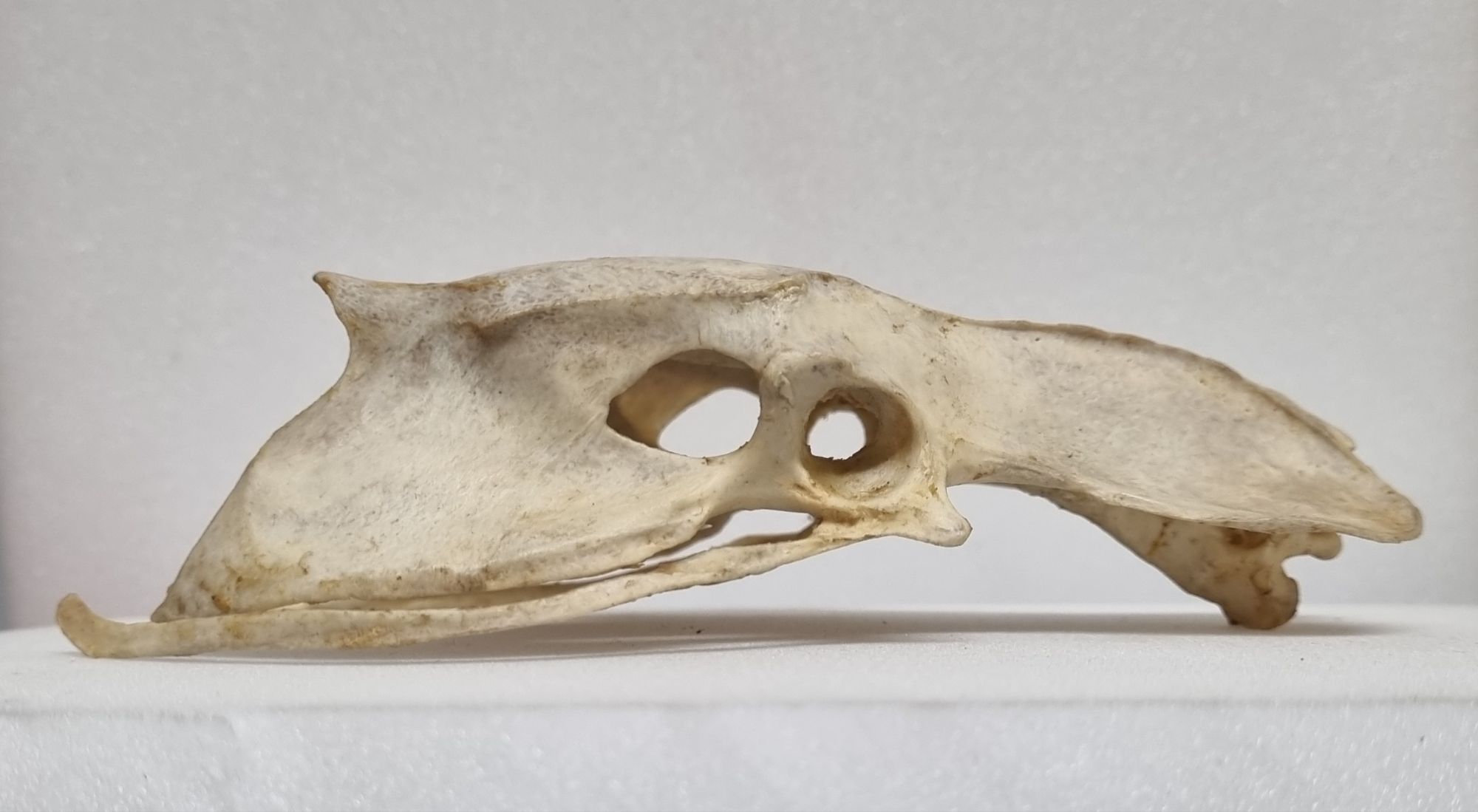
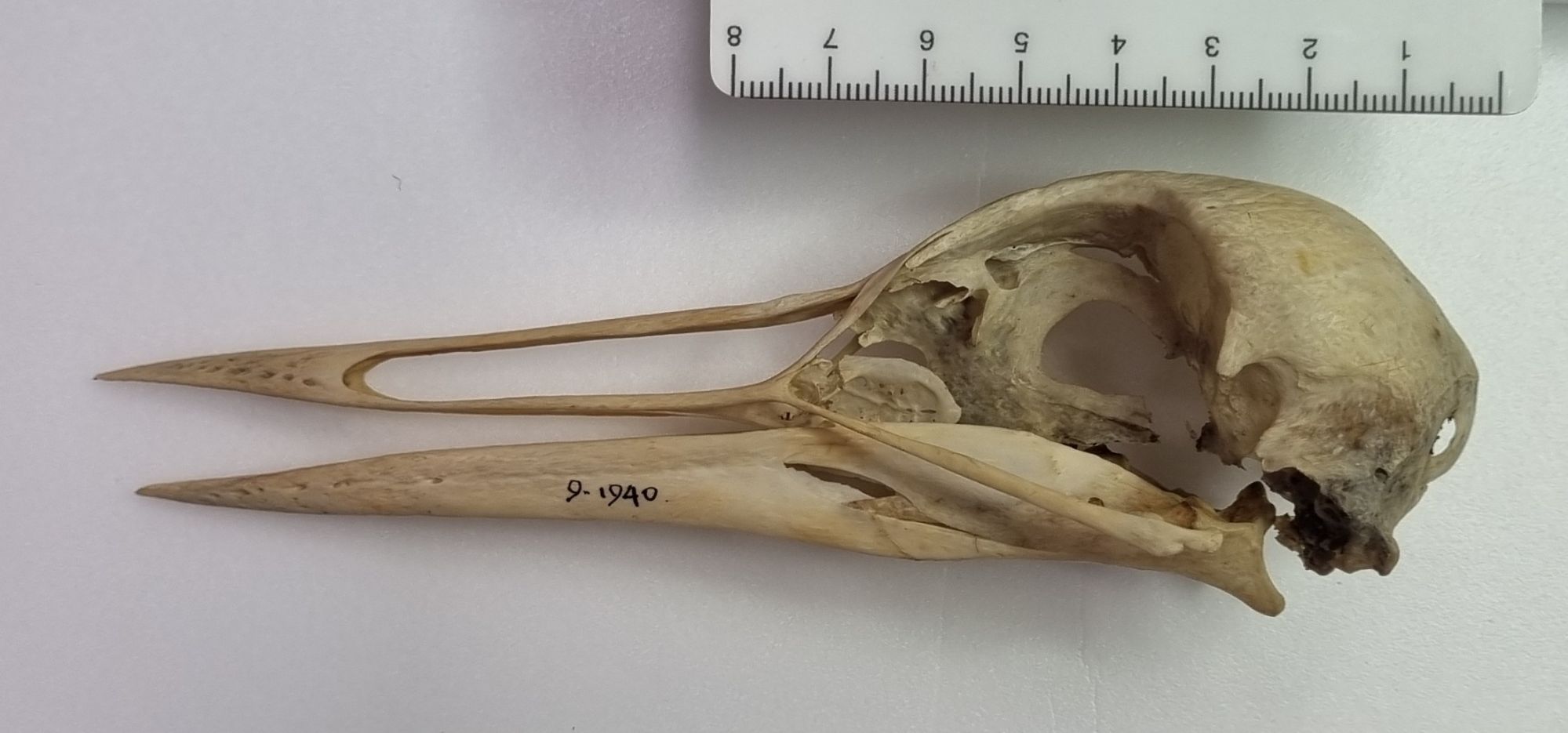
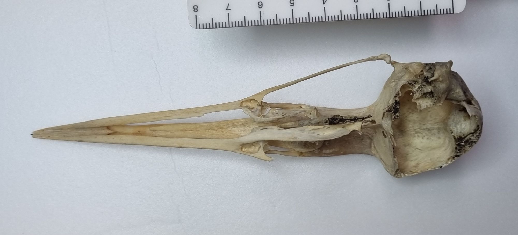
Last week I gave you this skull from the collections of the Dead Zoo to have a go at identifying:
Bird skull identification can take a bit of work, until you get your eye in on things like the bill morphology (especially without the clues provided by the keratin sheath). Resources like the excellent Skullsite.com certainly help a lot, by providing a huge range of images of different species for comparison, and with tools to help narrow down options based on skull size and bill morphology.
As it turns out, Adam Yates certainly had his eye in, and he was first to comment with a correct identification for this specimen. It’s a Common or European Crane Grus grus (Linnaeus, 1758).
I chose this specimen as it’s one that we recently put on display in the Dead Zoo Lab as part of a community curated project called Our Irish Natural History. Eight community groups involved with iCAN (the Irish Community Archive Network) contributed to the work, which was coordinated by Adriana Ballinger – a fantastic postgraduate humanities researcher who has been working with us for the past year on a project with a focus on the wider cultural context surrounding natural history collections. The community groups involved each explored a different areas of interest, illustrating and exploring some of the connections between objects and local communities.
These sorts of projects, that connect our ostensibly scientific objects back into local communities through a cultural link are a fantastic way to broaden the relevance and interest in our collections. As a scientist it can be easy to focus on one aspect of an object – but every item we look after can be viewed from multiple perspectives – all of which add value and relevance.
I look forward to working on similar projects in the future, while hopefully taking the opportunity to share more of the collection here, for those of you with an interest in identification!
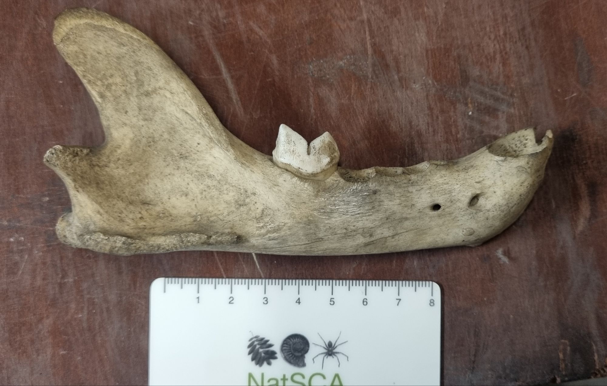
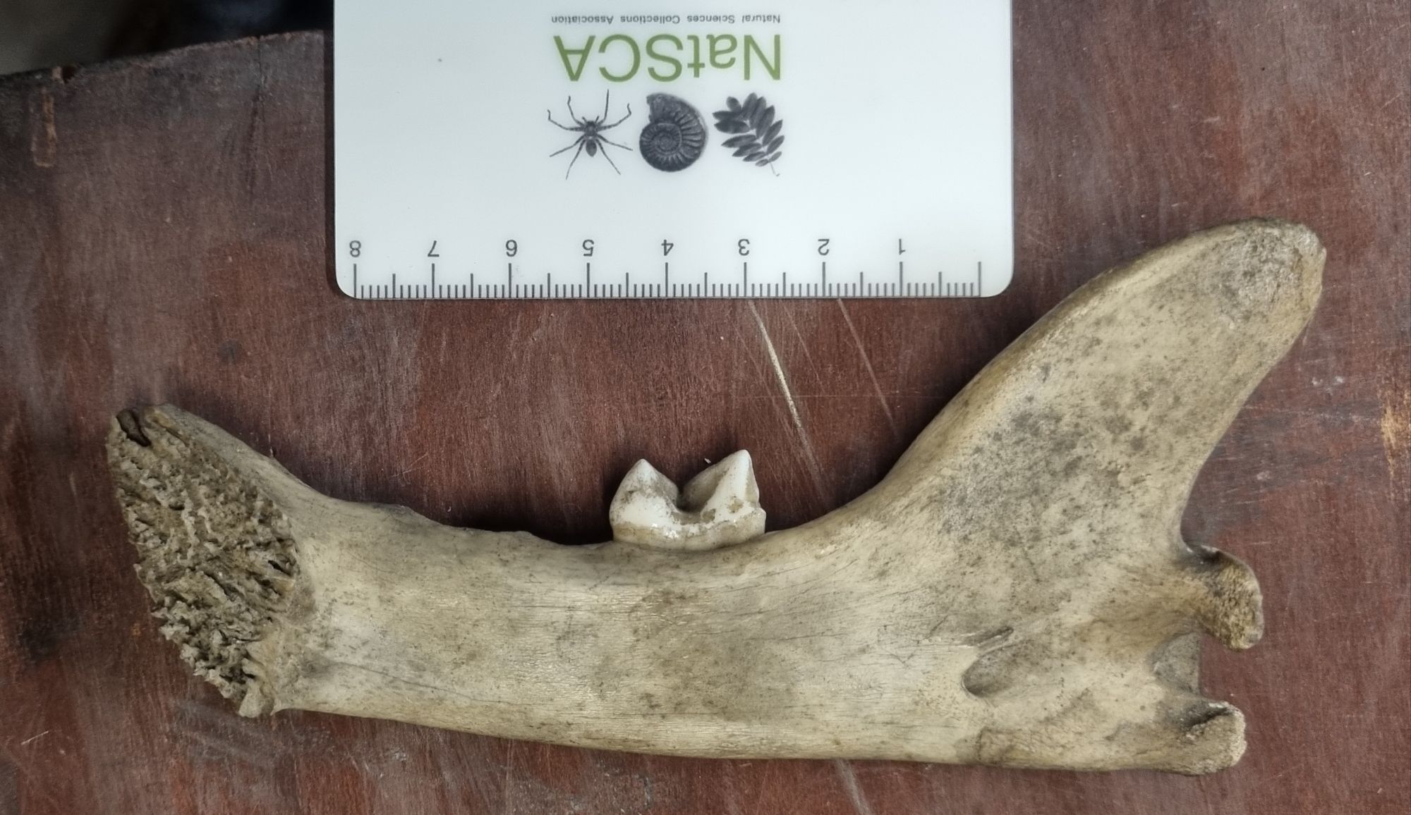
Last week I gave you this mystery mandible to help me identify:
It was found in a box of mixed shells and bony fragments, with no label, and nothing to offer a clue to its origin. This is very unusual, since these sorts of boxes of miscellany were mainly addressed years ago, but this one was well tucked away and somehow escaped being dealt with.
It’s fairly easy to recognise this as being the right mandible of some kind of cat. The Felidae have a reduced tooth count, with those hyper-carnivorous bladed premolars forming a meat-shearing carnassial row that’s very distinctive. So that’s the easy bit done.
The hard bit comes next, since the cats all share this same dental configuration, making the specific kitty in question a bit harder to narrow down. However, the size is a useful clue. This is much too big to be something like a Domestic Cat, Serval or Ocelot, but it’s also too small to be one of the big Big Cats – like the Lion or Tiger:
Tiger skulls facing off – the scale of these skulls is much greater than the mystery mandible
This leaves some of the medium-large Big Cats and perhaps a very large Small Cat (i.e. the Cheetah). Let’s start by ruling out that last one – the size is still a bit on the big side, but also the Cheetah has a more gracile coronoid process that curves, and a relatively shorter toothrow. Pumas also have a more rounded coronoid process.
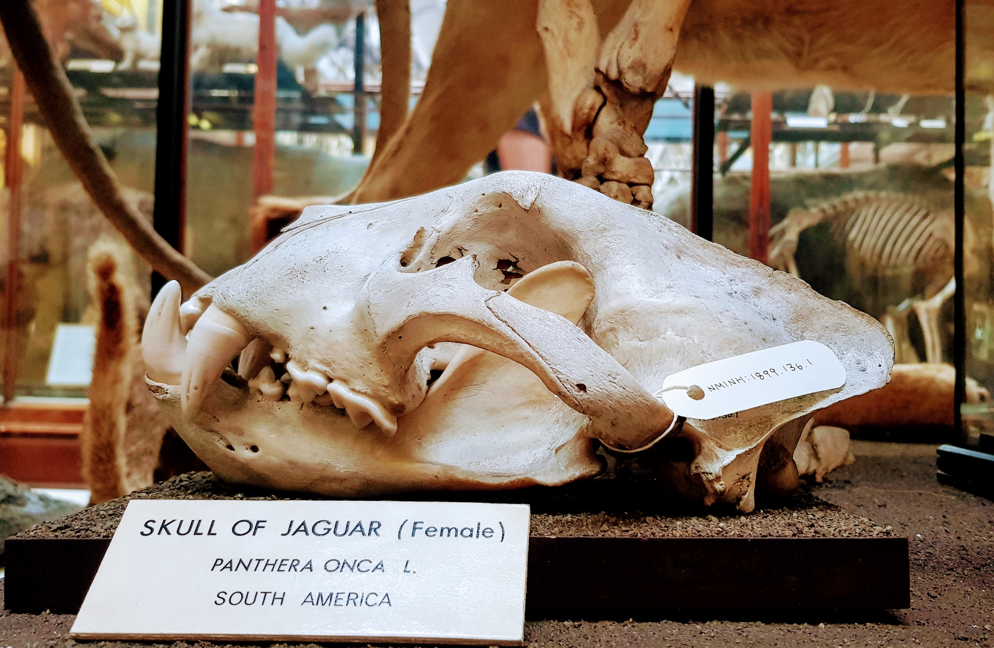
The Jaguar also has a more rounded coronoid process, and has a more robust mandible, that’s just built thicker to deal with the high bite forces these cats generate:
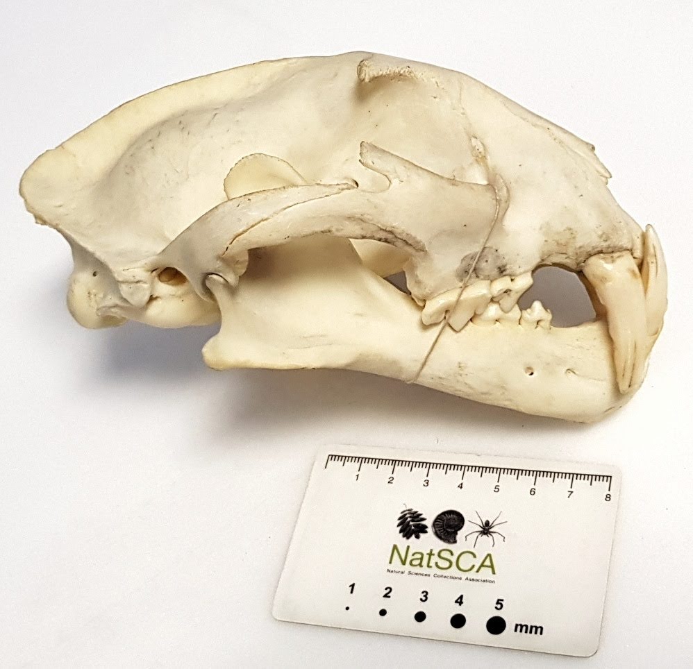
That leaves us with the Snow Leopard and the not-snow-Leopard, both of which have the more pointed coroid process that we see in the mystery object. However, the Snow Leopard has downwards inflected angular process, that’sI suspect may relate to the increased gape needed to use the excessively long canine teeth in this species:
Snow Leopard skull, showing the hypertrophied canines normal in this species.
So, by a process of elimination, we’re left with Leopard Pathera pardus (Linneaus, 1758). This species varies significantly in size and the morphology can be quite variable, since the species has a wide distribution, from Eastern Russia to as far West as Senegal.
This actually reminds me of a very similar mystery object from the Grant Museum of Zoology that I shared 10 years ago, so I’ hope that provided something useful for reference so I’m glad I trawled through some of my past posts to help solve this one. My thanks to everyone who offered their thoughts on this one – it’s great to see so many of you come to the same conclusion!
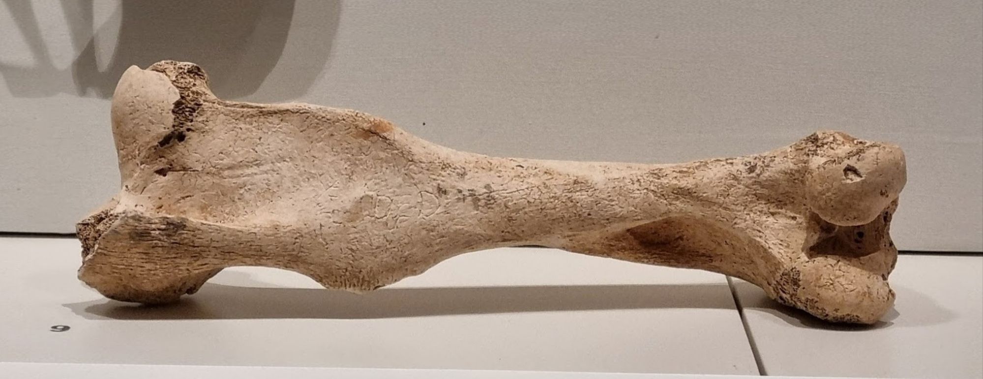
Last week I presented this mystery bone from the Words on the Wave exhibition at the National Museum of Ireland on Kildare Street:
The bone is over a thousand years old, so it’s unsurprising that a few of the processes that could be useful for identification have been worn down a little:
That said, there was plenty of information remaining to allow several of you to identify the animal, bone type and side of the animal this is from. Adam Yates was first to the comments and he also left a very helpful Toot on Mastodon, detailing the diagnostic features:
For me, the supracondylar fossa on the lateral caudal surface is the instant give-away. For those who don’t speak anatomese, that’s the bit that flares out from the side of the shaft of the bone (which is actually missing a bit in this example).
This is a feature I always associate with the Perissodactyla (the tapirs, rhinos and horses), and this one is nowhere near robust enough for anything other than one of the horses. It’s also fairly small (although the lack of a scale bar doesn’t make this obvious).
This a left femur that I suspect is from a Horse Equus ferus caballus Linnaeus, 1758, but it could also be from a Donkey Equus africanus asinus Linnaeus, 1758. It can be tricky to tell these species apart, especially when the bone is a little worn down and you only have a few photos to work from – so best to err on the side of caution and leave it at an identification of Equus sp.
If you’re in Ireland and wanted to take a look at this object – and a selection of remarkable mediaeval manuscripts from the Abbey of St Gall, Switzerland, some of which are returning to Ireland for the first time in 1000 years – you have until the 24th of this month, I’d definitely recommend it!